


India’s elderly population, constituting about 7.7% of the total demographic, faces significant oral health challenges, exacerbated by limited access to dental care, cognitive impairments, and financial constraints. Among common issues is denture stomatitis, often resulting from poor denture hygiene. Green tea (Camellia sinensis), known for its antioxidant and antimicrobial properties, offers a promising, cost-effective natural alternative for maintaining oral hygiene. Historically used for medicinal purposes across Asia, green tea contains polyphenols particularly EGCG that exhibit antibacterial, antifungal, antiviral, anti-inflammatory, and anticancer properties. These bioactive compounds effectively combat pathogens like Candida albicans, Streptococcus mutans, and Porphyromonas gingivalis, while also neutralizing volatile sulfur compounds associated with halitosis. Moreover, green tea catechins aid in managing periodontitis and show potential in suppressing oral squamous cell carcinoma. However, current evidence remains largely restricted to in vitro and formulation-level investigations; extensive clinical validation and methodological standardization are essential to enable translational implementation in geriatric oral healthcare. This review highlights green tea’s therapeutic potential as a denture cleanser and oral health adjunct, especially for elderly individuals in resource-limited settings, promoting accessible, natural solutions for comprehensive oral care.
Green Tea, Oral Health, Elderly Population, Denture Stomatitis, Antimicrobial Properties
India’s elderly population, comprising approximately 7.7% of the total demographic (about 76 million individuals),1 faces a considerable oral health burden associated with aging and prosthetic dependence. Functional limitations, comorbidities, and restricted access to dental services collectively increase the risk of infection, inflammation, and poor denture maintenance in this group.2-4 One of the primary concerns is the lack of access to professional dental care, particularly in rural regions where the dentist-to-population ratio is alarmingly low.5 Elderly individuals often depend on caregivers or family members for assistance, and financial dependency adds another layer of complexity, as many cannot afford regular oral healthcare or specialized prosthodontic services.4 Consequently, oral health declines quickly, which not only hampers chewing ability but also affects nutrition, speech, self-confidence, and overall quality of life.
According to the National Oral Health Survey, a considerable proportion of individuals aged 65-74 depend on prosthetic appliances to restore oral function. Specifically, 10.2% and 11.1% of this age group use prostheses in the upper and lower arches, respectively, with complete dentures being the most common.6 However, despite the high prevalence of prosthetic use, many elderly individuals are either unaware of or unable to maintain optimal denture hygiene, thereby increasing the risk of prosthesis-related complications such as denture stomatitis. Denture stomatitis is a prevalent condition among complete denture wearers and is characterized by inflammation of the oral mucosa beneath the denture base. Reports suggest its prevalence ranges from 11% to 67% among denture users.7 This condition is primarily linked to poor denture hygiene and microbial plaque accumulation. Over time, colonization by organisms such as Candida albicans can exacerbate the inflammatory response, leading to persistent mucosal irritation, burning sensations, and difficulty in eating or speaking. To mitigate such complications, rigorous cleaning and maintenance of dentures are essential. Regular denture hygiene not only helps in maintaining mucosal health but also improves the longevity and comfort of prosthetic devices. Various methods exist for denture cleaning, including mechanical brushing, chemical cleansers, ultrasonic devices, and natural products.8 Chemical agents like sodium hypochlorite and chlorhexidine are widely used, but they may cause adverse effects such as discoloration, surface roughness, or allergic reactions over long-term use. Moreover, the limited affordability and accessibility of commercial denture cleansers, especially in rural and economically constrained populations, necessitate exploration of natural alternatives that are both safe and cost-effective.
Given these limitations in accessibility and hygiene maintenance, there is a pressing need to explore cost-effective, accessible, and biocompatible alternatives for denture hygiene. Green tea (Camellia sinensis) has emerged as a scientifically characterized phytochemical system with well-defined pharmacognostic properties and multifactorial bioactivity. Its catechin derivatives, particularly epigallocatechin gallate (EGCG), demonstrate antimicrobial, antioxidant, and anti-inflammatory mechanisms that directly modulate microbial adhesion, oxidative stress, and mucosal inflammation-pathways critically implicated in denture-associated infections. The mechanistic substantiation of these effects provides a rational framework for investigating green tea as a sustainable, evidence-based adjunct for geriatric oral hygiene. Green tea is produced through minimal oxidation processes, preserving its rich antioxidant content, especially polyphenols such as catechins.9 The minimal processing also retains its antimicrobial and anti-inflammatory potential, which makes it a suitable option for oral health applications. Green tea contains various bioactive compounds, including epigallocatechin gallate (EGCG), which has demonstrated antibacterial efficacy against oral pathogens and biofilm-forming microbes.10 Studies have indicated that green tea extracts inhibit the growth of Candida albicans, one of the main etiological agents in denture stomatitis.11 Furthermore, green tea is well-tolerated, has a pleasant taste, and lacks the adverse chemical effects of synthetic cleansing agents. These attributes make it an attractive natural adjunct for denture hygiene, especially for elderly populations with limited access to conventional products. In addition to its antimicrobial properties, green tea exhibits anti-inflammatory and wound-healing capabilities, which can further benefit denture wearers by reducing mucosal irritation and supporting tissue regeneration.12 Its potential to serve as a preventive as well as therapeutic agent in oral health settings aligns with the broader goals of geriatric dentistry, which seeks to provide safe, effective, and patient-friendly interventions for aging populations.
In summary, India’s aging population is encountering severe challenges in maintaining oral health, primarily due to a complex interplay of systemic, behavioral, and infrastructural barriers. Denture-related conditions like stomatitis are widespread, but largely preventable through proper hygiene practices. Natural, accessible agents such as green tea present a promising, sustainable solution to bridge the gap in denture hygiene management among the elderly. Thus, incorporating green tea into oral care routines may serve as a culturally acceptable, low-cost, and effective strategy to improve the oral health outcomes and quality of life in this vulnerable demographic. Consequently, this review transitions from highlighting the epidemiological need to exploring green’s tea scientific rationale – it’s historical background, phytochemical composition, and therapeutic mechanisms relevant to denture hygiene and oral health.
History
The earliest pharmacological references to green tea (Camellia sinensis) appear in ancient Chinese literature (ca. 2737 BCE), identifying it as a therapeutic preparation used to promote digestion and physiological balance. Early medical documentation in traditional Chinese medicine further described it as a restorative agent capable of detoxification and alleviating fatigue-related ailments. Its application gradually expanded to neighboring regions, notably Japan and Korea, where it became incorporated into both therapeutic and cultural practices.
As centuries progressed, during the Tang (618-907 CE) and Ming (1368-1644 CE) dynasties, significant advances occurred in the processing and preparation of tea. The shift from compressed tea cakes to loose-leaf infusions enhanced catechin retention, thereby augmenting its pharmacological efficacy. This refinement represented one of the earliest documented instances of pharmacognostic advancement influencing bioactive stability in plant-derived preparations (Tables 1 and 2).
Table (1):
Inhibitory Effects of Green Tea Catechins on Microbial Enzymes in Denture Biofilm Control
Enzyme Type |
Function in Microbial Physiology |
Effect of Green Tea Catechins (EGCG) |
Oral Health Impact |
|---|---|---|---|
Proteases |
Break down proteins into peptides and amino acids; essential for microbial nutrition and tissue invasion. |
EGCG inhibits protease activity, limiting nutrient availability and protecting salivary proteins, mucosa, and denture material from degradation. |
Maintains denture integrity and reduces mucosal inflammation in edentulous patients. |
Lipases |
Hydrolyze lipids to fatty acids and glycerol; supports microbial energy and biofilm lipid matrix formation. |
EGCG suppresses lipase activity, restricting biofilm lipid synthesis and microbial metabolism. |
Reduces malodor, discoloration, and microbial adhesion on denture surfaces. |
Glycosidases |
Degrade polysaccharides (e.g., starch, glycogen) into fermentable sugars; fuels acidogenesis and biofilm formation. |
Catechins block glycosidase function, limiting carbohydrate metabolism and biofilm acid production. |
Minimizes acidogenic biofilm buildup, preventing inflammation and secondary infections. |
Table (2):
Historical timeline of green tea development and its medicinal significance
Historical Period |
Geographical Context |
Key Developments |
Medicinal and Cultural Significance |
|---|---|---|---|
Ancient China (~2737 BCE) |
China (Emperor Shen Nong era) |
Discovery of green tea; consumed as an herbal remedy. |
Used for digestion, detoxification, and enhancing vitality; revered as a healing tonic. |
Tang Dynasty (618-907 CE) |
China |
Rise of tea culture; popularity among aristocrats and monks. |
Used as a digestive aid and diuretic; integrated into spiritual and health rituals. |
Heian Period (9th Century) |
Japan (via Buddhist monks from China) |
Introduction of green tea seeds and brewing techniques. |
Adopted in Zen monasteries for meditation; symbol of purity and spiritual focus. |
Ming Dynasty (1368-1644 CE) |
China |
Innovation in tea processing from compressed cakes to loose leaves. |
Enhanced flavor and medicinal potency; used for fatigue, digestion, and cognition. |
Qing Dynasty (1644-1912 CE) |
China and Europe |
Global spread of green tea as a common beverage. |
Prescribed for fever, lethargy, digestive issues; recognized in herbal pharmacopoeias. |
Modern Era (20th Century – Present) |
Global |
Scientific validation of green tea’s bioactive compounds (e.g., EGCG). |
Proven antioxidant, anti-inflammatory, antimicrobial, and anticancer properties; widely used in wellness and functional medicine. |
Subsequently, by the late medieval and early modern periods, green tea’s medicinal attributes gained recognition beyond Asia. European pharmacopoeias described its use as an antipyretic, digestive stimulant, and restorative beverage, reflecting the early diffusion of ethnopharmacological knowledge. Subsequent centuries witnessed its integration into traditional medicinal systems worldwide, where it was prescribed for managing inflammation, infection, and metabolic disorders.
In the modern era, biomedical research has substantiated many of these empirical claims. Advances in phytochemistry and molecular pharmacology have identified catechins-particularly epigallocatechin gallate (EGCG) as the principal bioactive constituents responsible for antioxidant, antimicrobial, and anti-inflammatory functions. These discoveries establish a direct scientific linkage between ancient therapeutic knowledge and contemporary biomedical relevance. Overall, the progression of green tea from traditional ethnomedicine to evidence-based pharmacology exemplifies its enduring therapeutic significance and translational potential in oral healthcare.13
Chemical composition of green tea and its activities
Bioactive Phyto-constituents of Green tea
The chemical composition of green tea is complex and diverse, consisting of a wide range of bioactive compounds that contribute to its unique health benefits and sensory properties.14,15 Despite extensive research, the full spectrum of these compounds remains only partially understood, highlighting the need for continued scientific exploration into green tea’s intricate phytochemical makeup. Green tea leaves contain an array of micronutrients, macronutrients, and organic substances that together define its chemical profile.
Among the macronutrients, proteins constitute approximately 15%-20% of the dry weight of green tea leaves. These proteins include various enzymes and structural proteins essential for the plant’s growth and metabolism. Amino acids, another key macronutrient, represent about 1%-4% of the dry weight. These amino acids, including theanine, contribute significantly to the flavor and calming effects attributed to green tea. Carbohydrates account for 5%-7% of the dry weight, primarily serving as energy reserves for the plant. The lipid fraction is relatively minor but noteworthy, containing essential fatty acids such as linoleic and α-linolenic acids, which play vital roles in cellular functions and possess health-promoting properties. Additionally, sterols such as stigmasterol are present and contribute to membrane structure and potential cholesterol-lowering effects. In terms of micronutrients, green tea is rich in vitamins and minerals essential for human health. Vitamins B, C, and E are prominent components, each offering antioxidant and metabolic benefits. Vitamin C, for example, helps neutralize free radicals, while vitamin E supports cell membrane integrity. The tea also contains xanthine bases, primarily caffeine and theophylline, which contribute to its stimulant effects. The presence of pigments like chlorophyll and carotenoids gives green tea its characteristic color and antioxidant properties. Furthermore, volatile compounds such as aldehydes, alcohols, esters, lactones, and hydrocarbons contribute to green tea’s distinctive aroma and flavor profile (Figure 1).
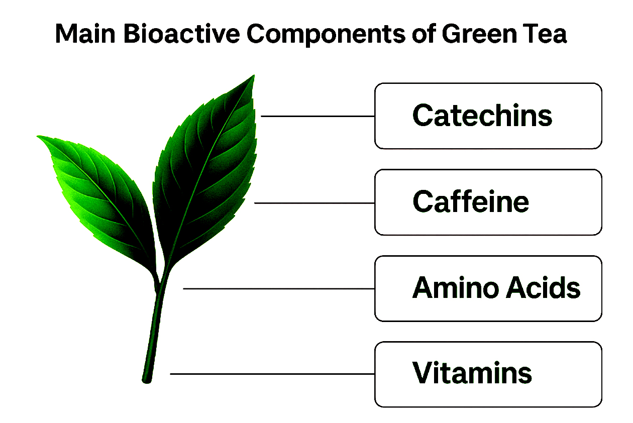
Figure 1. Main Bioactive components of Green Tea
From a biochemical perspective, the composition of green tea reveals a precise alignment between molecular structure and oral therapeutic function. The polyphenolic catechins, particularly epigallocatechin gallate (EGCG), exhibit strong electron-donating capacity that underlies their antimicrobial, antioxidant, and anti-biofilm activity against oral pathogens. Flavonols reinforce these effects by modulating oxidative stress and inflammatory signaling within the mucosa, promoting tissue homeostasis. Alkaloids such as caffeine and theanine enhance salivary stimulation, thereby improving the oral clearance of debris and microbial substrates. Meanwhile, amino acids and trace minerals contribute to enzymatic balance and epithelial repair. Collectively, this biochemical synergy defines the multifactorial efficacy of green tea in maintaining oral hygiene and prosthetic surface health.
Antimicrobial spectrum of catechins
Approximately 5% of the dry weight of the body comprises minerals and trace elements, including calcium, magnesium, iron, zinc, copper, chromium, manganese, molybdenum, selenium, phosphorus, sodium, potassium, cobalt, strontium, nickel, fluorine, and aluminum. These elements are critical for various physiological functions and contribute to the nutritional value of green tea. Among the many compounds found in green tea, polyphenols stand out as the most significant due to their abundance and potent biological activity. Polyphenols, particularly flavonoids, are key contributors to the health benefits connected with green tea consumption. The predominant flavonoids in green tea are catechins, a subgroup of flavan-3-ols. The four primary catechins are epigallocatechin-3-gallate (EGCG), epigallocatechin (EGC), epicatechin-3-gallate (ECG), and epicatechin (EC). EGCG is the most abundant, making up about 59% of total catechins, followed by EGC at 19%, ECG at 13.6%, and EC at 6.4%. These catechins exhibit strong antioxidant, anti-inflammatory, and anti-carcinogenic properties, which underpin many of green tea’s proposed health effects, including cardiovascular protection, cancer prevention, and metabolic regulation. Therefore, green tea’s chemical composition is marked by a rich diversity of macronutrients, micronutrients, and bioactive compounds, especially polyphenolic catechins. Although significant progress has been made in characterizing these constituents, ongoing research is necessary to fully elucidate the complex interactions and health implications of green tea’s phytochemical matrix (Figure 1).
Antibacterial, antifungal and antiviral effect of green tea
Green tea catechins exhibit multiple biological activities (Figure 2), including antibacterial, antifungal, and antiviral effects that contribute to their oral health benefits. The green tea catechins, which are water-soluble, exert a profound influence on cell membrane functions, modulating signaling pathways, cell cycle regulation, and mitochondrial activity. Notably, research has demonstrated the bactericidal effects of catechins on various microorganisms, including E. coli, S. salivarius, and S. mutans.16 The production of hydrogen peroxide by epigallocatechin gallate (EGCG) has been identified as a key factor in its bactericidal activity, as confirmed through chemiluminescent and ESR measurements.17 Furthermore, the hydrogen peroxide generated by EGCG has been shown to damage the bacterial cytoplasmic membrane, thereby exerting its antimicrobial effects. In addition to hydrogen peroxide-mediated oxidative stress, EGCG also exerts direct physicochemical effects on microbial membranes and enzyme systems. Its amphipathic polyphenolic structure allows insertion into lipid bilayers, increasing permeability and causing cytoplasmic leakage in pathogens such as Streptococcus mutans and Candida albicans. Moreover, EGCG chelates metal ions (Fe2+, Zn2+) essential for microbial enzymatic activity, thereby inhibiting key metabolic enzymes like glucosyltransferases and proteases involved in biofilm matrix synthesis. The compound additionally interferes with cell-surface proteins and quorum-sensing signals, disrupting adhesion and coordinated virulence expression. Collectively, these molecular interactions underpin EGCG’s broad-spectrum antimicrobial and anti-biofilm potency within oral microbial ecosystems.
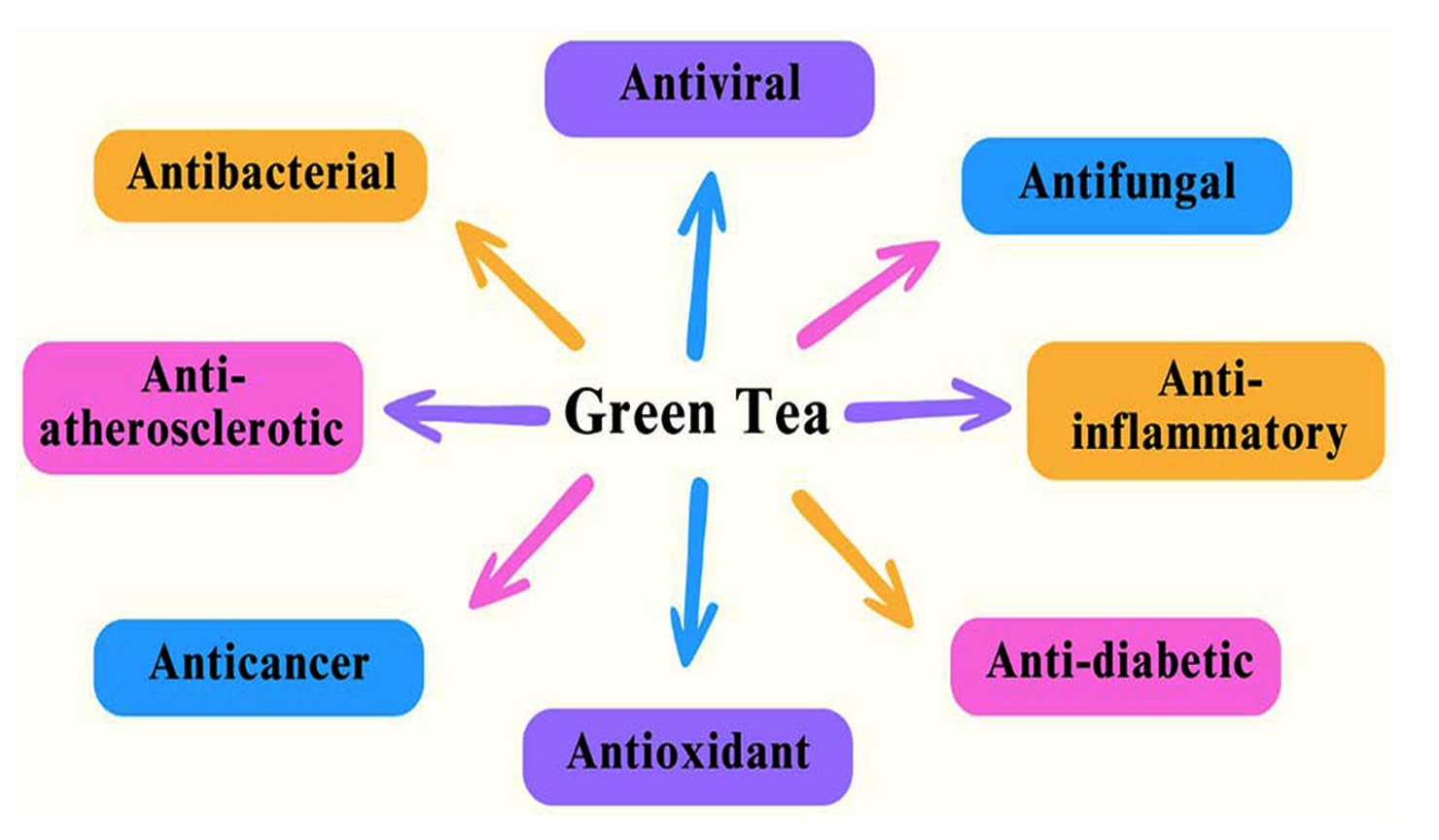
Figure 2. Key therapeutic properties of green tea catechins
Beyond its antibacterial actions, green tea also exhibits antiviral and antifungal activities. The polyphenols present in green tea have been shown to inhibit enzymes that damage cell membranes and prevent viruses from entering host cells.18 This antiviral mechanism is particularly relevant in preventing oral viral diseases. Specifically, EGCG has been found to block influenza virus infections by binding to viral hemagglutinin, thereby preventing the virus from attaching to cellular receptors.19 Moreover, green tea has been shown to be effective against HIV-1, herpes simplex virus, and adenoviruses.20 Regarding its antifungal properties, research has demonstrated that green tea exhibits synergistic antifungal activity when combined with antimycotics against Candida albicans.21 Specifically, the combined usage of EGCG and low-dosage amphotericin B has been shown to inhibit the growth of C. albicans, with fungicidal action. These findings exemplify the therapeutic potential of green tea as a natural antifungal agent, supporting its use in the prevention and management of oral infections.
Effect of green tea in halitosis
Halitosis, commonly known as bad breath, is a widespread oral health issue primarily caused by dental caries and poor oral hygiene. The underlying cause of halitosis is often the accumulation of volatile sulfur compounds (VSCs), such as methanethiol and hydrogen sulfide, produced by anaerobic bacteria in the oral cavity. These compounds are responsible for the characteristic foul odor associated with the condition, making halitosis a socially and personally distressing problem. Recent evidence has emphasized the efficacy of natural bioactive substances in controlling halitosis, with particular attention given to the bioactive compounds found in green tea. Green tea, derived from the leaves of Camellia sinensis, contains catechins-potent polyphenolic antioxidants known for their antimicrobial and deodorizing effects. These catechins have been shown to neutralize specific VSCs, including methanethiol and other allium-derived sulfur compounds, effectively reducing oral malodor.22 Supporting this, Rassameemasmaung’s research demonstrated the practical benefits of green tea in oral care through a study involving patients suffering from gingivitis. The study revealed that regular use of green tea as a mouthwash significantly decreased the concentration of VSCs in the oral cavity, thereby mitigating halitosis.23 This suggests that green tea not only has a deodorizing effect but may also contribute to controlling oral bacterial populations that produce these sulfur compounds. Further reinforcing the therapeutic potential of green tea, Sharma conducted a comparative study between a 5% Camellia sinensis mouth rinse and a commercially available 0.2% chlorhexidine mouth rinse, which is a gold standard antimicrobial agent in dentistry. The findings indicated that the green tea mouth rinse was equally effective in reducing plaque accumulation, tongue coating, and the severity of oral halitosis.24 This equivalency underscores the potential of green tea as a natural alternative to conventional chemical mouthwashes, which are often associated with side effects like tooth staining and altered taste sensation. Collectively, these studies establish green tea catechins as a natural, safe, and effective adjunct in managing halitosis and enhancing overall oral health. The ability of green tea to target VSCs directly, coupled with its antimicrobial properties, makes it a valuable candidate for integration into daily oral hygiene practices. Moreover, its accessibility and minimal side effects provide an attractive option for individuals seeking natural remedies for bad breath. (Figure 3). Hence, green tea, with its catechin content, offers a scientifically supported and sustainable approach to improving oral freshness and hygiene, with future research likely to reinforce its clinical value in halitosis management.
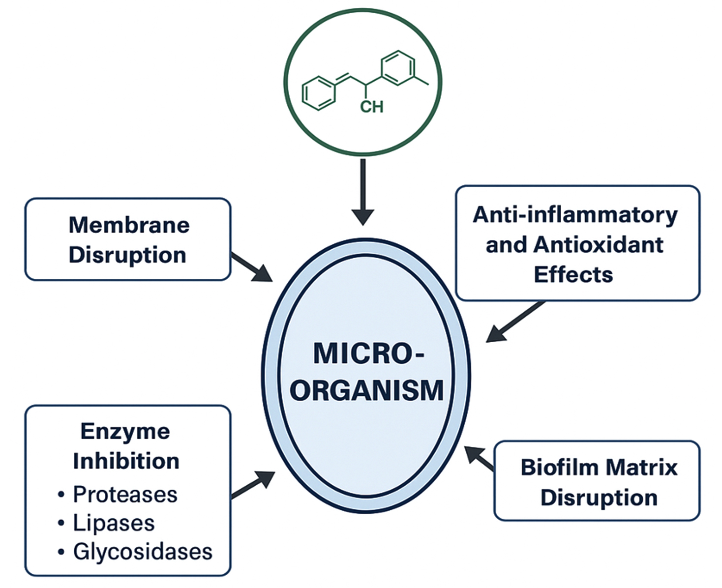
Figure 3. Schematic diagram of green tea catechin mechanisms of action
Green tea and periodontitis
Periodontitis is a long-term inflammatory condition marked by the proliferation of pathogenic microorganisms in the deepened gingival crevice, the space separating the teeth from the surrounding gum tissue. This microbial overgrowth leads to the destruction of supporting periodontal tissues, ultimately resulting in tooth loss if left untreated. Among the key bacterial culprits are periodontopathogenic species such as Porphyromonas gingivalis and Prevotella, which are known to trigger and sustain the inflammatory response that drives disease progression. Recent research has highlighted the promising role of green tea catechins in managing periodontitis due to their potent antimicrobial properties. Catechins, which are bioactive polyphenols found abundantly in Camellia sinensis, have demonstrated effective inhibition of key periodontopathogenic bacteria, including Porphyromonas gingivalis and various Prevotella species.25 By suppressing these bacterial populations, green tea catechins can help reduce the microbial load within periodontal pockets, a critical step toward halting the disease’s progression. In addition to their antimicrobial action, localized application of green tea catechins has been shown to improve clinical periodontal health outcomes. Studies have reported that targeted delivery of catechins to affected periodontal sites leads to a significant reduction in bacterial counts alongside improvements in gum inflammation and tissue healing.26 This suggests that green tea catechins may serve as a valuable adjunctive treatment alongside conventional periodontal therapies, enhancing their efficacy.
Beyond direct antimicrobial effects, green tea also exerts beneficial influences on the host’s immune and inflammatory responses. Catechins have been found to modulate inflammatory pathways, reducing excessive cytokine production and oxidative stress that contribute to tissue damage in periodontitis. Furthermore, green tea supports the health and integrity of gingival cells, promoting the maintenance of a resilient and functional periodontal barrier. This dual action-combating harmful bacteria while strengthening host defenses-positions green tea as a multifunctional agent in periodontal care. Overall, the integration of green tea catechins in periodontitis management offers a natural and effective strategy to both reduce pathogenic bacterial loads and enhance periodontal tissue health. Their antimicrobial and anti-inflammatory properties, combined with the ability to support gingival cell function, highlight green tea’s therapeutic potential in preventing and mitigating the chronic damage caused by periodontitis. Future clinical applications may see green tea-based formulations becoming a regular adjunct to standard periodontal treatments, improving patient outcomes through a safer and more holistic approach.
Green tea and oral malignancy
Oral squamous cell carcinoma (OSCC) represents a common and highly invasive malignancy of the head and neck region, contributing substantially to global morbidity and mortality. The disease is characterized by uncontrolled growth and invasion of malignant cells in the oral cavity, often leading to severe complications and poor prognosis. Conventional treatments, including surgery, radiation, and chemotherapy, can be effective but are frequently accompanied by substantial side effects and limited long-term success (Figure 4). Recent research has uncovered promising antitumor properties of green tea polyphenols in the context of OSCC. These bioactive compounds, especially epigallocatechin gallate (EGCG), have demonstrated the capability to selectively target cancer cells while sparing normal healthy cells. Green tea polyphenols exert their effects by inducing apoptosis, a programmed cell death mechanism that helps eliminate malignant cells. Additionally, they promote cell cycle arrest, thereby halting the uncontrolled proliferation of OSCC cells.27 More specifically, EGCG has been shown to inhibit the invasive behavior of OSCC cells, preventing their spread to surrounding tissues. This inhibition of cell invasion is a crucial therapeutic target, as tumor metastasis significantly worsens patient outcomes.
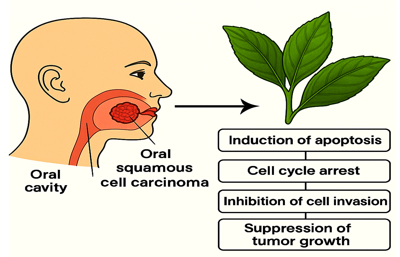
Figure 4. Green tea catechins target oral cancer through multiple cellular mechanisms
Experimental studies using SCC-induced mouse models have further validated these findings, demonstrating that EGCG treatment results in dose-dependent suppression of tumor growth. These preclinical models highlight the potential of EGCG as a powerful natural compound capable of reducing tumor burden in oral cancer.27 The multifaceted mechanisms of green tea polyphenols in combating OSCC-ranging from apoptosis induction and cell cycle regulation to invasion suppression-suggest that they may serve as effective adjuncts to current cancer therapies. Their natural origin and relatively low toxicity profile offer an advantage over some conventional treatments that often cause adverse side effects. Incorporating green tea-derived compounds into therapeutic regimens could enhance treatment efficacy while potentially improving patient quality of life.
Hence, OSCC remains a challenging malignancy with significant health impacts. The antitumor activities of green tea polyphenols, particularly EGCG, present a promising avenue for complementary cancer management. Continued research and clinical evaluation may establish green tea compounds as valuable tools in the fight against oral cancer, offering hope for more effective and less harmful treatment options.
Green tea as denture cleanser
Mechanism and Regulatory Perspective
Proper care and maintenance of dentures and the surrounding mucosal tissue in edentulous individuals are essential for preserving overall oral health and preventing complications such as mucosal inflammation and infections. Poor denture hygiene is a leading cause of oral mucosal irritation, highlighting the importance of consistent and effective cleaning and disinfection routines. Denture cleansers play a vital role by removing plaque, bacteria, and other microorganisms from denture surfaces, thereby supporting optimal oral hygiene. Various types of denture cleansers exist, including chemical cleansers containing agents like sodium hypochlorite, alkaline peroxides, and acidic peroxides, which chemically break down deposits. Enzymatic cleansers employ enzymes such as proteases, lipases, and amylases to target protein, lipid, and carbohydrate residues, while ultrasonic cleansers use high-frequency sound waves to mechanically dislodge biofilms. Natural denture cleansers, derived from essential oils, herbs, and plant extracts, have gained popularity due to their antimicrobial properties. Some cleansers combine these approaches to enhance cleaning effectiveness.28 As shown in Figure 5, green tea-based solutions are gaining interest as natural alternatives for denture disinfection.
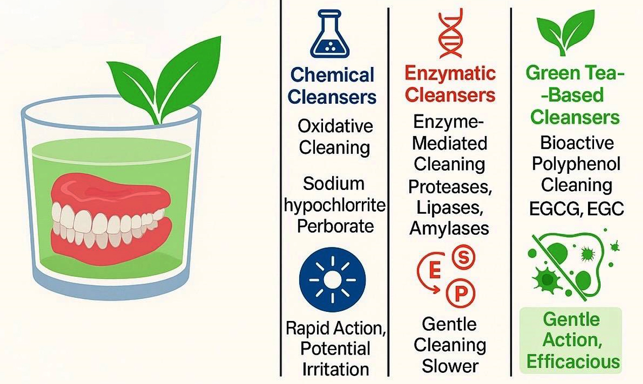
Figure 5. Mechanistic and Comparative overview of denture cleansers
Green tea-based denture cleansers, though compositionally biocompatible, must adhere to regulatory frameworks governing safety, biocompatibility, and product quality for oral-use medical devices (Class I, 21 CFR 872.3520). While current in vitro and formulation-level studies substantiate their antimicrobial efficacy and material compatibility, comprehensive toxicological validation and stability profiling remain prerequisites for clinical approval. Compliance with ISO 10993 biocompatibility standards and structured human trials is essential to confirm long-term mucosal safety and therapeutic reliability. Establishing such validated evidence will facilitate regulatory acceptance and support the clinical translation of catechin-based denture cleansers as safe and sustainable alternatives to conventional chemical formulations.
Among natural options, Camellia sinensis has gained considerable interest due to its potential health benefits, especially its antimicrobial and antifungal effects attributed to bioactive polyphenols. A study by Yoda demonstrated that green tea extracts inhibit bacterial strains such as Streptococcus pyogenes and Staphylococcus aureus, with inhibitory effects increasing proportionally to extract concentration.29 Green tea is rich in catechins, including epigallocatechin gallate (EGCG), epigallocatechin (EGC), epicatechin gallate (ECG), and epicatechin (EC), which exert antimicrobial activity through multiple mechanisms. These include disruption of microbial cell membranes by interacting with lipid bilayers, altering membrane fluidity and permeability, which causes leakage of essential nutrients and eventual microbial death. Catechins also inhibit microbial growth by interfering with metabolic enzymes, such as those involved in glucose metabolism, and induce apoptosis, or programmed cell death, in microorganisms, further reducing microbial load on dentures.30 Additionally, green tea catechins inhibit microbial adhesion, disrupt biofilm formation, interfere with quorum sensing (microbial communication), and provide antioxidant effects that protect oral tissues. Together, these properties make green tea an effective natural agent for maintaining denture hygiene and promoting oral health (Table 3).
Table (3):
Green tea: Balancing benefits and drawbacks
Parameter |
Advantages |
Disadvantages |
|---|---|---|
Microbiological efficacy |
Demonstrates strong antimicrobial properties effective against a wide range of microorganisms—including Gram-positive and Gram-negative bacteria, fungi, and viruses—thus helping to minimize the risk of infections associated with denture use |
Demonstrates limited efficacy against certain microbial strains or biofilm-embedded microorganisms, potentially compromising its effectiveness in complex oral environments |
Cytocompatibility |
Displays a favorable cytocompatibility profile, with minimal cytotoxic effects on mammalian cells, suggesting its potential for safe use in oral care applications |
High concentrations or prolonged exposure may induce cytotoxicity, underscoring the need for careful formulation and dosing |
Biofilm modulation |
Exhibits biofilm-disrupting properties, potentially mitigating the formation of complex microbial communities on denture surfaces |
May not completely eradicate mature biofilms, highlighting the need for further research into its biofilm-modulating effects |
Physicochemical stability |
Can be formulated into stable and shelf-stable products, ensuring consistent delivery of its antimicrobial properties |
Susceptible to degradation of active compounds over time, potentially impacting its efficacy and shelf-life |
Economic feasibility |
Offers a cost-effective and readily available alternative to conventional denture cleansers, with potential for widespread adoption |
Variability in quality and composition of green tea extracts may affect its efficacy and consistency |
Patient acceptability |
May be more appealing to patients seeking natural and non-invasive treatments, potentially enhancing patient compliance |
Patient compliance may be influenced by factors such as taste, staining, or allergic reactions, highlighting the need for careful patient selection and monitoring |
Clinical evidence base |
Emerging evidence supports its potential as a denture cleanser, warranting further investigation into its clinical applications |
Limited clinical trials and long-term studies are available, underscoring the need for rigorous clinical evaluation to fully elucidate its efficacy and safety profile |
Hence, maintaining proper denture hygiene is critical to prevent oral complications in edentulous patients. The use of denture cleansers, especially those based on natural ingredients like green tea, offers a promising and safe approach due to their demonstrated antimicrobial efficacy and additional health benefits. Incorporating green tea-based products into denture care regimens could enhance cleaning outcomes and reduce the risk of oral infections. A comparative analysis of green tea as a denture cleanser with conventional denture cleansers highlights the potential benefits and drawbacks of each approach (Table 4).
Table (4):
Comparative analysis: Green tea vs. Conventional denture cleansers
Parameter |
Green tea as a denture cleanser |
Conventional denture cleansers |
|---|---|---|
Toxicity |
Generally considered non-toxic and safe for use in oral care, with minimal cytotoxic effects on mammalian cells (LD50 > 2000 mg/kg) |
May exhibit cytotoxicity, depending on the type and concentration of active ingredients (e.g., sodium perborate, LD50 = 1200 mg/kg) |
Antimicrobial spectrum |
Exhibits broad-spectrum antimicrobial activity against bacteria (Gram-positive and Gram-negative), fungi, and viruses |
Exhibits narrow-spectrum antimicrobial activity, targeting specific microorganisms (e.g., sodium hypochlorite effective against bacteria, but less effective against fungi and viruses) |
Environmental impact |
Biodegradable and environmentally friendly, with minimal ecological footprint |
May contain harsh chemicals that can harm aquatic life and contaminate waterways (e.g., sodium perborate can release hydrogen peroxide, a toxic compound) |
Advanced applications of green tea catechins in oral health
Recent research highlights the synergistic antimicrobial benefits of green tea catechins, particularly epigallocatechin gallate (EGCG), when combined with other plant-derived compounds. These combinations often result in broader-spectrum antimicrobial activity, reduced effective dosages, and minimized cytotoxic risks, making them promising candidates for denture hygiene applications. Natural agents such as curcumin (derived from Curcuma longa), neem (Azadirachta indica) extract, and clove (Syzygium aromaticum) oil have shown particularly strong synergistic effects with EGCG. For instance, EGCG and curcumin together significantly enhance the inhibition of matrix metalloproteinases (MMPs), which play a critical role in periodontal tissue degradation. Neem extract has been found to augment EGCG’s antifungal action against Candida albicans, while clove oil contributes to improved biofilm disruption and interference with bacterial quorum sensing. Additionally, combinations with propolis extract expand EGCG’s antimicrobial range and support the destabilization of biofilms (Table 5). These synergistic interactions underscore the potential of developing multi-herbal formulations for oral hygiene that are both effective and biocompatible for long-term use.
Table (5):
Synergistic Combinations of Green Tea Catechins with Other Phytochemicals
Combination |
Target Organism(s) |
Observed Effect |
Source |
|---|---|---|---|
EGCG + Curcumin |
Porphyromonas gingivalis |
Enhanced inhibition of MMPs, anti-inflammatory synergy |
In vitro and in silico models |
EGCG + Neem Extract |
Candida albicans |
Increased antifungal efficacy, reduced MIC values |
Laboratory synergy studies |
EGCG + Clove Oil |
Mixed oral flora |
Stronger biofilm degradation, quorum sensing inhibition |
Pilot microbiological study |
EGCG + Propolis Extract |
Streptococcus mutans, C. albicans |
Improved antimicrobial spectrum and biofilm destabilization |
Comparative formulation test |
Pharmacokinetic Challenges and Stability Considerations in Oral Formulations
Despite its therapeutic promise, the application of EGCG in oral products is limited by its poor chemical stability and low bioavailability in aqueous and physiological environments. Factors such as salivary pH fluctuations, enzymatic breakdown, and binding to oral mucosa affect its retention and efficacy (Table 6). Although EGCG demonstrates a strong affinity for oral epithelial cells, allowing for localized activity, it remains sensitive to degradation by salivary esterases, pH variation, and oxidative exposure. Addressing these challenges has led to the exploration of formulation strategies such as nanoencapsulation, liposomal carriers, and mucoadhesive delivery systems to protect EGCG and extend its functional lifespan in oral care products.
Table (6):
Factors Affecting EGCG Stability and Bioavailability in Oral Products
Factor |
Impact on EGCG |
Potential Solution |
|---|---|---|
Salivary Esterases |
Degradation of EGCG |
Enzyme inhibitors, protective coatings |
pH Variation (5.5-8.0) |
Epimerization and reduced efficacy |
Buffer systems to maintain optimal pH |
Light and Oxygen |
Oxidative breakdown |
Amber packaging, antioxidants (vitamin E) |
Aqueous Exposure |
Hydrolysis over time |
Dry-powder or tablet-based formulations |
Nanotechnology-enhanced delivery systems for denture hygiene
To overcome the inherent limitations of EGCG in conventional oral formulations, nanotechnology-based carriers offer promising solutions. Nanoparticles composed of materials like chitosan, β-lactoglobulin, or zein provide controlled release, enhanced mucoadhesion, and improved local bioavailability. Nanoemulsions utilizing lipid-based carriers and surfactants increase EGCG’s stability against gastric and environmental degradation, enabling prolonged antimicrobial action in rinses or soak solutions. Similarly, nanoliposomes have demonstrated significant retention of EGCG and improved diffusion into denture crevices. Double emulsions offer long-term formulation stability and are particularly well-suited for sustained-release denture tablets. Collectively, these systems enhance EGCG’s therapeutic potential by improving mucosal retention, reducing bitterness, and targeting biofilms in difficult-to-clean regions of dental prostheses (Table 7).
Table (7):
Nanotechnology strategies to enhance EGCG stability and bioavailability in denture cleansers
Nano-Delivery System |
Carrier Material |
Encapsulation Features |
Benefits for EGCG |
Potential Denture Hygiene Relevance |
|---|---|---|---|---|
Nanoparticles |
Chitosan, β-lactoglobulin, zein |
~50–300 nm; mucoadhesive; controlled release |
Improved intestinal/mucosal stability; increased bioavailability |
Sustained antimicrobial activity; adherence to denture surface |
Nanoemulsions |
Soy/sunflower oil, surfactant (Tween 80) |
~100–280 nm; O/W format; high entrapment efficiency |
28.6% ↑ in bioavailability; protection against gastric degradation |
Prolonged release in effervescent soak or rinse solutions |
Nanoliposomes |
Phosphatidylcholine, cholesterol |
~180–200 nm; bilayer structure |
Up to 47.7% EGCG retained vs. 3.4% in free form |
Enhanced diffusion into denture crevices; reduces EGCG degradation |
Double Emulsions |
Glycerol mono-stearate, polyglycerol esters |
~500 nm; W/O/W structure; long-term stability |
1.93× ↑ absorption; 6-month formulation stability |
Useful in encapsulated denture tablets; controls burst release |
Clinical implications and future directions
The integration of nano-delivered EGCG into denture hygiene products may revolutionize patient care by offering sustained antimicrobial effects against pathogens like Streptococcus mutans and Candida albicans. These systems not only maintain bioactivity over extended periods but also increase patient compliance by reducing unpleasant sensory attributes such as bitterness. Importantly, nanoformulations can be designed to remain active in the oral cavity even after rinsing, owing to mucoadhesive properties. Future product innovations could focus on combining EGCG with other natural agents in stable, low-dose formulations that simultaneously deliver antimicrobial and antioxidant benefits while addressing the mechanical and biological challenges of denture maintenance.
Role in xerostomia management
Beyond antimicrobial applications, EGCG has demonstrated therapeutic value in the management of xerostomia (dry mouth). Clinical evaluations, including a Phase II placebo-controlled trial, have reported significant increases in both unstimulated and stimulated salivary flow following the use of catechin-based lozenges in patients with dry mouth, including those with Sjogren’s syndrome. These findings suggest that green tea derivatives may offer a multifunctional approach in oral health care, supporting both antimicrobial defense and salivary function enhancement (Table 8).
Table (8):
Clinical Trial Outcomes for Green Tea-Based Oral Interventions
Study |
Population |
Intervention |
Primary Outcome |
|---|---|---|---|
Tiwari et al.,31 (India) |
90 hostel residents (adults) |
5% Camellia sinensis rinse vs. 0.2% CHX |
Comparable reductions in plaque, tongue coating, and halitosis |
Rao et al.,32 (India) |
100 adults with mild gingivitis |
Green tea & neem extract mouth rinse |
Significant decrease in bleeding on probing (BOP) and plaque index |
Kim et al.,33 (Korea) |
75 orthodontic patients |
Green tea gel (2%) applied twice daily |
Reduced gingival inflammation and Streptococcus mutans counts |
Akhtar et al.,34 (Pakistan) |
80 denture wearers |
EGCG-based denture cleanser (tablet formulation) |
Improved denture hygiene; reduced Candida albicans load |
Yamada et al.,35 (Japan) |
60 xerostomia patients (21–74 yrs) |
Green tea catechin lozenges (MighTeaFlow®) |
↑ Salivary flow: 3.8× (unstimulated), 2.1× (stimulated) from baseline |
Singh et al.,36 (India) |
120 dental patients |
Nano-EGCG mouthwash vs. placebo |
Enhanced plaque control; longer-lasting antimicrobial effect |
Kwon et al.,37 (South Korea) |
45 elderly care-home residents |
Warm vs. cold green tea rinse |
Warm rinse is most effective in reducing plaque, halitosis, and inflammation |
Nguyen et al.,38 (Vietnam) |
60 patients with dry mouth symptoms |
Green tea–honey lozenges twice daily |
Improved subjective comfort and salivary viscosity |
Prevention of biofilm formation
Green tea catechins have garnered increasing scientific interest due to their potent antimicrobial and anti-biofilm properties, which are particularly beneficial for individuals who wear dentures. Biofilm formation on denture surfaces is a common and persistent issue, contributing significantly to oral infections, inflammation, and discomfort in edentulous patients. Biofilms are complex groups of microorganisms that attach to surfaces and become embedded within a self-produced extracellular matrix, providing protection against antimicrobial agents and physical removal. Once established, biofilms are difficult to eradicate, necessitating proactive measures to prevent their development in the first place. Green tea, derived from Camellia sinensis, is rich in polyphenolic compounds known as catechins. The major catechins include epigallocatechin gallate (EGCG), epigallocatechin (EGC), epicatechin gallate (ECG), and epicatechin (EC), all of which exhibit significant antimicrobial activities. One of the most important actions of these catechins is their ability to prevent biofilm formation on denture surfaces. They achieve this through several interconnected mechanisms that disrupt the microbial lifecycle at various stages.
Firstly, green tea catechins inhibit microbial adhesion, which is the initial and most critical step in biofilm development. Without firm attachment to the denture surface, microbial cells are unable to establish a stable colony. Studies have shown that EGCG can alter the surface properties of both microbes and substrata, reducing hydrophobic interactions and thereby hindering microbial attachment. By interfering with this adhesion process, green tea catechins effectively prevent the initial colonization of microbes on denture surfaces, which is key to halting biofilm formation at its earliest stage. Secondly, these catechins are capable of disrupting the extracellular polymeric matrix that encases mature biofilms. This matrix, composed of polysaccharides, proteins, and nucleic acids, offers protection to the microbial community, shielding it from antimicrobial agents and host immune responses. Green tea catechins can destabilize this matrix by breaking down its structural components, leading to enhanced susceptibility of the microbial cells to cleaning agents and physical disruption. The weakening of the biofilm’s integrity facilitates its removal during routine denture cleaning, thereby promoting better oral hygiene.
Another crucial mechanism involves the interference of green tea catechins with microbial quorum sensing. Quorum sensing is a form of cell-to-cell communication that bacteria use to regulate collective behaviors, including biofilm maturation and virulence factor production. By disrupting quorum sensing signals, green tea catechins prevent bacteria from coordinating the complex actions required to maintain and expand a biofilm. This interruption of microbial communication impairs the structural and functional development of the biofilm, rendering it less stable and more vulnerable to disruption. In addition to these direct antimicrobial effects, green tea catechins also exhibit antioxidant properties that support the health of oral tissues, particularly in individuals who may experience inflammation or irritation due to biofilm presence. By reducing oxidative stress and microbial load simultaneously, green tea provides a dual benefit in denture hygiene management. In summary, the catechins present in green tea offer a natural, effective strategy for preventing biofilm formation on denture surfaces. Through the inhibition of microbial adhesion, disruption of the biofilm matrix, and interference with quorum sensing, green tea catechins significantly reduce microbial colonization and contribute to improved oral hygiene. These properties highlight the potential of green tea-based products as a safe and beneficial adjunct in the daily care routine of denture wearers.30
Inhibition of microbial enzymes
Green tea catechins, particularly those found in Camellia sinensis, are recognized for their extensive antimicrobial activity, which extends beyond direct microbial killing to include inhibition of key enzymatic processes necessary for microbial growth and biofilm formation. The enzymes produced by microorganisms play a critical role in their survival, colonization, and biofilm development on denture surfaces. These enzymes, including proteases, lipases, and glycosidases, enable microbes to break down host-derived and environmental nutrients, aiding their proliferation and contributing to the degradation of oral structures and prosthetic materials. Green tea catechins, especially epigallocatechin gallate (EGCG), have been shown to interfere with these enzymatic pathways, thereby disrupting microbial homeostasis and reducing the likelihood of persistent colonization. One of the notable actions of green tea catechins is their ability to inhibit microbial proteases. Proteases are enzymes that catalyze the breakdown of proteins into peptides and amino acids, which microorganisms utilize as a nutrient source. In the oral cavity, microbial proteases can degrade salivary proteins, mucosal tissues, and even components of dental prosthetics. The inhibition of these proteases by catechins not only limits the availability of essential nutrients for microbes but also protects denture materials and the underlying mucosal tissues from enzymatic degradation. This dual action helps maintain the structural integrity of dentures while minimizing inflammation and irritation in edentulous patients. In addition to protease inhibition, green tea catechins are effective suppressors of microbial lipases. Lipases are enzymes that hydrolyze lipids into fatty acids and glycerol, facilitating microbial energy production and contributing to the formation of lipid-rich biofilm matrices. By inhibiting lipase activity, green tea catechins reduce the availability of these essential lipid components, thereby hindering microbial growth and weakening the structural cohesion of biofilms. This is particularly beneficial for denture hygiene, as lipid degradation can lead to malodor, surface discoloration, and increased microbial adhesion on denture bases. Furthermore, green tea catechins have been shown to inhibit glycosidases, which are critical for the degradation of carbohydrates such as starch and glycogen. Glycosidases enable bacteria to access and metabolize polysaccharides, leading to acid production, microbial proliferation, and biofilm maturation. By blocking glycosidase activity, catechins significantly reduce carbohydrate metabolism in oral microbes, thereby decreasing acidogenesis and microbial colonization. This mechanism not only helps in preventing denture-associated biofilm buildup but also contributes to lowering the risk of oral infections and tissue inflammation.
Collectively, these inhibitory effects on microbial enzyme activity position green tea catechins as powerful agents for enhancing oral hygiene in denture wearers. By targeting essential enzymes involved in protein, lipid, and carbohydrate degradation, catechins disrupt fundamental microbial processes that support biofilm development and survival. This enzymatic inhibition, when coupled with green tea’s antioxidant and anti-inflammatory properties, underscores its potential as a natural and effective adjunct in the prevention of denture-related oral complications and in the promotion of a healthier oral environment.30
Potential formulations
Green tea catechins, known for their potent antimicrobial, antioxidant, and anti-biofilm properties, have been increasingly explored for incorporation into various denture hygiene formulations. Their adaptability and compatibility with different delivery systems make them highly suitable for modern denture cleansers aimed at improving oral hygiene among edentulous patients. By integrating green tea catechins into commonly used cleansing formats, such as dissolvable tablets, powders, gels, pastes, and mouthwashes, manufacturers can offer a natural yet effective alternative to chemical-based cleansers, while enhancing the ease and comfort of denture maintenance routines. One of the most practical and widely accepted formulations for denture cleansing is the use of dissolvable tablets or powders. These products are designed to be dissolved in warm water, producing a cleansing solution into which dentures can be immersed. The inclusion of green tea catechins in these tablets or powders ensures that the solution not only mechanically loosens debris but also exerts an antimicrobial action that targets bacteria, fungi, and biofilms. The catechins, particularly epigallocatechin gallate (EGCG), function by disrupting microbial cell membranes, interfering with enzyme activity, and inhibiting microbial adhesion – all of which contribute to reducing plaque accumulation and odor-causing organisms on denture surfaces. The effervescence in these formulations further enhances cleansing by increasing fluid movement and penetration, making it easier to reach intricate areas of the denture base and fitting surfaces.
In addition to immersion products, green tea catechins can be effectively incorporated into gels or pastes intended for direct application to dentures. These formulations allow for targeted mechanical cleaning while maximizing surface contact with the active catechins. When applied using a soft brush, the gel or paste can help dislodge biofilms and deposits while simultaneously delivering the antimicrobial benefits of green tea compounds. This format is particularly useful for individuals who prefer manual cleaning or who require an additional step in their hygiene routine to ensure the physical removal of debris and microbial colonies. Another convenient and popular delivery system for green tea catechins is the mouthwash. A green tea-based mouth rinse not only refreshes the oral cavity but also contributes to denture hygiene by rinsing residual debris and microbial particles from the mouth and denture surfaces. Unlike chemical antiseptics, which may cause mucosal irritation or discoloration of denture materials with prolonged use, green tea mouthwashes are generally well tolerated and gentle on both soft tissues and prosthetic appliances. Furthermore, they provide sustained antimicrobial action due to the lingering presence of catechins in the oral cavity after rinsing. The versatility of green tea catechins in various denture hygiene formulations offers both practical and therapeutic advantages. Their integration into dissolvable cleansing solutions, direct-application gels or pastes, and mouthwashes provides denture wearers with flexible, effective, and natural options for maintaining oral cleanliness. These formulations not only promote the removal of microbial biofilms and food residues but also support long-term oral health by preventing infections and reducing inflammation. As research continues to validate the efficacy of green tea polyphenols in oral care, their role in next-generation denture cleansers is likely to expand, aligning with the growing demand for safe, plant-based alternatives in personal hygiene products.30
Limitations
Despite the promising antimicrobial, antioxidant, and anti-biofilm properties of green tea catechins, the application of green tea extracts in denture cleansers presents several challenges that must be addressed before their widespread adoption. One of the primary limitations lies in the stability of catechins, particularly epigallocatechin gallate (EGCG), which is known to degrade rapidly when exposed to light, heat, oxygen, and varying pH levels. This instability can reduce the overall efficacy of the cleanser over time, especially in aqueous formulations like mouthwashes or dissolvable tablets, where long-term stability is crucial for shelf life and user confidence. Another significant concern is the potential for staining. Although green tea is milder than coffee or black tea in this regard, prolonged exposure to green tea extracts may lead to discoloration of denture materials, especially if the formulation is not carefully balanced or if the dentures are made from more porous polymers. This aesthetic drawback could discourage consistent use among patients who prioritize the appearance of their prosthetics. Furthermore, achieving the optimal concentration of catechins in a formulation that is both effective and safe poses a formulation challenge (Table 9). High concentrations may enhance antimicrobial efficacy but can also increase the risk of mucosal irritation or affect the structural integrity of denture materials. Additionally, variability in the composition of green tea extracts due to differences in cultivar, harvest time, and extraction methods can impact consistency in product performance. These limitations underscore the need for continued research and innovation to improve the formulation, stability, safety, and user acceptability of green tea-based denture cleansers.30
Table (9):
Limitations and considerations
Limitation |
Description |
|---|---|
Lack of standardized formulations |
Absence of consensus regarding optimal concentration, pH, and preparation methods limits reproducibility and clinical adoption. |
Limited clinical evidence |
Current evidence is largely preclinical; comprehensive, multicenter human trials are needed to validate safety and efficacy. |
Staining potential |
Polyphenols, particularly tannins, may cause extrinsic discoloration of denture bases with prolonged exposure. |
Short shelf life |
Green tea extracts are susceptible to degradation by light, oxygen, and temperature, reducing product stability and effectiveness over time. |
Batch-to-batch variability |
Natural extracts often suffer from phytochemical variability due to differences in cultivation, harvesting, and processing, leading to inconsistent therapeutic outcomes. |
Lack of regulatory approval |
Green tea-based denture cleansers have yet to receive regulatory endorsement for clinical use by major dental or pharmacological bodies. |
Green tea catechins offer significant potential as natural agents for enhancing denture hygiene due to their well-documented antimicrobial, anti-inflammatory, and antioxidant properties. Their ability to inhibit microbial growth, enzyme activity, and biofilm formation supports their effectiveness in maintaining oral cleanliness and preventing denture-related infections. Moreover, the versatility of green tea catechins allows their incorporation into various user-friendly formulations such as dissolvable tablets, gels, pastes, and mouthwashes. However, despite these promising benefits, certain limitations such as catechin instability, potential staining, and formulation challenges must be addressed through further research and development. Standardizing extraction methods, improving formulation stability, and ensuring biocompatibility will be essential for optimizing the use of green tea in oral care products. With continued innovation, green tea-based denture cleansers could become a safe, effective, and environmentally friendly alternative to traditional chemical disinfectants, aligning with the growing demand for natural healthcare solutions.
ACKNOWLEDGMENTS
The authors would like to thank Vinayaka Mission’s Research Foundation (DU) for their support.
CONFLICT OF INTEREST
The authors declare that there is no conflict of interest
AUTHORS’ CONTRIBUTION
All authors listed have made a substantial, direct and intellectual contribution to the work, and approved it for publication.
FUNDING
This study was supported by the Seed money Project funded by VMRF-DU [VMRF/Seed Money/AY-2023-24/VMSDC/2]
DATA AVAILABILITY
All datasets generated or analyzed during this study are included in the manuscript.
ETHICS STATEMENT
This article does not contain any studies on human participants or animals performed by any of the authors.
- Pendyala G, Joshi S, Choudhary S. The ageing nation. Indian Journal of Community Medicine, 2014;39(1):3-7.
Crossref - Ettinger RL. Oral health and the aging population. J Am Dent Assoc. 2007;138(Suppl.):5S-6S.
Crossref - Wu B, Plassman BL, Crout RJ, Liang J. Cognitive function and oral health among community-dwelling older adults. J Gerontol A Biol Sci Med Sci. 2008;63(5):495-500.
Crossref - Kujur D, Ekka RP. Socio-Economic status of elderly people in India. Int Referred Res J. 2010;2(15):3-6.
- Pasricha, Neeta, Sidana V. Evaluation of awareness and knowledge about denture cleansers among dental professionals. The Journal of Indian Prosthodontic Society. 2014;14(4):400-407
- National Oral Health Survey and Fluoride Mapping. An Epidemiological Study of Oral Health Problems and Estimation of Fluoride Levels in Drinking Water. Dental Council of India; New Delhi. 2004.
- Naik AV, Pai RC. A study of factors contributing to Denture Stomatitis in a North Indian community. Int J Dent. 2011;2011:589064.
Crossref - Mylonas P, Milward P, McAndrew R. Denture cleanliness and hygiene: an overview. Br Dent J. 2022;233(1):20-26.
Crossref - Ramadon D, Pramesti SS, Anwar E. Formulation, stability test and in vitro penetration study of transethosomal gel containing green tea (Camellia sinensis L. Kuntze) leaves extract. Int J Appl Pharm. 2017;9(5):91-96.
Crossref - Chacko SM, Thambi PT, Kuttan R, Nishigaki I. Beneficial effects of green tea: A literature review. Chin Med.2010;5:13.
Crossref - Wu CD, Wei GX. Tea as a functional food for oral health. Nutrition. 2002;18(5):443-444.
Crossref - Taylor PW, Miller JMTH, Stapleton PD. Antimicrobial properties of green tea catechins. Food Sci Technol Bull. 2005;2:71-81.
Crossref - A History of tea in china – Sharyn Johnston Australian Tea Masters.
- Cabera C, Artacho R, Gimenez R. Beneficial effects of green tea-A review. J Am Coll Nutr. 2006;25(2):79-99.
Crossref - McKay DL, Blumberg JB. The role of tea in human health: An update. J Am Coll Nutr. 2002;21(1):1-3.
Crossref - Rasheed A, Haider M. Antibacterial activity of Camellia sinensis extracts against dental caries. Arch Pharm Res. 1998;21(3):348-352.
Crossref - Arakawa H, Maeda M, Okubo S, Shimamura T. Role of hydrogen peroxide in bactericidal action of catechin. Biol Pharm Bull. 2004;27(3):277-281.
Crossref - Friedman M. Overview of antibacterial, antitoxin, antiviral, and antifungal activities of tea flavonoids and teas. Mol Nutr Food Res. 2007;51(1):116-34.
Crossref - Nakayama M, Suzuki K, Toda M, Okubo S, Okubo S, Hara Y, Shimamura T. Inhibition of infectivity of influenza virus by tea polyphenols. Antiviral Res. 1993;21(4):289-299.
Crossref - Savi LA, Barardi CR, Simoes CM. Evaluation of antiherpetic activity and genotoxic effects of tea catechin derivatives. J Agric Food Chem. 2006;54(7):2552- 2557.
Crossref - Hirasawa M, Takada K. Multiple effects of green tea catechin on the antifungal activity of antimycotics against Candida albicans. J Antimicrob Chemother. 2004;53(2):225-229.
Crossref - Yasuda H and Arakawa T. Deodorizing mechanism of (-)-epigallocatechin gallate against methyl mercaptan. Biosci Biotechnol Biochem. 1995;59 1232-6.
Crossref - Rassameemasmaung S, Phusudsawang P, Sangalungkarn V. Effect of green tea mouthwash on oral malodor. ISRN Prev Med. 2013;2013(1):975148.
Crossref - Sharma P, Chandrashekar BR, Mruthunjaya K, Bhaskar V. Evaluation of the effectiveness of green tea mouth rinse on oral halitosis, tongue coating, and plaque accumulation in comparison with 0.2% chlorhexidine mouth rinse – A double blind randomized control trial. J Indian Soc Periodontol. 2023;27(3):308-14.
Crossref - Sakanaka S, Aizawa M, Kim M, Yamamoto T. Inhibitory effects of green tea polyphenols on growth and cellular adherence of an oral bacterium, Porphyromonas gingivalis. Biosci Biotechnol Biochem. 1996;60(5):745-749.
Crossref - Hirasawa M, Takada K, Makimura M, Otake S. Improvement of periodontal status by green tea catechin using a local delivery system: A clinical pilot study. J Periodontal Res. 2002;37(6):433-438.
Crossref - Chen PN, Chu SC, Kuo WH, Chou MY, Lin JK, Hsieh YS. Epigallocatechin-3 gallate inhibits invasion, epithelial-mesenchymal transition, and tumor growth in oral cancer cells. J Agric Food Chem. 2011;59(8):3836-3844.
Crossref - Council on Dental Materials, Instruments, and Equipment. “Denture cleansers.” J Am Dent Assoc. 1983;106:77-79.
- Yoda Y, Hu ZQ, Shimamura T, et al. Different susceptibilities of Staphylococcus and Gram- negative rods to epigallocatechin gallate. J Infect Chemother. 2004;10(1):55-58.
Crossref - Reygaert WC. Green Tea Catechins: Their Use in Treating and Preventing Infectious Diseases. Biomed Res Int. 2018;2018(1):9105261.
Crossref - Tiwari RP, Bharti SK, Kaur HD, Dikshit RP, Hoondal GS. Synergistic antimicrobial activity of tea & antibiotics. Indian Journal of Medical Research. 2005;122(1):80.
- Rao Deepika PC, Saxena RM. Comparison of glycosylated hemoglobin levels in severe periodontitis patients and healthy controls: a study in an Indian population. Quintessence International. 2013;44(4).
Crossref - Kim S, Park TH, Kim WI, Park S, Kim JH, Cho MK. The effects of green tea on acne vulgaris: A systematic review and meta-analysis of randomized clinical trials. Phytotherapy Research. 2021;35(1):374-83.
Crossref - Akhtar A, Khan A, Qadeer A, Khan S. Xerostomia and its effects on the removable denture’s properties and oral functions in elderly patients: a cross-sectional study. Journal of Khyber College of Dentistry. 2021;11(04):27-31.
Crossref - Yamada Y, Yurikusa T, Furukawa K, Tsubosa Y, Niihara M, Mori K, Asoda S, Kawana H, Kitagawa Y, Nakagawa T. The effect of improving oral hygiene through professional oral care to reduce the incidence of pneumonia post-esophagectomy in esophageal cancer. The Keio Journal of Medicine. 2018;68(1):17-25.
Crossref - Singh O, Reddy VK, Pradhan D, Sharma L. Comparative evaluation of green tea and chlorhexidine mouthwashes on gingivitis: A randomized controlled trial. Journal of Indian Association of Public Health Dentistry. 2019;17(4):269-74.
Crossref - Kwon SR, Lee S, Oyoyo U, Wiafe S, De Guia S, Pedersen C, Martinez K, Rivas J, Chavez D, Rogers T. Oral health knowledge and oral health related quality of life of older adults. Clinical and experimental dental research. 2021;7(2):211-8.
Crossref - Nguyen CT, MacEntee MI, Mintzes B, Perry TL. Information for physicians and pharmacists about drugs that might cause dry mouth: a study of monographs and published literature. Drugs & aging. 2014;31(1):55-65.
Crossref
© The Author(s) 2025. Open Access. This article is distributed under the terms of the Creative Commons Attribution 4.0 International License which permits unrestricted use, sharing, distribution, and reproduction in any medium, provided you give appropriate credit to the original author(s) and the source, provide a link to the Creative Commons license, and indicate if changes were made.