


ISSN: 0973-7510
E-ISSN: 2581-690X
 , Mardiastuti Wahid1,2, Yeva Rosana1,2 and Ika Ningsih1,2
Candida krusei is a non-albicans Candida species that has been reported to exhibit intrinsic resistance to several antifungal agents, making infections caused by this organism challenging to control and highlighting the need for effective antiseptic alternatives. The exploration of plants with antifungal activity became an important area of research because of the increasing demand for effective and safe natural antiseptic alternatives. In this study, we evaluated the potential of extracts of Moringa oleifera and Centella asiatica extracts as antiseptic agents, either individually or in combination, against C. krusei. The antifungal activity of the extracts was evaluated using a time-kill assay at different concentrations. Fungal viability was assessed at 1, 2, and 5 minutes after exposure and expressed as the percentage of fungal killing. The antifungal activities of the individual and combined extracts were compared at each contact time. Both M. oleifera and C. asiatica extracts exhibited concentration- and time-dependent fungicidal activity against C. krusei. The C. asiatica extract showed greater antifungal activity than the M. oleifera extract at the tested concentrations. The combination of M. oleifera (300 mg/mL) and C. asiatica (250 mg/mL) extracts showed rapid fungicidal activity against C. krusei, with killing rates of 80.0% at 1 minute, 90.0% at 2 minutes, and 97.0% at 5 minutes. Phytochemical analysis showed that C. asiatica had higher total phenolic and flavonoid contents than M. oleifera. The total phenolic content was approximately 16.6 mg GAE/g for M. oleifera and 22.1 mg GAE/g for C. asiatica, while the total flavonoid content was approximately 1.6 mg QE/g and 6.6 mg QE/g, respectively. The individual and combined extracts demonstrated rapid fungicidal activity against C. krusei in vitro, with the combined extracts showing greater killing activity than the individual extracts at the tested concentrations. Further studies are needed to elucidate the mechanisms of action, determine the optimal formulation, and evaluate the safety and efficacy of these extracts for antiseptic applications.
C. krusei, M. oleifera, C. asiatica, Natural antiseptic, Time-kill Assay
Candida species are well documented and recognized as common opportunistic fungal pathogens in humans. Infections caused by these opportunistic pathogens may vary in severity, ranging from mild infections to severe and potentially life-threatening infections. Candida albicans has long been recognized as the most common and prevalent species causing the majority of Candida infections.1,2 However, the epidemiology of candidiasis has changed in recent decades, with an increasing proportion of infections caused by non-albicans Candida species. Among these, Candida krusei has attracted particular attention because of its intrinsic resistance to several commonly used antifungal agents particularly fluconazole.3,4 Resistance to these antifungal agents can have serious clinical consequences, including increased morbidity, mortality, and healthcare costs, particularly in vulnerable populations such as immunocompromised individuals, critically ill patients, and those with prolonged hospital stay or antifungal prophylaxis.5
The clinical significance of C. krusei is becoming more apparent because of its ability to adhere to and colonize the mucosal surface and medical devices, produce biofilms, and survive in adverse environmental conditions.6 C. krusei infections involving biofilms are particularly difficult to treat because biofilms can protect fungal cells from antifungal agents and host immune responses.7 As the number of C. krusei infections is increasing, effective strategies to prevent colonization and transmission are needed, particularly at sites such as the skin, mucosal surfaces, and medical devices where antiseptic interventions may be beneficial.
Antiseptics have been used extensively to prevent infection, manage contaminated wounds, and reduce microbial contamination of surfaces because of their antimicrobial properties. However, the prolonged or improper use of synthetic antiseptics has been associated with adverse effects, including side effects, which include tissue cytotoxicity, skin irritations, and the promotion of microbial tolerance and resistance.8,9 This has led to increased interest in natural antimicrobial agents, which are considered potentially useful because of their biocompatibility and environmental advantages.10
Medicinal plants have been used for centuries in traditional medicine and are recognized as a rich source of bioactive compounds with a wide range of pharmacological effects. Numerous phytochemicals, such as flavonoids, phenolic acids, tannins, alkaloids, and terpenoids, have been reported to possess antimicrobial activity through different mechanisms, including disrupting cell membranes, inhibiting enzyme function, interfering with nucleic acid synthesis, and inducing oxidative stress in microbial cells.11,12 The variety of these mechanisms may provide multiple antimicrobial targets, making plant-derived compounds promising candidates for further investigation as potential antiseptic agents.
Moringa oleifera Lam. (M. oleifera), a plant cultivated in tropical and subtropical regions, has become a topic of considerable scientific interest due to its diverse biological activities, including antimicrobial, anti-inflammatory, and antioxidant activities, as well as its capacity to accelerate wound healing.13 Various plant extracts, particularly leaf extracts, contain phenolic compounds, flavonoids, and other secondary metabolites, which are responsible for the inhibitory activities exhibited by the plant towards different bacterial and fungal strains.14,15 Several studies suggest that the antifungal property of the plant is attributed to membrane disruption, intracellular leakage, and inhibition of fungal growth and morphogenesis.16,17 Such properties of the plant suggest its potential as a natural antiseptic, particularly against fungal pathogens with limited therapeutic options.
Likewise, Centella asiatica (L.) Urb. (C. asiatica) is an important medicinal plant that has been used traditionally for the healing of wounds, regeneration of the skin, and the management of inflammatory disorders. Phytochemical investigations have revealed that the major bioactive compounds of C. asiatica include triterpenoids, particularly triterpenoids, including asiaticoside and madecassoside, along with flavonoids and phenolic acids, which may contribute to its antimicrobial and antifungal activities. Various studies have shown that the extracts of C. asiatica antifungal activity against pathogenic fungi, supporting their potential for further investigation in topical antimicrobial applications.18,19 Apart from its antifungal activity, the plant has also been associated with enhanced collagen synthesis and wound-healing activity, further supporting its potential relevance to topical applications.
Although evidence of the antifungal potential of M. oleifera and C. asiatica is increasing information about the antifungal efficacy of the extracts against C. krusei is still limited. In addition, many previous studies have primarily evaluated antifungal activity using endpoint-based methods, such as agar diffusion or broth dilution assays.20 Although the methods are effective in the initial screening of the extracts, they provide limited information regarding the kinetics of microbial killing over time and may not fully characterize the rate of fungal cell death.
Time-kill assays provide a dynamic assessment of antimicrobial activity by measuring the number or proportion of viable microbial cells at defined intervals following exposure to an antimicrobial agent.21 This approach provides information on the rate and extent of microbial killing and is therefore useful for evaluating agents intended for applications in which rapid antimicrobial activity is desirable. This is particularly important in the development of antiseptics, as it is essential to have agents that are active in a short time.
Thus, the main objective of the present study is to evaluate the in vitro killing activity of M. oleifera and C. asiatica extracts against C. krusei. Specifically, the study evaluated the concentration- and time-dependent killing activity of M. oleifera and C. asiatica extracts at different concentrations and exposure times using a time-kill assay. The study also evaluated the antifungal activity of the extracts individually and in combination against C. krusei. The results of the study are expected to provide preliminary evidence for the potential use of these plant extracts as natural antimicrobial agents and support further investigation of their safety, formulation, and efficacy as potential antiseptic agents against C. krusei.
Study design
This study employed an in vitro time-kill assay to evaluate the antifungal activity of Moringa oleifera and Centella asiatica extracts against Candida krusei. The antifungal activity of the extracts was evaluated individually and in combination at predetermined concentrations and contact times.
The concentrations evaluated in the main experiment were selected based on a preliminary study conducted to identify concentrations that produced measurable antifungal activity against C. krusei. Based on the preliminary findings, different concentration ranges were selected for M. oleifera and C. asiatica and subsequently evaluated using the time-kill assay.
Contact times of 1, 2, and 5 minutes were selected because the study was intended to assess the rapid antifungal activity of the extracts for their potential application as antiseptic agents. Antiseptic preparations are expected to exert antimicrobial activity within a relatively short contact period. Therefore, contact times longer than 5 minutes were not included in this study.
The extracts were first evaluated individually, followed by evaluation of a selected combination of the two extracts based on the findings of the individual-extract experiments.
Microorganisms and culture conditions
The strain of the fungus used in the study was C. krusei (ATCC 6538), which was obtained from the Department of Clinical Microbiology, Faculty of Medicine, University of Indonesia. The strain was re-cultured in Sabouraud Dextrose Agar (SDA) medium and incubated for 24-48 hours at 35-37 °C. The identification of the strain was performed using the Vitek 2 Compact Automated Identification System with YST ID (BioMerieux Inc., France), which was utilized for the confirmation of the purity and viability of the culture prior to the test. Suspensions of the fungus were prepared in sterile saline solution, which was adjusted according to the turbidity of the 0.5 McFarland standard solution (1-5 x 10⁶ CFU/mL) according to the CLSI.22
Plant extract preparation
Fresh leaves of M. oleifera and C. asiatica were collected from authentic sources. The plant materials were identified using taxonomic identification carried out at the Biopharmaceutical Garden, Bogor Agricultural University (IPB), Bogor, Indonesia. The method of extraction of the plant and the study of the active compounds were carried out at the Department of Pharmacy, Faculty of Medicine, University of Indonesia. Only healthy plant materials without any kind of damage were used for the method of extraction.
For the plant extracts, the dried plant materials, containing the leaves of the plant M. oleifera and C. asiatica, were used for the study. The plant materials were extracted using the maceration method with ethanol as a solvent. The filtrate obtained was then evaporated using a rotary evaporator under reduced pressure, which resulted in a viscous substance. For the extracts, the dried extracts containing the plant materials were dissolved in a solvent to obtain a stock solution. The stock solution was then diluted with a sterile broth to obtain the desired concentration.
The dried leaves of M. oleifera with 15.3% moisture content and 84.7% purity and C. asiatica with 21.35% moisture content and 78.65% purity were used to perform the extraction procedure. Because ethanol was used as the solvent for the extracts in the antifungal assay, 70% ethanol was includes as a solvent control to account for the potential antifungal effect of the solvent itself. The stock solution was prepared by dissolving 1.18 g of the extracts M. oleifera in 1 mL sterile distilled water and 1.27 g of the C. asiatica extracts in 1 ml sterile water. The solution was then sterilized by filtration with a 0.22 µm filter to remove any impurities present in the extracts.
Selection of extract concentrations
The concentrations used in the main experiment were determined based on a preliminary study performed before the main experiment.
The preliminary study was conducted to identify concentrations that demonstrated measurable antifungal activity against C. krusei and were therefore suitable for subsequent evaluation using the time-kill assay. Based on the preliminary study, the concentrations selected for M. oleifera were 200, 300, 400, 500, and 600 mg/mL, whereas the concentrations selected for C. asiatica were 100, 200, 300, 400, and 500 mg/mL. These concentrations were subsequently evaluated at 1, 2, and 5 minutes of contact time.
Time-kill assay for individual extracts
A time kill test was conducted to determine the antifungal efficacy against C. krusei at contact intervals of 1, 2, and 5 minutes. The test was conducted by mixing 1 mL of C. krusei suspensions (~106 CFU/mL) with the extract at the predetermined concentration. An aliquot was obtained after the set time interval, and the number of viable fungal cells was determined by plating the suspensions on Sabouraud Dextrose Agar (SDA), incubated at 35-37 °C for 24-48 hrs, followed by colony counting. This was performed simultaneously on both treated (extract) and untreated (without extract) specimens. Each assay was performed in triplicate. The fungal killing percentage was calculated relative to the treated and untreated specimens and ethanol was also used as a control.
Time-killing assay for combination extracts
In the case of the combination assay, the sub-inhibitory concentration was determined based on the activity of the individual extracts. The extracts had an activity of 300 mg/mL for Moringa and 250 mg/mL for Centella. The combined solution was prepared by mixing the sterile stock solution of the extracts and then subjecting the solution to homogenization and re-sterilization by filtration. The C. krusei suspension was then treated with the combined extracts for 1, 2, and 5 minutes, and the viable cells were counted by the same method as mentioned earlier.
Determination of fungal killing
The percentage kill was calculated as the difference between the number of colonies in the control and the number of colonies in the extract, divided by the number of colonies in the control and multiplied by 100%. This calculation is done for each contact time. The result is declared effective if the value obtained is ³90%.22,23 The 70% ethanol solvent control was evaluated using the same calculation.
Determination of total flavonoid content
The total flavonoid content of M. oleifera and C. asiatica extracts was determined using a colorimetric method with quercetin as the reference standard.24
A 1000 ppm quercetin stock solution was prepared by dissolving 10 mg of quercetin standard in 10 mL of 96% ethanol. Working standard solutions were prepared at concentrations of 0, 1.56, 3.125, 6.25, 12.5, 25, 50, 100, and 200 ppm.
For sample preparation, 10 mg of each plant extract was dissolved in methanol and brought to volume in a 10 mL volumetric flask. Subsequently, 1 mL of each working standard or sample solution was transferred into a test tube, followed by the addition of 3 mL of 96% ethanol, 0.2 mL of 10% aluminum chloride (AlCl3), and 0.2 mL of 1 M potassium acetate. Distilled water was added to obtain a final volume of 10 mL. The mixture was homogenized using a vortex mixer and incubated for 15 minutes in a closed container.24
The absorbance was measured using a UV–Vis spectrophotometer at 376 nm. A calibration curve was constructed using the quercetin standard solutions, and the resulting linear regression equation was used to calculate the total flavonoid content of the extracts.
Determination of total phenolic content
The total phenolic content of M. oleifera and C. asiatica extracts was determined using the Folin–Ciocalteu colorimetric method, with gallic acid as the reference standard.24
A 1000 ppm gallic acid stock solution was prepared by dissolving 10 mg of gallic acid in 10 mL of methanol. Working standard solutions were prepared at concentrations of 0, 1.56, 3.125, 6.25, 12.5, 25, 50, and 100 ppm.
For sample preparation, 10 mg of each plant extract was dissolved in methanol and brought to volume in a 10 mL volumetric flask. An aliquot of 0.5 mL of each working standard or sample solution was transferred into a test tube, followed by the addition of 2.5 mL of 2% Folin–Ciocalteu reagent and 2.5 mL of distilled water.
The mixture was homogenized using a vortex mixer and incubated for 10 minutes in a closed container. Subsequently, 0.2 mL of 7.5% sodium carbonate (Na2CO3) was added. The mixture was homogenized and incubated for a further 15 minutes.24
The absorbance was measured using a UV–Vis spectrophotometer at 725 nm. A calibration curve was constructed using the gallic acid standard solutions, and the resulting linear regression equation was used to calculate the total phenolic content of the extracts.
Data analysis
The antifungal activity of the individual extracts was evaluated based on the percentage of fungal killing at each concentration and contact time. The results of M. oleifera and C. asiatica extracts were compared to identify the concentrations that demonstrated the most effective killing activity within the evaluated contact period. For the combination assay, the killing activity of the combined extracts was compared with that of the corresponding individual extracts at each contact time.
Time-kill curves were plotted to illustrate the relationship between contact time and percentage of fungal killing. All experimental results are presented as mean ± standard deviation (SD) based on three independent replicates.
The antifungal efficacy of M. oleifera and C. asiatica extracts against C. krusei was determined using the time-kill assay method at concentrations ranging from 200-600 mg/mL for Moringa and 100-500 mg/mL for Centella. The results showed that the fungicidal activities of M. oleifera and C. asiatica extracts are highly concentration- and time-dependent.
Table 1. Time-kill assay of M. oleifera extract against C. krusei
| Concentration (mg/mL) | Time kill (Percentage) | ||
|---|---|---|---|
| 1 min (%) | 2 min (%) | 5 min (%) | |
| 200 | 35.0 ± 2.5 | 50.0 ± 2.3 | 65.0 ± 2.0 |
| 300 | 55.0 ± 2.2 | 70.0 ± 2.0 | 85.0 ± 1.8 |
| 400 | 70.0 ± 2.0 | 85.0 ± 1.8 | 92.0 ± 1.2 |
| 500 | 80.0 ± 1.8 | 90.0 ± 1.2 | 95.0 ± 1.0 |
| 600 | 91.0 ± 1.0 | 95.5 ± 0.8 | 99.0 ± 0.5 |
| Positive control (Ethanol) | 99.4 ± 0.2 | 99.7 ± 0.1 | 99.2 ± 0.3 |
For the M. oleifera extract (Table 1), a concentration of 200 mg/mL resulted in a fungal mortality rate of 35.0 ± 2.5% at 1 minute, increasing to 50.0 ± 2.3% at 2 minutes and 65.0 ± 2.0% at 5 minutes. Raising the concentration to 300 mg/mL further enhanced antifungal activity, with mortality rates of 55.0 ± 2.2%, 70.0 ± 2.0%, and 85.0 ± 1.8% at 1, 2, and 5 minutes, respectively. This upward trend continued at 400 mg/mL and 500 mg/mL, with the mortality rate reaching 92.0 ± 1.2% at 5 minutes for the 400 mg/mL concentration. At the highest tested concentration (600 mg/mL), C. krusei viability decreased by 91.0 ± 1.0% at 1 minute, rose to 95.5 ± 0.8% at 2 minutes, and reached 99.0 ± 0.5% at 5 minutes.
In the case of the C. asiatica extract, it was observed that it exhibited relatively stronger antifungal activity. At a concentration of 500 mg/mL, the fungal kill rate was found to be 92.0 ± 1.0% at 1 minute. This value further increased to 96.0 ± 0.8% at 2 minutes. At 5 minutes, the kill rate was found to be 99.0 ± 0.5%. At lower concentrations of 100-300 mg/mL, it was observed that the kill rate continued to increase with an increase in contact time. For example, at 300 mg/mL concentration, the kill rate was found to be 68.0 ± 2.0% at 1 minute. This value further increased to 90.0 ± 1.2% at 5 minutes. It was observed that the positive control, i.e., ethanol, exhibited >90% kill rate at all contact times. This indicates that the experimental procedure is reliable (Table 2).
Table 2. Time-kill assay of C. asiatica extract against C. krusei
| Concentration (mg/mL) | Time kill (Percentage) | ||
|---|---|---|---|
| 1 min (%) | 2 min (%) | 5 min (%) | |
| 100 | 32.0 ± 2.5 | 48.0 ± 2.3 | 60.0 ± 2.0 |
| 200 | 50.0 ± 2.2 | 68.0 ± 2.0 | 80.0 ± 1.8 |
| 300 | 68.0 ± 2.0 | 82.0 ± 1.8 | 90.0 ± 1.2 |
| 400 | 78.0 ± 1.8 | 88.0 ± 1.2 | 95.0 ± 1.0 |
| 500 | 92.0 ± 1.0 | 96.0 ± 0.8 | 99.0 ± 0.5 |
| Positive control (Ethanol) | 99.4 ± 0.2 | 99.7 ± 0.1 | 99.2 ± 0.3 |
Figures 1 and 2 illustrate the killing curves of the fungal cells over time for both the extracts. It can be observed that there is a significant reduction in the cell count within the first minute of incubation, especially at the highest concentrations. After this initial reduction, the curves continue to show a steady decline in the viability of the fungal cells. It can be observed that the fungal cells have been almost completely eliminated after 5 minutes. It can be observed that the killing curve of Centella at 500 mg/mL is steeper than that of Moringa at 600 mg/mL, visually depicting the potency of the antifungal properties of the plant.
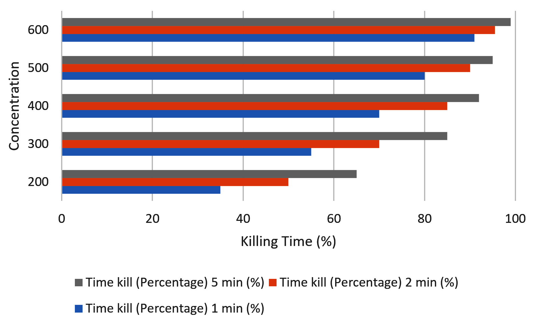
Figure 1. Time-kill assay of M.oleifera extract against C. krusei
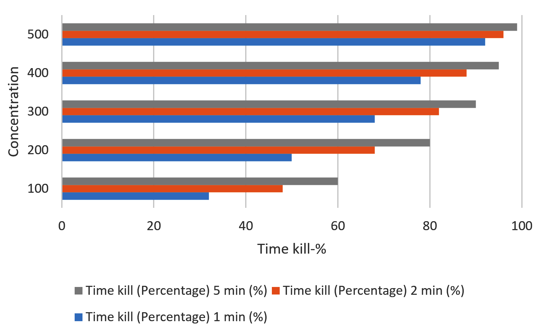
Figure 2. Time-kill assay of C. asiatica extract against C. krusei
Overall, the results indicated that both extracts have the potential to inhibit the viability of C. krusei within 5 minutes. The Centella extract was found to have 2-3 times higher activity than epicatechin, particularly at lower concentrations. The rapid killing of the pathogen may be useful in endorsing the use of these extracts for the implementation of antiseptic agents. Overall, it was found that both extracts have the potential to rapidly kill the C. krusei pathogen with significant percentages within 5 minutes. The activity of the extracts was found to be higher for the Centella extract, particularly at lower concentrations. The rapid killing effect of these extracts endorses their use as candidate antiseptic agents.
From the data presented in Table 3, the three treatments demonstrated increased antifungal activity as the exposure time increased. At 1 minute of exposure, the Moringa plant extract at 300 mg/mL demonstrated a killing rate of 55.0 ± 2.2%, while the Centella plant extract at 250 mg/mL demonstrated a slightly higher rate of 55.0 ± 2.0%. However, when the mixture of the two plant extracts at 300 + 250 mg/mL was used, the effect was more pronounced, with a killing rate of 80.0 ± 1.5%. This indicates that the mixture of the plant extracts has the potential of acting in a synergistic manner.
Table 3. Time-kill assay for combination extracts
Time (min) |
Moringa 300 mg/mL (%) |
Centella 250 mg/mL (%) |
Combination 300 + 250 mg/mL (%) |
|---|---|---|---|
1 |
55.0 ± 2.2 |
55.0 ± 2.0 |
80.0 ± 1.5 |
2 |
70.0 ± 2.0 |
70.0 ± 2.0 |
90.0 ± 1.0 |
5 |
85.0 ± 1.8 |
82.5 ± 1.8 |
97.0 ± 0.5 |
Positive Control (Ethanol) |
99.4 ± 0.2 |
99.4 ± 0.2 |
99.4 ± 0.2 |
By the second minute, it was observed that the antifungal activity of both extracts was even higher. The Moringa extract was found to attain a killing rate of 70.0 ± 2.0%, whereas the Centella extract was found to attain the same rate. The mixed form of these two extracts was found to attain an even higher rate of 90.0 ± 1.0%. The increasing trend of these rates suggests that the antifungal activity is time-dependent.
After five minutes of exposure time, each extract showed activity close to its maximum effect. For the Moringa extract, it showed a killing rate of 85.0 ± 1.8%, while Centella showed 82.5 ± 1.8%. However, the combined effect of both extracts showed an even higher killing rate of 97.0 ± 0.5% compared to the positive control, which used ethanol at 99.4 ± 0.2%. This proved that the combined extract of Moringa-Centella showed nearly equivalent results.
In conclusion, the results proved that the combined extract of Moringa and Centella showed stronger antifungal activity compared to the individual extracts. Moreover, the activity shown by the extracts increased gradually with longer exposure time, as shown in Figure 3.
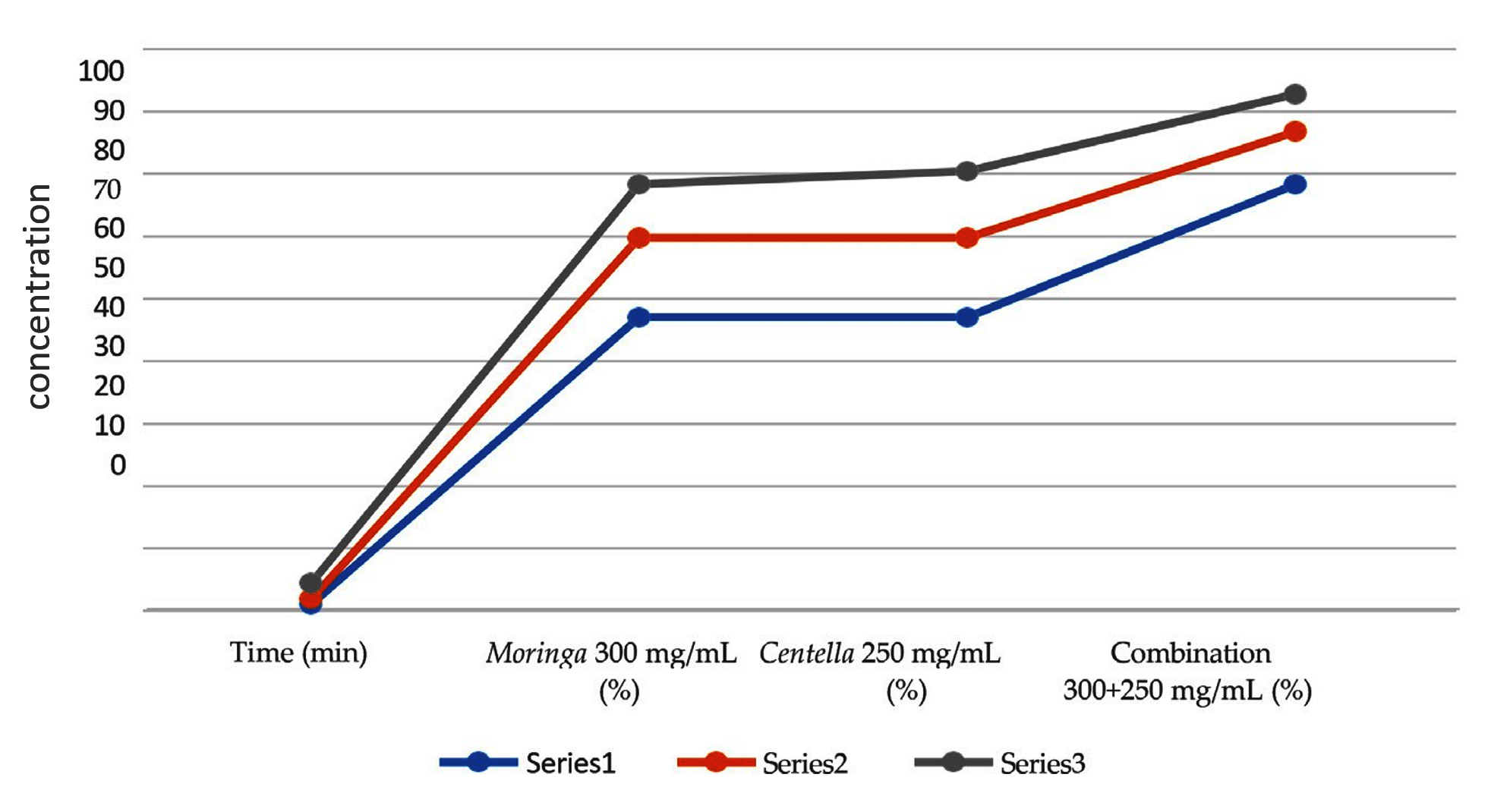
Figure 3. Time-kill assay for combination extracts
Series 1: 1 minute, Series 2: 2 minutes, Series 3: 5 minutes
Phenol and flavonoid content test
Analysis of the phenol and flavonoid content of M. oleifera extract (Figures 4 and 5) showed a total phenol content was 16.63 mg GAE/g, while flavonoid content of 1.61 mg QE/g. Meanwhile, the phenol content in C. asiatica was 22.18 mg GAE/g, while total flavonoid content was 6.60 mg QE/g. These results indicate that phenol content is more dominant than flavonoid content in both extracts and Centella showed a higher total phenol content than Moringa.
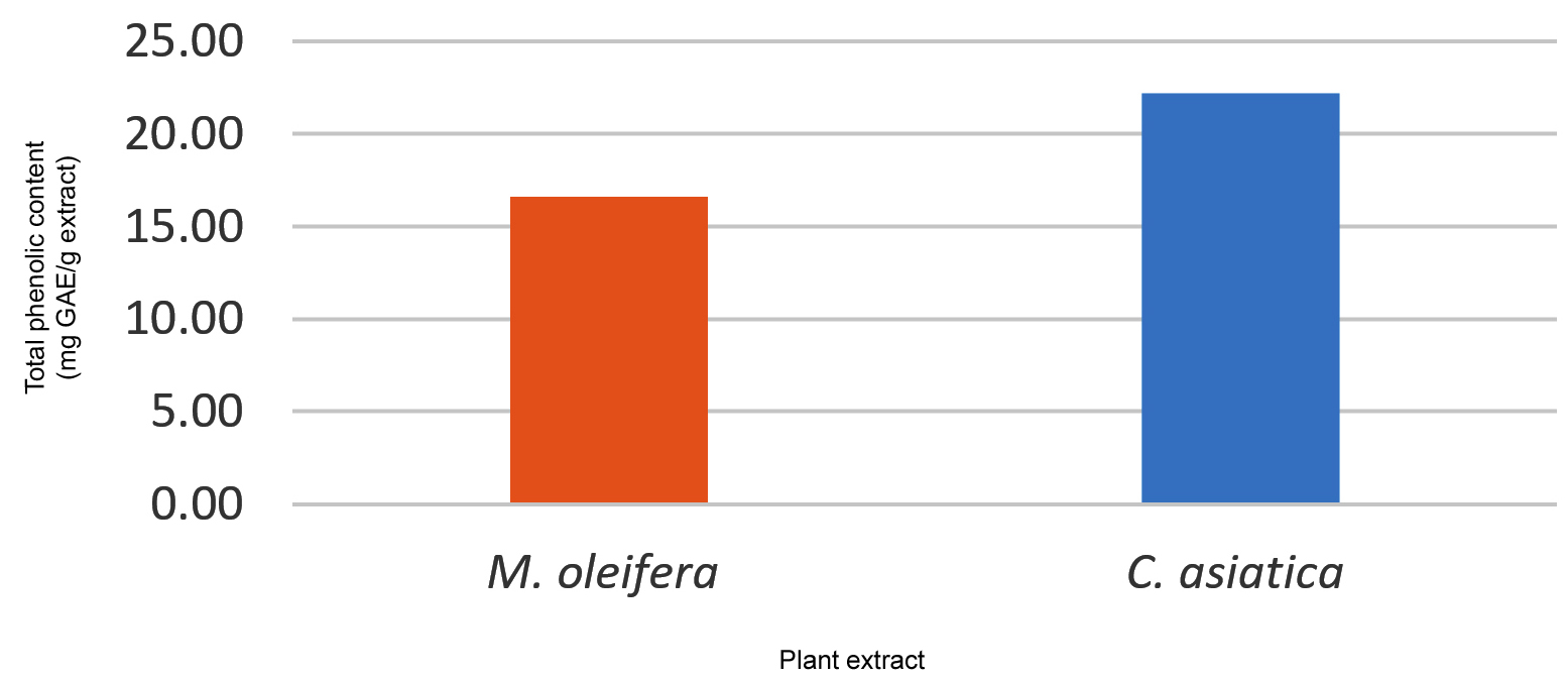
Figure 4. Total Phenolic content of M. oleifera and C. asiatica
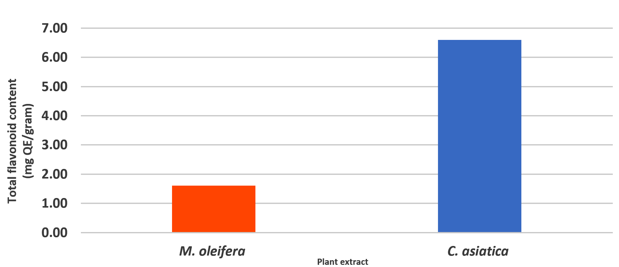
Figure 5. Total Flavonoid content of M. oleifera and C. asiatica
The results of the present study have shown that the extracts of M. oleifera and C. asiatica possess significant in vitro antifungal activity against C. krusei. The combined extract showed better fungicidal kinetics compared to the separate extracts. These results are in line with the emerging facts that phytochemicals of plant origin possess significant antimicrobial activity and have the potential for the development of effective antiseptic formulations.
Intrinsic resistance of C. krusei and the need for alternative agents
C. krusei is known to be intrinsically resistant to a variety of commonly used antifungal drugs, including fluconazole, as well as echinocandins in some instances. This, in turn, causes infections that do not respond to traditional antifungal treatments. This is one of the reasons why there is a need to improve therapeutic as well as antiseptic regimens. For this reason, there is a growing need for developing alternative antifungal drugs that use different modes of action.25
Mechanisms of antifungal activity of M. oleifera
The antifungal potential of M. oleifera has been well established in the recent literature, and various bioactive compounds are responsible for the antimicrobial activity of the compound. Phytochemical studies have established the presence of various bioactive compounds in the M. oleifera extracts, including phenolic compounds, flavonoids, sterols like β-sitosterol and stigmasterol, and small proteinaceous compounds like chitin-binding protein (CBP). The antifungal potential of the compound is attributed to the disruption of the cell structure and the metabolic pathways of the fungi. The compound CBP, isolated from Moringa seeds, has been established to interact with the cell membranes of the fungi, thereby enhancing the permeability of the membranes, leading to oxidative stress and disruption of the cell membranes of the fungi. The antifungal potential of the compound may thus be attributed to the disruption of the cell membranes of the fungi, thereby leading to the death of the fungi, which was evident from the results of the present study, as well as from the results of the previous studies.26
The total phenolic content of M. oleifera extract was approximately 16.6 mg GAE/g, whereas its total flavonoid content was approximately 1.6 mg QE/g, indicating that phenolic compounds were present at a higher level than flavonoids in the extract. Phenolic compounds in Moringa generally include phenolic acids and tannins, which may contribute to its antimicrobial properties. The relatively higher phenolic content may be related to the ability of ethanol to extract polar to semipolar compounds, including polyphenolic constituents that are abundant in Moringa. The presence of these phenolic compounds may contribute to the antifungal activity observed in the present study, although the individual contribution of each phytochemical component to the antifungal effect was not determined.27
Moreover, molecular docking studies of the phytochemicals present in Moringa were conducted, showing that there is potential interaction with key fungal enzymes like dihydrofolate reductase (DHFR), suggesting that these compounds can inhibit key cellular processes in addition to membrane damage. Although most of the studies carried out on these molecular targets were conducted using C. auris, it shows the potential of Moringa phytochemicals against drug-resistant fungal species.28
Bioactive ingredients and antifungal potential of Centella asiatica
C. asiatica is known to contain various bioactive compounds, especially pentacyclic triterpenoids like asiaticoside, madecassoside, and their aglycone forms like asiatic acid and madecassic acid. It is also known to contain flavonoids and phenolic compounds. Although most studies on C. asiatica have been conducted to investigate its wound-healing properties and anti-inflammatory activities, in vitro studies have shown that it possesses antimicrobial properties. Ethanol and methanol extracts of the plant were found to inhibit the growth of various fungal strains, including Candida. This is attributed to the fact that triterpenoids and phenolic saponins can disrupt the microbial cell membrane, thus interfering with the cell wall structure.29
In the present study, C. asiatica extract showed a higher total phenolic content (approximately 22.1 mg GAE/g) and total flavonoid content (approximately 6.6 mg QE/g) than M. oleifera extract. The higher levels of these phytochemical groups may contribute to the greater antifungal activity of C. asiatica observed in the present study. Phenolic and flavonoid compounds have been reported to affect microbial cell membranes and may also interfere with cellular oxidative processes. In addition, C. asiatica contains pentacyclic triterpenoids, including asiaticoside, madecassoside, asiatic acid, and madecassic acid, which have been associated with antimicrobial and antifungal activities. The coexistence of these bioactive constituents may contribute to the overall antifungal activity of the extract; however, the present study did not determine the contribution of individual compounds or their specific interactions.30
Apart from their direct action on the cell membrane, various studies carried out on triterpenoid derivatives like asiatic acid have shown that triterpenoids can affect the physiology of the fungal cell by various mechanisms. This includes the generation of reactive oxygen species, inhibition of drug efflux pumps, and prevention of morphological changes that are vital for the survival of the fungal cell. While most of these studies were carried out on C. albicans rather than C. krusei, as mentioned by Ali et al.31 it is likely that the same mechanisms might be responsible for the antifungal activity of C. asiatica extracts, as shown in the present study.
Enhanced antifungal effect of the combined extracts
The main finding of this study was the greater fungicidal activity observed when M. oleifera and C. asiatica extracts were used in combination compared with the individual extracts. The combined extract demonstrated faster killing kinetics and a greater reduction in fungal viability under the conditions tested. These findings suggest that combining the two plant extracts may enhance their overall antifungal activity against C. krusei. However, because a formal synergy analysis, such as determination of the fractional inhibitory concentration index (FICI), was not performed in the present study, the observed effect should not be interpreted as definitive evidence of synergism. Rather, the findings indicate an enhanced antifungal effect of the combination compared with the individual extracts.30
The enhanced activity of the combination may be related to the presence of different classes of bioactive compounds in the two plants that could potentially affect fungal cells through complementary mechanisms. Phenolic compounds and flavonoids present in M. oleifera may affect fungal membrane integrity and cellular oxidative balance, while triterpenoid compounds and phenolic constituents of C. asiatica may exert additional effects on membrane function, oxidative stress, and other cellular processes. The simultaneous presence of these compounds could therefore contribute to a greater overall antifungal effect than that observed with either extract alone. Nevertheless, this proposed interaction remains hypothetical, as the present study did not investigate the individual contributions or molecular interactions of specific phytochemicals.31
The faster reduction in fungal viability observed with the combined extracts may therefore reflect complementary or additive effects between their bioactive constituents. Such an interaction could be particularly relevant against C. krusei, which has limited therapeutic options because of its intrinsic resistance to fluconazole. Further studies using formal pharmacological interaction models, such as checkerboard assays with FICI determination, would be required to establish whether the interaction between M. oleifera and C. asiatica is truly synergistic, additive, or indifferent.32
Relevance to antiseptic development
The use of conventional antiseptics, including alcohol and chlorhexidine, is widely known. However, long-term use has shown side effects, such as irritation, cytotoxicity, and the potential development of resistance. Natural extracts, which have strong fungicidal activity, can be considered as an alternative for use as antiseptics, which can be used in the environment, on the skin, or on mucosal surfaces.33
In this regard, plant-based antiseptic agents have shown promising results and may offer additional benefits such as anti-inflammatory and antioxidant activity, which may simultaneously aid wound healing. However, their safety and stability need to be assessed before proceeding to clinical trials.34,35
Although the extracts demonstrated antifungal activity at the concentrations tested, the relatively high concentrations may limit their immediate practical application. Further optimization of the extract concentrations, particularly in combination, together with cytotoxicity, safety, and formulation studies, is required to establish their feasibility as an antiseptic alternative.
M. oleifera and C. asiatica extracts exhibited rapid antifungal activity in a concentration-dependent manner. The combination of both extracts exhibited a greater fungicidal activity than the individual extracts. The mechanism of antifungal activity of these extracts can be explained by the combined action of several bioactive phytochemicals, which target the fungal cell membrane, intracellular pathways, and stress response, thus exerting antifungal activity even at lower concentrations. The antifungal activity of the combination of M. oleifera and C. asiatica extracts makes them a promising natural antiseptic substance for practical use.
ACKNOWLEDGMENTS
The authors sincerely thank the Department of Microbiology, Faculty of Medicine, Universitas Indonesia, for providing the facilities and laboratory equipment used in this study. We also express our gratitude to the Department of Pharmaceutical Medicine, Faculty of Medicine, Universitas Indonesia, for supplying the extracts used in this research. We would like to thank the laboratory analysts from the Department of Medical Chemistry, Faculty of Medicine, University of Indonesia, for their analysis of the extract compounds, and the Occupational & Environmental Health Research Center, Faculty of Medicine, University of Indonesia, for the facilities provided. Additionally, we thank the Research Cluster on the Diagnosis and Therapy of Fungal Infections, Faculty of Medicine, Universitas Indonesia, Jakarta, Indonesia, for their support.
CONFLICT OF INTEREST
The authors declare that there is no conflict of interest.
AUTHORS’ CONTRIBUTION
All authors listed have made a substantial, direct and intellectual contribution to the work, and approved it for publication.
FUNDING
None.
DATA AVAILABILITY
The datasets generated and analyzed during the current study are available from the corresponding author on reasonable request.
ETHICS STATEMENT
Not applicable.
- Pfaller MA, Diekema DJ. Epidemiology of invasive candidiasis: a persistent public health problem. Clin Microbiol Rev. 2007;20(1):133-163.
Crossref - Lass-Florl C, Kanj SS, Govender NP, Thompson GR 3rd, Ostrosky-Zeichner L, Govrins MA. Invasive candidiasis. Nat Rev Dis Primers. 2024;10(1):20.
Crossref - Pappas PG, Kauffman CA, Andes DR, et al. Clinical practice guideline for the management of candidiasis: 2016 update by the Infectious Diseases Society of America. Clin Infect Dis. 2016;62(4):e1-e50.
Crossref - Perfect JR. The antifungal pipeline: a reality check. Nat Rev Drug Discov. 2017;16(9):603-616.
Crossref - McDonnell G, Russell AD. Antiseptics and disinfectants: activity, action, and resistance. Clin Microbiol Rev. 1999;12(1):147-179.
Crossref - Cowan MM. Plant products as antimicrobial agents. Clin Microbiol Rev. 1999;12(4):564-582.
Crossref - Daglia M. Polyphenols as antimicrobial agents. Curr Opin Biotechnol. 2012;23(2):174-181.
Crossref - Pareek A, Pant M, Gupta MM, et al. Moringa oleifera: an updated comprehensive review of its pharmacological activities, ethnomedicinal, phytopharmaceutical formulation, clinical, phytochemical, and toxicological aspects. Int J Mol Sci. 2023;24(3):2098.
Crossref - Leone A, Fiorillo G, Criscuoli F, et al. Nutritional characterization and phenolic profiling of Moringa oleifera leaves grown in Chad, Sahrawi refugee camps, and Haiti. Int J Mol Sci. 2015;16(8):18923-18937.
Crossref - Brinkhaus B, Lindner M, Schuppan D, Hahn EG. Chemical, pharmacological and clinical profile of the East Asian medicinal plant Centella asiatica. Phytomedicine. 2000;7(5):427-448.
Crossref - Min DH, Yu YB, Kim TH, Kim H, Lee S. Pharmacological effects of pentacyclic triterpenoids isolated from Centella asiatica. Hortic Environ Biotechnol. 2024;65(2):189-197.
Crossref - Taghizadeh M, Jalili S. Phytochemical content, antioxidant properties, and antibacterial activities of Centella asiatica L. Nat Prod Res. 2024;38(20):3693–3698.
Crossref - Balouiri M, Sadiki M, Ibnsouda SK. Methods for in vitro evaluating antimicrobial activity: a review. J Pharm Anal. 2016;6(2):71-79.
Crossref - Tóth Z, Forgács L, Kardos T, et al. Relative Frequency of Paradoxical Growth and Trailing Effect with Caspofungin, Micafungin, Anidulafungin, and the Novel Echinocandin Rezafungin against Candida Species. J Fungi. 2020;6(3):136.
Crossref - Carton JD, de-la-Fuente I, Sevillano E, et al. In vitro assessment of fluconazole and cyclosporine A antifungal activities against Candida species. J Fungi. 2025;11(2):133.
Crossref - Lundvik H, Santini R, Altintop TU, Özenci V, Andersson DI, Fatsis-Kavalopoulos N. Fast detection of synergy and antagonism in antifungal combinations used against Candida albicans clinical isolates. Sci Rep. 2025;15(1):36103.
Crossref - Filatov VA, Ilin EA, Kulyak OY, Kalenikova EI. Development and validation of a gas chromatography-mass spectrometry method for the analysis of the novel plant-based substance with antimicrobial activity. Antibiotics. 2023;12(10):1558.
Crossref - Klepser ME, Wolfe EJ, Jones RN, Nightingale CH, Pfaller MA. Antifungal pharmacodynamic characteristics of fluconazole and amphotericin B tested against Candida albicans. Antimicrob Agents Chemother. 1997;41(6):1392-1395.
Crossref - Cho E, Acosta K, Henkin J, Abzalimov R, Raskin I. Synergistic antifungal effects of botanical extracts against Candida albicans. PLoS One. 2026;21(1):e0340665.
Crossref - Pankey GA, Sabath LD. Clinical relevance of bacteriostatic versus bactericidal mechanisms. Clin Infect Dis. 2004;38(6):864-870.
Crossref - Adelakun AO, Awosika A, Adabanya U, Omole AE, Olopoda AI, Bello ET. Antimicrobial and synergistic effects of Syzygium cumini, Moringa oleifera, and Tinospora cordifolia against different Candida infections. Cureus. 2024;16(1):e52857.
Crossref - Floyd HEE, Kavanagh AM, Lowe GJ, et al. Standardisation of high throughput microdilution antifungal susceptibility testing for Candida albicans and Cryptococcus neoformans. Sci Rep. 2024;14(1):23407.
Crossref - Anwar F, Latif S, Ashraf M, Gilani AH. Moringa oleifera: a food plant with multiple medicinal uses. Phytother Res. 2007;21(1):17-25.
Crossref - Alomari AA. Ultrasound-assisted extraction of phenolic, flavonoid and antioxidant compounds from Dodonaea viscosa and its green synthesis of silver nanoparticles by aqueous extract. Orient J Chem. 2020;36(1):179-188.
Crossref - James JT, Dubery IA. Pentacyclic triterpenoids from the medicinal herb, Centella asiatica (L.) Urban. Molecules. 2009;14(10):3922-3941.
Crossref - Tjampakasari CR, Kiranasari K, Baswara RMK. Potential of Moringa oleifera leaf extract as an antiseptic against Candida albicans using percentage kill test. Tradit Med J. 2025;30(2):211-217.
Crossref - Santos LMM, Silva PM, Moura MC, et al. Anti-Candida activity of water soluble lectin from Moringa oleifera seeds (WSMoL). J Med Mycol. 2021;31(1):101074.
Crossref - Sahu BK, Patra SK, Sahu MC, Panda SK. Natural product based approaches to overcome Candida glabrata and emerging AMR threats. Front Antibiot. 2026;5:1767032.
Crossref - You ZL, Sun L, Wang LX, et al. Inhibition mechanism of the fungal β-1,3-glucan synthases by triterpenoid antifungal drugs. Nat Commun. 2026;17(1):2347.
Crossref - Wizrah MSI, Yahia ZA, Al-Omari H, et al. Comprehensive molecular docking and antifungal activity analysis of Moringa extracts targeting Candida auris dihydrofolate reductase (8CRH). Sci Rep. 2025;15(1):44185.
Crossref - Ali K, Kumar S, Khan MF. A concise review on bioactive pentacyclic triterpenoids of Centella asiatica (Gotu Kola). Discov Chem. 2025;2:54.
Crossref - Dantas-Medeiros R, Zanatta AC, de Souza LBFC, et al. Antifungal and antibiofilm activities of B-type oligomeric procyanidins from Commiphora leptophloeos used alone or in combination with fluconazole against Candida spp. Front Microbiol. 2021;12:613155.
Crossref - Woo S, Marquez L, Crandall WJ, Risener CJ, Quave CL. Recent advances in the discovery of plant-derived antimicrobial natural products to combat antimicrobial resistant pathogens: insights from 2018–2022. Nat Prod Rep. 2023;40(7):1271–1290.
Crossref - Fakhim H, Mohamadi B, Gharibi S, et al. Antifungal activity of polyphenolic compounds against fluconazole-susceptible and -resistant Candida species. Iran J Microbiol. 2025;17(2):342-347.
Crossref - Khursheed T, Khalil AA, Akhtar MN, et al. Ultrasound-assisted solvent extraction of phenolics, flavonoids, and major triterpenoids from Centella asiatica leaves: a comparative study. Ultrason Sonochem. 2024;111:107091.
Crossref
© The Author(s) 2026. Open Access. This article is distributed under the terms of the Creative Commons Attribution 4.0 International License which permits unrestricted use, sharing, distribution, and reproduction in any medium, provided you give appropriate credit to the original author(s) and the source, provide a link to the Creative Commons license, and indicate if changes were made.