

ISSN: 0973-7510
E-ISSN: 2581-690X
 , Ahmad Luay Osman Hashim1 and Sara Mohammed Ali1
Vulvovaginal candidiasis (VVC) is a common infection among women of reproductive age; however, data on vaginal yeast species and Candida albicans genotypes in the United Arab Emirates (UAE) are limited. This cross-sectional laboratory study determined the species distribution of vaginal yeast isolates and characterized confirmed C. albicans isolates by 25S rDNA ABC genotyping. High vaginal swabs from patients with suspected VVC attending Thumbay Hospital, Ajman, between 2020 and 2021 were cultured on Sabouraud dextrose agar and blood agar. The yeast isolates were examined microscopically and identified using conventional phenotypic tests and the API 20C AUX system. The confirmed C. albicans isolates were subjected to DNA extraction, PCR amplification of the 25S rDNA intron region, and agarose gel electrophoresis. Among the 60 vaginal yeast isolates, 50 (83.33%) were C. albicans, 7 (11.67%) were Saccharomyces cerevisiae, a non-Candida yeast, and 3 (5.00%) were Candida guilliermondii, the only non-albicans Candida species identified. Most isolates were recovered from patients aged 11-49 years (50/60, 83.33%). ABC genotyping of the C. albicans isolates identified genotype A as the most frequent profile (66.67%), followed by genotypes C (25.00%) and B (8.33%). C. albicans was therefore the predominant vaginal yeast species, and genotype A had the predominant 25S rDNA intron profile. These findings provide local baseline data for species-level diagnoses and molecular epidemiological surveillance in the UAE.
Vulvovaginal Candidiasis, High Vaginal Swab, 25S rDNA Intron, Molecular Epidemiology, Non-albicans Candida, Saccharomyces cerevisiae, API 20C AUX
Vulvovaginal candidiasis (VVC) is a mucosal fungal infection affecting women worldwide. Clinical estimates suggest that approximately 70%-75% of women experience at least one episode of VVC, and 5%-10% have recurrent VVC, commonly defined as repeated symptomatic episodes within 1 year.1-4 Although not usually life-threatening, VVC causes vulvar pruritus, burning, dyspareunia, and abnormal discharge, significantly impairing quality of life.1,2 Most cases are due to Candida albicans, a yeast that may exist as a commensal organism in the vaginal microbiota but can become pathogenic when host, microbial, or hormonal conditions shift.4-7 However, epidemiologic studies show a gradual rise in non-albicans Candida (NAC) species, including C. glabrata, C. tropicalis, and C. krusei, especially among recurrent or complicated cases.8-10 NAC species often exhibit reduced susceptibility to azole drugs, complicating treatment and supporting the need for laboratory-guided diagnosis.9-14
Multiple host factors predispose to VVC. Pregnancy, estrogen-rich states, broad-spectrum antibiotic use, uncontrolled diabetes mellitus, immunosuppression, and changes in the vaginal microbiome can disrupt mucosal homeostasis and favor yeast overgrowth.2,7,15,16 Oral contraceptives, sexual behaviors, and previous antimicrobial exposure have also been implicated in some populations.2,3,17 These risk factors contribute to the high global burden of VVC and recurrent VVC, which imposes substantial healthcare costs, lost productivity, and psychosocial stress.1,2,4 Despite its ubiquity, epidemiological data on VVC vary widely by region and diagnostic methods, underscoring the need for locale-specific studies.1,8,18
Given the predominance of C. albicans, many uncomplicated VVC episodes are managed empirically; however, shifting species patterns, recurrent disease, and emerging antifungal resistance support attention to species-level diagnosis and epidemiology.8,9,12,14,19 Moreover, C. albicans possesses unique virulence traits, including surface adhesins, hyphal differentiation, biofilm formation, and secreted hydrolytic enzymes, that facilitate mucosal invasion and recurring infection.3,5-7 These pathogenic mechanisms partly explain why C. albicans continues to dominate even as NAC species increase in frequency.5,7
Molecular epidemiology provides tools for tracking C. albicans strain diversity and comparing local isolates with regional and international patterns. One widely used method is ABC genotyping, which amplifies the group I intron region of the 25S rDNA. This PCR yields a ~450-bp fragment for genotype A when the intron is absent, an ~840-bp fragment for genotype B when the intron is present, and both fragments for genotype C when intron-containing and intron-free rDNA copies coexist.20-22 These A/B/C labels are intron-based molecular profiles within C. albicans and are not separate species. They are also distinct from the historical type I/type II designations of C. stellatoidea; type I isolates can share the genotype B band pattern; hence, ABC PCR alone does not establish classification as C. stellatoidea.23,24 Genotype A is frequently reported among clinical isolates, although the relative frequency of genotypes A, B, and C varies by geography, infection site, sample size, and method.20-22,25 While ABC typing is less discriminatory than multilocus sequence typing, microsatellite analysis, or whole-genome sequencing, it is rapid and cost-effective for descriptive epidemiology.21,22 Few studies have applied C. albicans genotyping to vaginal isolates, and published data from the Gulf region remain scarce.
Data on VVC prevalence and Candida epidemiology in the United Arab Emirates (UAE) and the broader Middle East are sparse. Recent UAE fungal-burden mapping highlighted that local data on fungal diseases remain limited, and the estimated recurrent VVC is one of the most common fungal conditions in the country.18 Regional studies from neighboring or broadly comparable settings have also reported that Candida species distribution and antifungal susceptibility can differ by population and clinical context.10,11,15 However, no recent surveillance of vaginal Candida species distribution or C. albicans genotypes has been published for Ajman or neighboring emirates. This gap is noteworthy given the differences in regional climate, healthcare, population mobility, and diagnostic practices that may influence VVC epidemiology.
Rationale
Improved knowledge of the local distribution of vaginal yeast species can support clinical diagnosis and epidemiological surveillance. Therefore, this study aimed to determine the species distribution among vaginal yeast isolates recovered from high vaginal swabs in Ajman and to characterize confirmed C. albicans isolates using 25S rDNA ABC genotyping.
Study design and setting
This cross-sectional laboratory study was conducted from January 2020 to December 2021 at Thumbay Hospital, affiliated with the Gulf Medical University in Ajman, UAE. The laboratory serves patients from Ajman and surrounding emirates. High vaginal swab specimens from women presenting with symptoms suggestive of VVC, such as vaginal discharge, pruritus, irritation, burning sensation, or clinical suspicion of fungal vaginitis, were subjected to fungal culture as part of routine diagnostic care. Ethical approval was obtained from the relevant institutional ethics committee, and all data were handled anonymously without recording patient identification information.
The study included culture-positive vaginal yeast isolates available for species identification and, where applicable, molecular analysis. Isolates with unclear species identification, mixed bacterial contamination preventing purification, or insufficient growth for downstream DNA extraction were excluded from molecular testing. Demographic data were limited to age groups and laboratory isolate information, allowing the study to describe local species distribution and genotype patterns while maintaining patient confidentiality.
Sample collection and culture
High vaginal swabs were collected using sterile cotton swabs during clinical examination and promptly transported to the microbiology laboratory for processing. Each swab was inoculated onto Sabouraud Dextrose Agar (SDA) and Brain Heart Infusion (BHI) Blood Agar plates to support yeast recovery and a preliminary evaluation of colony morphology. Plates were incubated aerobically at 37 °C for 24-48 hrs and examined for yeast-like colonies. Suspected colonies were sub-cultured to obtain pure growth before identification. Colony appearance, including creamy, smooth, and white to off-white colonies, together with Gram staining showing Gram-positive budding yeast cells with or without pseudohyphae, was used as a preliminary confirmation of yeast growth.
Phenotypic species identification
Isolates were identified to the species level using the API 20C AUX yeast identification system (bioMérieux, France), according to the manufacturer’s instructions. Pure yeast colonies were suspended in sterile saline to the recommended turbidity level and inoculated into API 20C AUX strip wells. The strips were incubated at 30 °C for 48-72 hrs, and carbohydrate assimilation reactions were recorded. The final species identification was performed using the API web database. C. albicans identification was supported by colony morphology, microscopic findings, germ tube testing, where applicable, and the corresponding API biochemical profiles.
DNA extraction using QIAamp DNA Mini Kit (Qiagen)
Genomic DNA was extracted from the confirmed C. albicans isolates using the QIAamp DNA Mini Kit (Qiagen, Hilden, Germany) according to the manufacturer’s instructions, with minor modifications to improve yeast cell lysis. Fresh colonies were suspended in phosphate-buffered saline and pelleted via centrifugation. The pellet was lysed using Buffer ATL and proteinase K, followed by incubation to promote cellular disruption and protein digestion. Buffer AL and ethanol were then added to facilitate the binding of DNA to the silica membrane of the spin column. The column was sequentially washed with the kit wash buffers to remove proteins, salts, and other PCR inhibitors. DNA was eluted in Buffer AE and stored at -20 °C until PCR analysis. The use of purified genomic DNA was essential to ensure reliable amplification of the 25S rDNA region during ABC genotyping.
DNA template standardization
The DNA concentration was determined using a NanoDrop 2000 spectrophotometer (Thermo Fisher Scientific, USA) to standardize the template input used for ABC genotyping PCR.
ABC genotyping PCR
ABC genotyping of C. albicans was performed using PCR amplification of the 25S rDNA region. The primers used were CA-INT-L (forward) and CA-INT-R (reverse) as previously described20:
CA-INT-L: 5′-ATAAGGGAAGTCG GCAAAATAGATCCG-3′
CA-INT-R: 5′-CTTGGCTGTGGTTTC GCTAGATAGTAGAT-3′
These primers flanked the group I intron insertion sites. PCR reactions were prepared in 0.2 mL tubes (total volume 25 µL) containing: 12.5 µL of 2× PCR Master Mix (which includes Taq DNA polymerase, 1.5 mM MgCl₂, 200 μM each dNTP, PCR buffer; e.g. Promega GoTaq Green or equivalent), 0.5 µL of each primer (10 µM stock, final concentration 0.2 µM), 2 µL of template DNA (~50 ng), and 9.5 µL of nuclease-free water. If a master mix with premixed primers was available, it could be used accordingly.
Thermocycling was performed on a thermal cycler (e.g., Applied Biosystems SimpliAmp) under the following conditions: an initial denaturation at 95 °C for 5 min; followed by 35 cycles of 95 °C for 30 sec, 55 °C for 30 sec, and 72 °C for 60 sec; and a final extension at 72 °C for 5 min. The annealing temperature (55 °C) was chosen based on primer melting temperatures and literature precedents.20 PCR products were stored at 4 °C until analysis.
Expected amplicon sizes were ~450 bp for genotype A (group I intron absent), ~840 bp for genotype B (group I intron present), and both ~450 and ~840 bp for genotype C (intron-containing and intron-free rDNA copies coexist).20 These classifications describe 25S rDNA intron profiles within the confirmed C. albicans isolates and do not represent separate species.
Agarose gel electrophoresis and interpretation
The PCR products were resolved by electrophoresis on a 1.5% agarose gel in 1× TAE buffer. Ethidium bromide (0.5 μg/mL) or a safe nucleic acid stain (e.g., SYBR Safe) was added to the gel for visualization. Five microliters of each PCR reaction was mixed with loading dye and loaded into the gel wells; a 100 bp DNA ladder (Thermo or similar) was included. Electrophoresis was performed at ~5-8 V/cm for ~60 min. The gels were photographed under UV illumination. The band sizes were estimated by comparison with a ladder.
Genotypes were assigned based on band patterns: a single ~450-bp band = genotype A, a single ~840-bp band = genotype B, and two bands (~450 and ~840 bp) = genotype C.20 For quality control, reference strains or previously genotyped isolates (if available) were run in parallel. Because the ~840-bp product may also occur in isolates historically designated C. stellatoidea type I, this band was recorded as genotype B and was not considered sufficient for classification as C. stellatoidea.23,24
Data analysis
The data were summarized using descriptive statistics. Frequencies and percentages were calculated for the age groups, vaginal yeast species distribution, and ABC genotype categories among the confirmed C. albicans isolates. Because this was a descriptive cross-sectional laboratory study, inferential statistical testing was not performed.
A total of 60 vaginal yeast isolates were recovered from high vaginal swab samples. The distribution of isolates by age group revealed that most isolates were recovered from women of reproductive age. The highest frequency was observed in women aged 11-49 years, accounting for 50 cases (83.33%). Patients aged ≥50 years accounted for 7 cases (11.67%), while patients aged ≤10 years accounted for 3 cases (5.00%). This pattern indicates that most vaginal yeast isolates were recovered from patients in the reproductive age group, which is consistent with the known influence of hormonal and physiological factors on vaginal yeast colonization and infection15 (Figure 1).
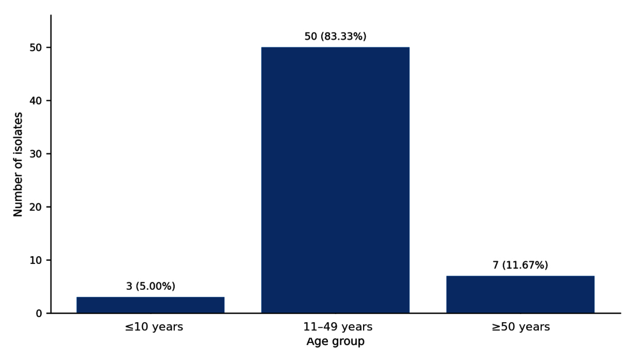
Figure 1. Distribution of vaginal yeast isolates by age group (n = 60)
All isolates exhibited morphological and staining characteristics consistent with yeast, including Gram-positive budding yeast cells. Biochemical identification using the API 20C AUX yeast identification system demonstrated the presence of 3 yeast species among the 60 isolates. C. albicans was the predominant species, representing 50 isolates (83.33%). Saccharomyces cerevisiae was identified in 7 isolates (11.67%) and represented a non-Candida yeast, whereas Candida guilliermondii was identified in 3 isolates (5.00%) and represented the only NAC species identified. Neither C. guilliermondii nor S. cerevisiae were classified as C. albicans, and neither was included in the C. albicans ABC genotype analysis (Figure 2).
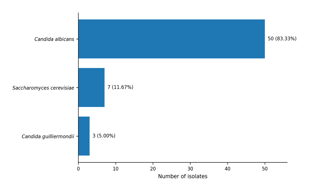
Figure 2. Species distribution of vaginal yeast isolates identified using the API 20C AUX system (n = 60)
ABC genotyping of the 50 C. albicans isolates was performed using PCR targeting the 25S rDNA intron region. Agarose gel electrophoresis revealed clear genotype-specific banding patterns. Genotype A, characterized by a single band of approximately 450 bp, was the most frequent genotype and was detected in 33 isolates (66.67%). Genotype C, characterized by the presence of both approximately 450 bp and 840 bp bands, was detected in 13 isolates (25.00%). Genotype B, characterized by a single band of approximately 840 bp, was the least frequent genotype and was detected in 4 isolates (8.33%). These results demonstrate a clear predominance of genotype A among C. albicans isolates obtained from high vaginal swab samples in Ajman, UAE, whereas genotypes B and C demonstrate variation in the 25S rDNA intron profile within the local isolate collection, rather than high-resolution genome-wide diversity (Figure 3).
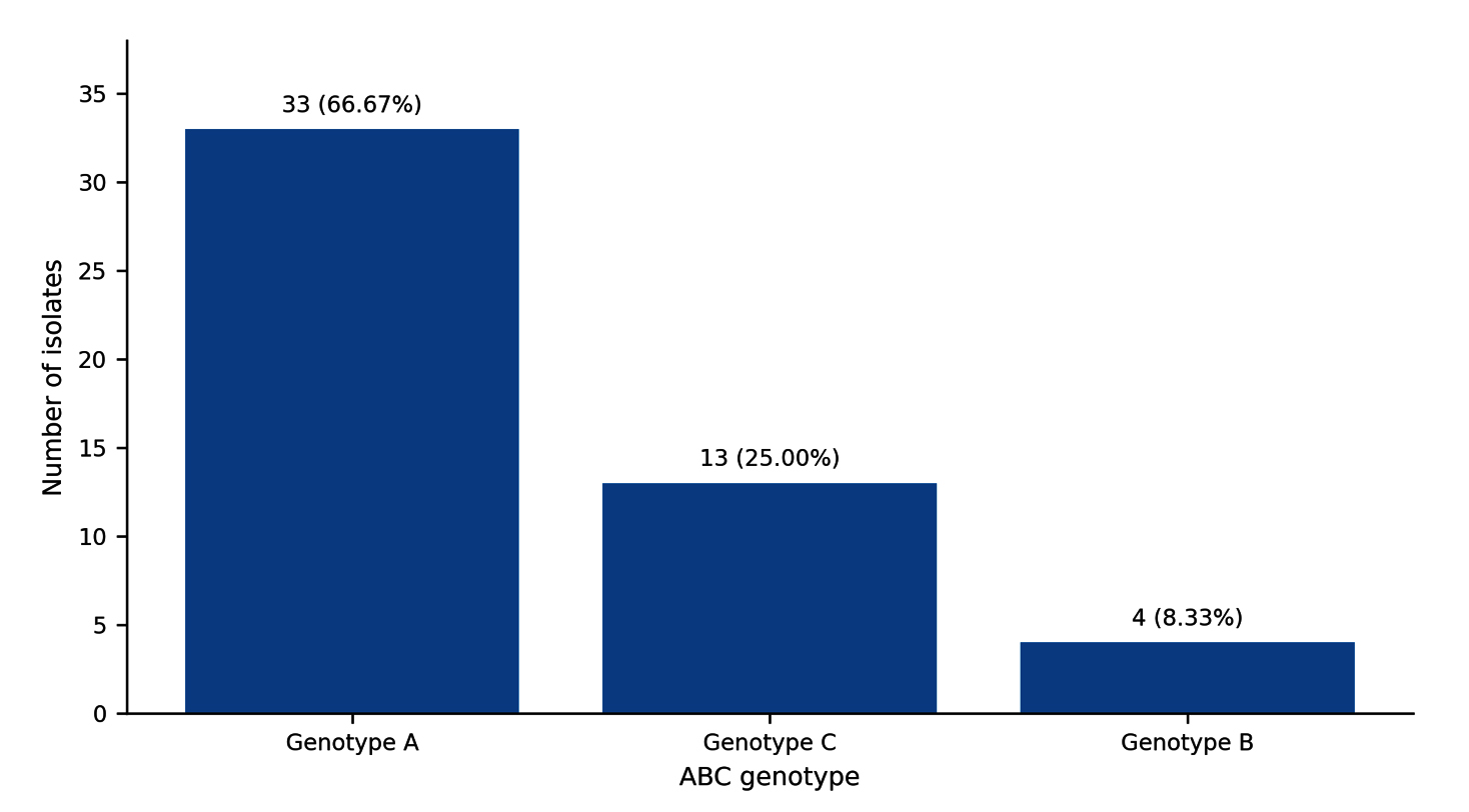
Figure 3. ABC genotype distribution among confirmed Candida albicans isolates (n = 50). Genotype A, approximately 450 bp (group I intron absent); genotype B, approximately 840 bp (group I intron present); genotype C, both fragments (intron-containing and intron-free rDNA copies)
In the ABC system, the labels A, B, and C describe the 25S rDNA intron profiles rather than separate species. The single ~840-bp genotype B pattern has also been reported in isolates historically designated C. stellatoidea type I. However, this PCR pattern alone is not sufficient to identify C. stellatoidea. Accordingly, the 4 isolates with the ~840-bp product were retained as C. albicans genotype B based on the phenotypic/API identification, and no separate C. stellatoidea classification was made.23,24
This study provides local laboratory-based data on the species distribution of vaginal yeast isolates and the ABC genotype profile of C. albicans isolates recovered from high vaginal swab samples in Ajman, UAE. These findings contribute to the limited regional evidence on VVC and molecular epidemiology of C. albicans. This study is particularly relevant because local data on vaginal yeast species and C. albicans genotypes remain scarce, and clinical management is often based on assumptions derived from international epidemiological patterns rather than UAE-specific evidence.
The predominance of Candida isolation among women aged 11-49 years is consistent with the epidemiology of VVC, which most commonly affects women during the reproductive years.1 Hormonal changes, estrogen exposure, pregnancy, contraceptive use, sexual activity, antibiotic exposure, and alterations in the vaginal microbiota may contribute to increased susceptibility in this age group.3,15 Estrogen-rich conditions can promote epithelial glycogen deposition and support Candida colonization, while disruption of lactobacilli-dominated vaginal flora may facilitate yeast overgrowth. Therefore, the lower number of isolates from younger and older age groups in this study is biologically plausible, although it may also reflect healthcare-seeking patterns and the distribution of the submitted samples.
Species identification showed that C. albicans was the predominant vaginal yeast species, accounting for 83.33% of the isolates. This finding is consistent with many previous reports that have identified C. albicans as the main cause of VVC.1,7,9,10 The remaining isolates represented two distinct taxonomic categories: C. guilliermondii was the only NAC species identified, and S. cerevisiae was a non-Candida yeast. Neither organism was regarded as C. albicans nor was it included in the ABC genotype analysis. Their recovery highlights the value of routine species-level identification, particularly because non-Candida yeasts such as S. cerevisiae may be recovered from clinical specimens and display distinct antifungal susceptibility patterns.26 These yeasts may differ in their pathogenic potential, recurrence pattern, and antifungal susceptibility profile, and they cannot be reliably distinguished by symptoms alone.9-11,13 Therefore, laboratory confirmation remains important, especially in persistent, recurrent, or treatment-unresponsive cases.14,19
Molecular analysis using ABC genotyping revealed that genotype A was the dominant genotype among C. albicans isolates in this study. This finding is consistent with studies reporting genotype A as a common ABC genotype in clinical C. albicans isolates, although some vaginal isolates have shown different genotype distributions.20-22,25 Because ABC typing evaluates only the presence or absence of the 25S rDNA group I intron, the predominance of genotype A describes the frequency of this intron profile in the present collection but does not by itself demonstrate enhanced vaginal adaptation, virulence, or antifungal resistance. Recent studies using higher-resolution genotyping and phenotypic analysis likewise indicate that genotype alone does not consistently predict azole resistance or pathogenicity.27,28 Although ABC genotyping is not as discriminatory as multilocus sequence typing, microsatellite analysis, or whole-genome sequencing, it remains a practical, low-cost method for describing broad genotype distributions in clinical isolate collections.21,22
The ABC designations should not be confused with species names or the historical type I/type II division of C. stellatoidea. Later molecular studies have generally treated C. stellatoidea as synonymous with C. albicans while recognizing a differentiated lineage. Type I isolates have been reported to produce the same single ~840-bp profile as C. albicans genotype B, whereas type II was historically described as a sucrose-negative C. albicans variant.23,24 These historical C. stellatoidea types are therefore not equivalent to ABC genotypes A, B, and C. In particular, not every genotype B isolate is C. stellatoidea, and the ABC assay alone cannot make this taxonomic distinction. The four genotype B isolates in this study should be interpreted as C. albicans genotype B, unless additional confirmatory phenotypic or high-resolution molecular testing is performed.
Genotype C represented one quarter of the C. albicans isolates, whereas genotype B was the least common. The presence of genotype C in 25.00% of the isolates indicates that the 25S rDNA intron profiles were not uniform across the local isolate collection; this should not be interpreted as a genome-wide measure of genetic diversity. Differences in genotype distribution between studies may reflect geographic variations, host population characteristics, infection sites, sample sizes, and methodological differences. The detection of all three major ABC genotypes supports the usefulness of molecular typing for establishing a baseline profile of 25S rDNA intron-profile distribution among C. albicans isolates in Ajman.
Although virulence factors and antifungal susceptibility testing were not evaluated in the present study, previous investigations have demonstrated that C. albicans genotypes and strain backgrounds may be associated with phenotypic traits, such as phospholipase activity, proteinase activity, biofilm formation, cell-surface hydrophobicity, and antifungal resistance.5,7,11,20,25,29-31 These traits are clinically important because they can influence persistence, recurrence, and response to treatment. Therefore, the genotype distribution reported here should be interpreted as a foundation for future studies, rather than as direct evidence of virulence or resistance. Future studies should combine ABC genotyping with antifungal susceptibility testing, biofilm assays, and hydrolytic enzyme profiling to determine whether the locally detected genotypes differ in clinically meaningful ways.11,13,25,29,30,32
The findings of this study emphasize the importance of incorporating molecular typing methods such as ABC genotyping into the epidemiological surveillance of C. albicans.20-22,25 In routine clinical practice, species identification is more immediately relevant for treatment decisions; however, genotyping adds epidemiological value by describing strain diversity and enabling comparison with findings from other regions. In settings such as the UAE, where published molecular data on vaginal Candida isolates are limited, basic genotyping can provide useful baseline information and support future multicenter surveillance.18
This study had several limitations. First, the sample size was moderate, and isolates were obtained from a single diagnostic center, which may limit the generalizability to all emirates or healthcare settings in the UAE. Second, only ABC genotyping was performed; this method is useful for broad classification but does not provide high-resolution strain typing.21,22 Because ABC genotyping generates categorical 25S rDNA intron banding profiles rather than nucleotide sequences or multilocus distance data, the present dataset does not support the construction of a valid phylogenetic tree. Establishing phylogenetic relationships among local isolates requires additional sequence-based analyses, such as multilocus sequence typing or whole-genome sequencing. ABC PCR cannot, by itself, distinguish genotype B C. albicans from isolates historically designated C. stellatoidea type I. Dedicated phenotypic and/or higher-resolution molecular testing would be required for that assignment.23,24 Third, antifungal susceptibility testing, virulence factor assays, and clinical outcome data were not included, preventing a direct correlation between genotype, pathogenicity, and treatment response.11,29-31 Finally, the study relied on available laboratory isolates and limited demographic information. Despite these limitations, this study provides useful preliminary local evidence and establishes a baseline for larger studies that incorporate multicenter sampling, antifungal susceptibility testing, and more discriminatory molecular typing methods.
This study investigated the species distribution of vaginal yeast isolates and the ABC genotype profile of confirmed C. albicans isolates recovered from high vaginal swab samples in Ajman, UAE. The findings showed that most isolates were recovered from women aged 11-49 years, supporting the known predominance of VVC in women of reproductive age.
Species identification confirmed C. albicans was the predominant species among 60 isolates tested. C. guilliermondii was the only NAC species identified, whereas S. cerevisiae is a non-Candida yeast species. Neither was regarded as C. albicans. Molecular analysis using ABC genotyping, which was restricted to the confirmed C. albicans isolates, demonstrated that genotype A was the most common genotype among the C. albicans isolates, followed by genotypes C and B. This genotype pattern is broadly consistent with previously published studies and indicates that genotype A is dominant in this local isolate collection, whereas genotypes B and C show variations in the 25S rDNA intron profile. The labels A, B, and C should be understood as 25S rDNA intron profiles rather than as separate species. Specifically, the genotype B pattern alone does not justify the reclassification of an isolate as C. stellatoidea.
Although virulence factors and antifungal susceptibility were not evaluated in this study, the results provide baseline molecular epidemiological data that may support future investigations. In particular, combining species identification, ABC genotyping, antifungal susceptibility testing, biofilm assessment, and virulence assays in larger cohorts would help to clarify whether local genotype patterns are associated with treatment response, recurrence, or pathogenic potential.
In conclusion, this study provides local baseline data on vaginal yeast species distribution and C. albicans ABC genotypes in Ajman, UAE. These results support the continued use of species-level identification in suspected VVC and demonstrate the value of simple molecular typing methods for regional surveillance. Future multicenter studies across the UAE are recommended to confirm these findings and strengthen evidence-based management of VVC.
ACKNOWLEDGMENTS
The authors express their gratitude to Gulf Medical University, the Gulf Medical University Hospital at Ajman for providing laboratory facilities and technical support throughout this research.
CONFLICT OF INTEREST
The authors declare that there is no conflict of interest.
AUTHORS’ CONTRIBUTION
All authors listed have made a substantial, direct and intellectual contribution to the work, and approved it for publication.
FUNDING
None.
DATA AVAILABILITY
All datasets generated or analyzed during this study are included in the manuscript.
ETHICS STATEMENT
This study was approved by the Gulf Medical Institutional Board of Research (Approval No. 2019MSCMLSM05; date 15 April 2021).
- Rautemaa-Richardson R, Sobel JD, Stone N, et al. State-of-the-art review: managing vulvovaginal candidiasis. Clin Infect Dis. 2026;82(3):371-382.
Crossref - De Seta F, Warris A, Roselletti E. New insights toward personalized therapies for vulvovaginal candidiasis and vaginal co-infections. Front Microbiol. 2025;16:1625952.
Crossref - David H, Solomon AP. Molecular association of Candida albicans and vulvovaginal candidiasis: focusing on a solution. Front Cell Infect Microbiol. 2023;13:1245808.
Crossref - Valentine M, Wilson D, Gresnigt MS, Hube B. Vaginal Candida albicans infections: host-pathogen-microbiome interactions. FEMS Microbiol Rev. 2025;49:fuaf013.
Crossref - Faustino M, Ferreira CMH, Pereira AM, et al. Candida albicans: the current status regarding vaginal infections. Appl Microbiol Biotechnol. 2025;109(1):91.
Crossref - Frois-Martins R, Lagler J, LeibundGut-Landmann S. Candida albicans virulence traits in commensalism and disease. Curr Clin Micro Rpt. 2024;11:231-240.
Crossref - Liu Z, Yang H, Huang R, Li X, Sun T, Zhu L. Vaginal mycobiome characteristics and therapeutic strategies in vulvovaginal candidiasis (VVC): differentiating pathogenic species and microecological features for stratified treatment. Clin Microbiol Rev. 2025;38(2):e00284-24.
Crossref - Subedi A, Upreti MK, Rana JC, Sapkota RP, Shrestha UT. Vulvovaginal candidiasis, an increasing burden to women in the tropical regions attending Bharatpur Hospital, Chitwan. J Med Mycol. 2024;34(4):101509.
Crossref - Benedict K, Smith DJ, Lyman M, Gold JAW. Vulvovaginal candidiasis culture results from a major national commercial laboratory, United States, 2019-2023. Am J Obstet Gynecol. 2024;231(3):e101-e104.
Crossref - Ali MAM, Edrees WH, Al-Shehari WAA, et al. Antifungal susceptibility pattern of Candida species isolated from pregnant women. Front Cell Infect Microbiol. 2024;14:1434677.
Crossref - Diaz-Navarro M, Irigoyen-von-Sierakowski A, Delcan I, et al. New insights in the role of Candida biofilm in the pathogenesis of recurrent vulvovaginal candidiasis: a prospective clinical study. Front Microbiol. 2025;16:1566171.
Crossref - Benedict K, Lyman M, Jackson BR. Possible misdiagnosis, inappropriate empiric treatment, and opportunities for increased diagnostic testing for patients with vulvovaginal candidiasis-United States, 2018. PLoS One. 2022;17(4):e0267866.
Crossref - Aboagye G, Waikhom S, Asiamah EA, et al. Antifungal susceptibility profiles of Candida and non-albicans species isolated from pregnant women: implications for emerging antimicrobial resistance in maternal health. Microbiol Spectr. 2025;13(7):e00787-25.
Crossref - Cornely OA, Sprute R, Bassetti M, et al. Global guideline for the diagnosis and management of candidiasis: an initiative of the ECMM in cooperation with ISHAM and ASM. Lancet Infect Dis. 2025;25(5):e280-e293.
Crossref - Hussen I, Aliyo A, Abbai MK, Dedecha W. Vaginal candidiasis prevalence, associated factors, and antifungal susceptibility patterns among pregnant women attending antenatal care at Bule Hora University Teaching Hospital, Southern Ethiopia. BMC Pregnancy Childbirth. 2024;24(1):619.
Crossref - Messina A, Mariani A, Brandolisio R, et al. Candidiasis in pregnancy: relevant aspects of the pathology for the mother and the fetus and therapeutic strategies. Trop Med Infect Dis. 2024;9(5):114.
Crossref - Sun Z, Ge X, Qiu B, et al. Vulvovaginal candidiasis and vaginal microflora interaction: microflora changes and probiotic therapy. Front Cell Infect Microbiol. 2023;13:1123026.
Crossref - Al Dhaheri F, Thomsen J, Everett D, Denning DW. Mapping the burden of fungal diseases in the United Arab Emirates. J Fungi. 2024;10(5):353.
Crossref jof10050353 - Centers for Disease Control and Prevention. Vulvovaginal candidiasis-STI treatment guidelines. Updated 2024. Accessed October, 2025. https://www.cdc.gov/std/treatment-guidelines/candidiasis.htm.
- Diaz-Huerta H, Garcia-Salazar E, Ramirez-Magana X, et al. Phenotypic and molecular characterization of Candida albicans isolates from Mexican women with vulvovaginitis. J Fungi. 2025;11(5):354.
Crossref - Amanloo S, Zanjani M, Serajian S, Ahmadi F, Kakavand F. Genotyping of Candida albicans isolates obtained from vulvovaginal candidiasis patients in Zanjan, Iran, based on ABC and RPS typing systems. Curr Med Mycol. 2022;8(4):9-14.
Crossref - Mahmoodi M, Nouraei H, Nasr R, Zomorodian K, Khodadadi H, Pakshir K. Phenotypes characterization and ABC genotypes distribution of clinical Candida albicans isolates. J Clin Lab Anal. 2023;37(7):e24888.
Crossref - McCullough MJ, Clemons KV, Stevens DA. Molecular and phenotypic characterization of genotypic Candida albicans subgroups and comparison with Candida dubliniensis and Candida stellatoidea. J Clin Microbiol. 1999;37(2):417-421.
Crossref - Jacobsen MD, Boekhout T, Odds FC. Multilocus sequence typing confirms synonymy but highlights differences between Candida albicans and Candida stellatoidea. FEMS Yeast Res. 2008;8(5):764-770.
Crossref - Mahmoodi M, Nouraei H, Nasr R, et al. Investigating the presence of phospholipase and secreted aspartyle proteinase gene family in different genotypes of Candida albicans species. BMC Res Notes. 2025;18(1):431.
Crossref - Yan Z, Fu Y, Tan X, et al. Isolate distribution and antifungal susceptibility of Saccharomyces cerevisiae in the national regional medical center of Southwest China for women and children during 2018-2023. BMC Microbiol. 2024;24(1):345.
Crossref - Morales-Lopez S, Villero Wolf Y, Torres Y, Lechuga D, Caicedo L, Garcia-Effron G. HWP1 gene sequence diversity and antifungal susceptibility patterns in Colombian vulvovaginal isolates of the Candida albicans species complex. Int J Microbiol. 2025;2025:8507361.
Crossref - Davari A, Liu J, Fortwendel JP, et al. Phenotypic and genotypic analysis of Candida albicans vaginal isolates reveals that ECE1 expression underpins pathogenicity. Infect Immun. 2026;94(7):26.
Crossref - Yan C, Zhang J, Yang Y, Zeng X, Xiao G. Virulence factors, biofilm formation and antifungal resistance in Candida albicans from recurrent vulvovaginal candidiasis patients: a comparative study. Sci Rep. 2025;15(1):37557.
Crossref - Bras G, Satala D, Juszczak M, et al. Secreted aspartic proteinases: key factors in Candida infections and host-pathogen interactions. Int J Mol Sci. 2024;25(9):4775.
Crossref - Pan Y, Sun Y, Chen L, et al. Candida causes recurrent vulvovaginal candidiasis by forming morphologically disparate biofilms on the human vaginal epithelium. Biofilm. 2023;6:100162.
Crossref - Clinical and Laboratory Standards Institute. Performance Standards for Antifungal Susceptibility Testing of Yeasts. 4th ed. CLSI supplement M60. Clinical and Laboratory Standards Institute. 2024.
© The Author(s) 2026. Open Access. This article is distributed under the terms of the Creative Commons Attribution 4.0 International License which permits unrestricted use, sharing, distribution, and reproduction in any medium, provided you give appropriate credit to the original author(s) and the source, provide a link to the Creative Commons license, and indicate if changes were made.