

ISSN: 0973-7510
E-ISSN: 2581-690X
Study the sequencing of phzM and phzS genes and the effect of their mutation on phenazine production. Pseudomonas aeruginosa has the ability to adapt and grow in a variety of environments and is owing to its wide genetic flexibility. This organism produces a diverse group of phenazine including pyocyanin, phenazine-1-carboxylic acid and phenazine-1-carboxamide. The Phenazine-1-carboxylic acid that converted to pyocyanin is mediated by phzM and phzS novel phenazine modifying genes. The two genes that convert the phenazine-1-carboxylic acid to pyocyanin encode the putative enzymes, methyltransferase and monooxygenase, respectively, however, the overexpression of gene (s) encoding biosynthesis of pyocyanin via increasing the copy number has not so far identified in P. aeruginosa. Molecular technique was used to detect phenazine modifying genes (phzM, phzS), detection of mutations in these genes by sequencing. The results revealed that the phzM and phzS gene were present in 21(80.76%), 14(53.84%) respectively. Sequencing of phzM and phzS from isolates of P.aeruginosa reveal that 9 mutations in 8 isolates (7), (2) mutations for both genes respectively with the identities (99%) with the standard strand in NCBI web site.
Phenazine modifying genes, P.aeruginosa, sequencing.
Pseudomonas is classified as a diverse genus of gram-negative aerobic bacilli of Gammaproteobacteria, belonging to the family Pseudomonadaceae. This bacterium is with more than 60 species displaying diverse lifestyles in various environments1. Pseudomonas aeruginosa has the ability to adapt and grow in a variety of environments and is owing to wide genetic flexibility. It is a common source of hospital-acquired infections, mainly burned victims or immunocompromised hosts. Using the whole-genome sequence of P. aeruginosa, chromosomal alterations have been investigated between clinical and environmental isolates of this microorganism and have postulated that variable regions some of which encode virulence genes are not preserved between clinical isolates2. It is a marine and soil organism and an opportunistic human pathogen able to infect different sites and tissues3. Although the organism is especially distinguished for the pathogenicity, the molecular studies have directed light into the distinctive feature of this bacterium to produce different secondary metabolites with the many biotechnological applications4. This organism produces a diverse group of phenazine including pyocyanin, phenazine-1-carboxylic acid and phenazine-1-carboxamide. The chloroform-soluble, blue-green phenazine pigment of P. aeruginosa has been recognized as antibiotic of wide-spectrum5. The biosynthetic genes of phenazine are organized in one operon, phz ABCDEFG in all Pseudomonas species with the exception of P.aeruginosa which keeps dual copies of these “core” genes responsible for biosynthesis of phenazine -1- carboxylic acid synthesis. The phenazine-1-carboxylic acid that converted to pyocyanin is mediated by phzM and phzS novel phenazine modifying genes. The two genes that convert phenazine-1-carboxylic acid to pyocyanin encode the putative enzymes methyltransferase and monooxygenase, respectively6. Phenazine-1-carboxylic acid is first represented by the enzyme PhzM, a methyltransferase, followed by the action of the PhzS, a FAD-dependent monooxygenase enzyme. However, the overexpression of gene (s) encoding biosynthesis of pyocyanin via increasing the copy number was not found in P. aeruginosa4. The study aimed to detect the genetic bases of phenazine pigment production by phzM and phzS genes and confirmed by sequencing analysis.
Patients
Out of 100 samples, 30 P. aeruginosa isolates were recovered from clinical and environmental sources. These samples include 21 isolates from burn wound, 9 isolates from the hospital environment (antiseptics and soaps in burn unit). All samples and patients were obtained at Al-Hilla Surgical Teaching Hospital in Al-Hilla City/Iraq.
Diagnosis of bacteria
All samples that collected from different sources were cultured on MacConkey agar and incubated overnight at 37°C of plates. Diagnosis of bacteria was carried out by biochemical methods (oxidase, IMVIC, and nitrate reduction), as well as using phenazine production according to previous studies7, 8.
DNA extraction
DNA of 26 bacterial isolates collected from 21 foot ulcer and 5 environmental samples was extracted according to the Geneaid manufacturer’s instructions (Presto TM Mini Gdna bacterial Kit).
Molecular detection of phenazine modifying genes by PCR
Primers, PCR program, and mixtures were used to detect phzM and phzS genes as following; each 25 µl of PCR mixture consists of forward and reverse primer (2.5 µl). DNA extraction in a concentration of 0.1µg/ml (5 µl) and mastermix (12.5 µl). The polymerase chain reaction product was detected by gel electrophoresis on 1.5 % agarose gel for 40 min. at 70V. Upstream and downstream primers for phzM and phzS genes were used according to [9, 10] respectively.
Sequencing of phzm and phzs genes and sequence alignment
Four isolates of P. aeruginosa that carry both genes phz M and phzS were subjected to sequencing in this study. According to the results of PCR products, the products were separated on a 2% gel electrophoresis agarose and visualized by UV light at (302 nm) after addition of Red Stain or ethidium bromide. Automatic sequencing was conducted in a biotechnology lab using DNA sequencer 3730XL, Applied Biosystems. The Basic Local Alignment Search Tool (BLAST) and BioEdit programs using the National Center Biotechnology Information (NCBI) at (http:// www.ncbi.nlm.nih.gov) were used to analyze this data.
E value and score
The expectation value is defined to give an estimate of the number of times expected to get the same similarity coincidental and the lower the value of E. This indicates that the degree of similarity was high between sequences which give greater confidence. The value of close to zero means that these sequences are identical and the bit score: a statistical analysis of the moral similarity and the higher value indicate that there is a high degree of similarity. If the value dropped from the class of 50 points, this similarity may not be shown.
Detection of phenazine modifying genes by PCR
The two novel genes, phzM and phzS which are responsible for pyocyanin pigment synthesis in P.aeruginosa. In the present study, the PCR technique was used to detect phzM and phzS genes through the use of pieces of DNA with a limited number of the oligonucleotide which acts as primer specialized virulence genes of P.aeruginosa. It was found that most of the clinical isolates were carried the genes responsible for pigment production with some exclusion. The results also found that two environmental isolates lacked both phenazine genes examined. Phenotypically, all the clinical isolates were found to be producing phenazine, but the environmental isolates were not be the producer. Regarding phzM, out of 26 P. aeuroginosa isolates, 21 (80.76%) isolates harbored this gene at 330 bp in PCR amplification while phzS gene represents 14(53.84%) with 664 bp, as shown in (Fig.1). The findings of the present study were comparable with that obtained by9, which initiates that phzM gene exists at a percentage of (84%) in P. aeruginosa isolates. However, similar findings were obtained by4, which referred to the presence of the two genes is essential to create phenazine in P.aeruginosa. Although, the phzM gene regulating phenazine synthesis was found in (80.76%) of present tested isolates, that may be due to the diversity of these isolates11. According to mutant strain analysis of these two genes, it was postulated that mutant strain demonstrates variable phenazine production4.
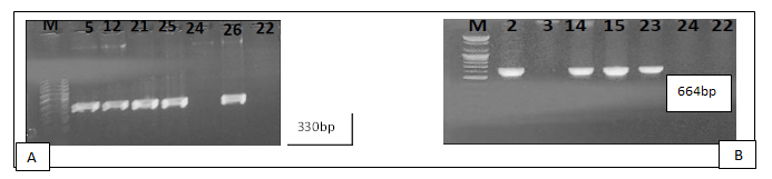
Fig. 1. A: Electrophoresis gel of PCR product of phzM genes. Lane L: DNA marker (100-1500bp); Lane (24) negative control; Lane (22) environmental isolate; Lane (5, 12, 21, 25, 26) clinical isolates give a positive result for this gene. B: Gel electrophoresis of PCR product of the phzS gene. Lane L: DNA marker (100-1500bp); Lane (24) negative control; Lane (3, 22) environmental isolates; Lane (2, 14, 15, 23) clinical isolates.
Sequencing of phzm and phzs genes
In the present study genotypic variation of environmental and clinical isolates of this bacterium was studied by sequencing of phzM and phzS genes. The relationships of genetic evolution of pseudomonas isolates were examined by comparing the analysis of the sequence with the NCBI. On the basis of the evidence by sequencing, it has been postulated that all the four isolates from clinical and environmental samples belong to the species Pseudomonas aeruginosa (Fig.2). The analysis of phylogenetic sequences has not revealed considerable diversity in environmental and clinical isolates (Fig.2). The findings of phzM gene sequence demonstrated that there were 7 mutations with 99% identities in 4 isolates as shown in (table 1) and (Fig.3), While for the phzS gene 2 mutations with identities 99% were detected by sequencing in the same isolates (table 2) and (Fig.4). The current results also appeared that P. aeruginosa isolates collected from environmental and clinical sources have a core genome which was highly conserved. However, these isolates tended to be variable in regards to the presence of regions involved in the phenazine phenotype.
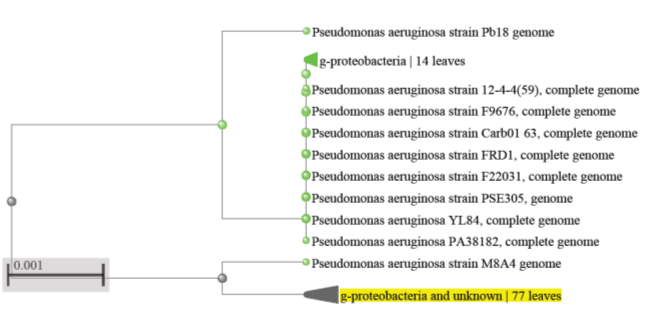
Fig. 2. Phylogenetic tree based on 16S rRNA gene sequence of 4 isolates of Pseudomonas aeruginosa compared with sequence available in the GenBank. Nodes indicate E value
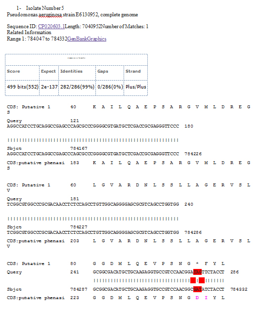
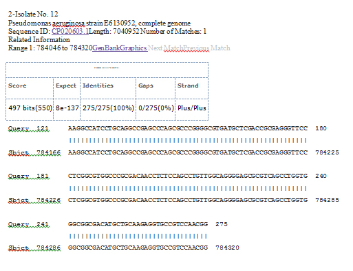
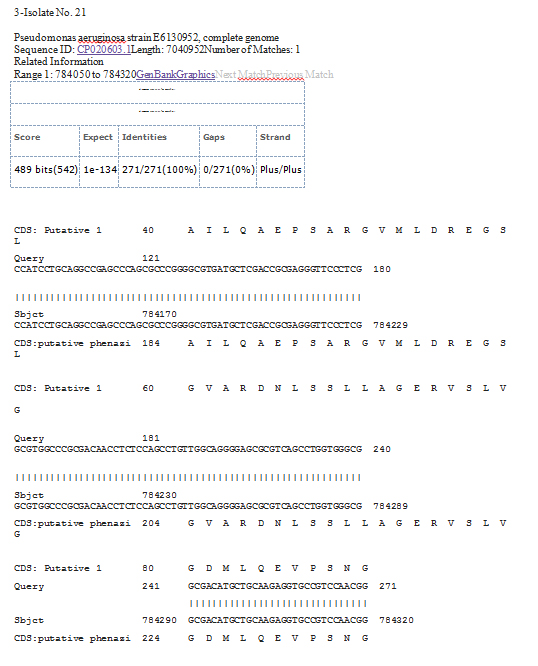
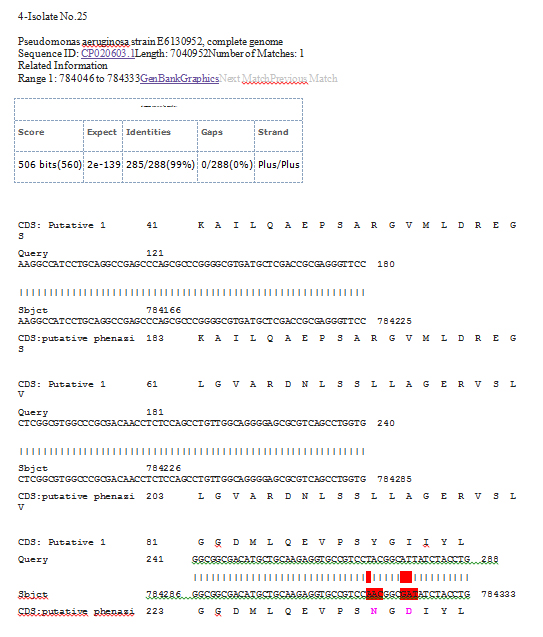
Fig. 3. Sequencing results of phzM genes of Pseudomonas aeruginosa in 4 isolates
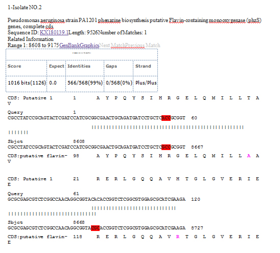
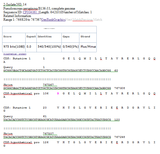
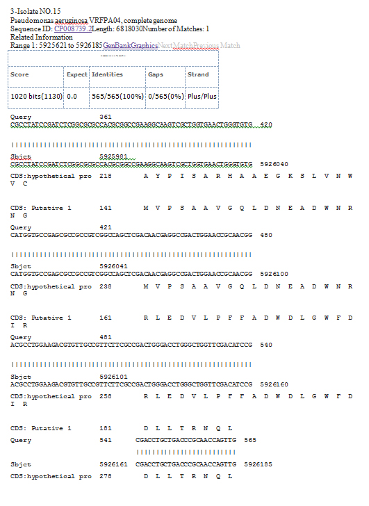
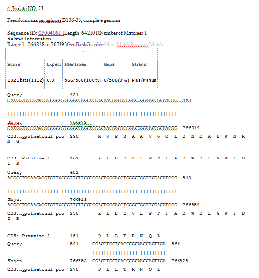
Fig. 4. Sequencing results of phzS genes of Pseudomonas aeruginosa in 4 isolates
Table (1):
Type of mutations in the phzM gene sequence of Pseudomonas aeruginosa in 4 isolates.
No. Of sample |
Wild-type |
Mutant type |
Type of mutation |
Location |
Amino acid change |
effect |
Sequence ID |
Identities |
|---|---|---|---|---|---|---|---|---|
5 |
GGC |
GGA |
Transversion |
784321 |
Glycine > Glycine |
nonsense |
ID: CP020603.1 |
99% |
GAT |
TAG |
Transvertion |
784322 |
Aspartic acid > Stop codons |
Missense |
|||
GAT |
TAG |
Transvertion |
784324 |
Aspartic acid > Stop codons |
Missense |
|||
ATC |
TTC |
Transvertion |
784342 |
Isoleucine> Phenylalanine |
Missense |
|||
12 |
ID: CP020603.1 |
100% |
||||||
21 |
ID: CP020603.1 |
100% |
||||||
25 |
AAC |
TAC |
Transvertion |
784316 |
Asparagine> Tyrosine |
Missense |
ID: CP020603.1 |
99% |
GAT |
ATT |
Transition |
784322 |
Aspartic acid > Isoleucine |
Missense |
|||
GAT |
ATT |
Transvertion |
784323 |
Aspartic acid > Isoleucine |
Missense |
Table (2):
Type of mutations in the phzS gene sequence of Pseudomonas aeruginosa in 4 isolates
No. Of sample |
Wild- type |
Mutant type |
Type of mutation |
Location |
Amino acid change |
effect |
Sequence ID |
Identities |
|---|---|---|---|---|---|---|---|---|
z2 |
GCC |
ACC |
Transition |
8660 |
Alanine> Threonine |
Missense |
ID: KX180139.1 |
99% |
CGC |
CAC |
Transition |
8697 |
Arginine> Histidine |
Missense |
|||
14 |
ID: CP004061.1 |
100% |
||||||
15 |
ID: CP008739.2 |
100% |
||||||
23 |
ID: CP004061.1 |
100% |
Table (3):
Effect of the type and percentage of mutation on phzM, phzS genes.
Effect of mutation |
number |
phzM (%) |
phzS (%) |
percentage |
|---|---|---|---|---|
nonsense |
1 |
1(11.11) |
0 (0.00) |
11.11 |
Missense |
8 |
6 (66.66) |
2 (22.22) |
88.88 |
Total |
9 |
7 |
2 |
99.99 |
Effect of mutations in phzM and phzS genes on phenazine production
To assess the impact of the biosynthesis modifying genes phzM and phzS on phenazine production in pseudomonas isolates with mutations in these genes. The isolates showed uncommon pigment phenotypes when they were cultured. While blue cultures of wild-type were attributable to the pyocyanin production, this finding was in agreement with the results conducted by4. It was interesting to mention that the existence of dual operons regulating biosynthesis of phenazine makes this bacterium more flexibility in modulating the number of phenazine compounds. It has been suggested that the variation in phenazine production might be ascribed to growth phase or in response to signals from the environment. Mutations affect the phzM and phzS genes through the creation of change in the gene sequence. The results in the table (1) showed that 1(11.11%) nonsense mutation which leads to change the code of amino acid stop codon, causing dysfunction of the protein. Furthermore, there is 8(88.88%) missense mutation in both genes, this type of mutation influences the phenotype because they lead to substitution of amino acids and thus in protein. An amino acid can be replaced with another amino acid that has similar chemical characteristics, therefore the protein could normally work. There is also an amino acid encoded by more than one code which could result in mutation. Nevertheless, this mutation does not produce any change in the translation.
From the present study, it seems that P. aeruginosa isolates of clinical and environmental sources have a very preserved genome core. The sequence of bacterial genome gives significant evidence to appreciate the regulatory and metabolic network that link chromosomal genes. Although the pseudomonas isolates have mutations in phzM and phzS genes, it has been proposed that these mutations have no role in the pathogenesis. In this site, the crucial part of P. aeruginosa virulence looks to be the regulation of gene expression instead of the existence or nonexistence of genes.
ACKNOWLEDGMENTS
I am thankful the Microbiology Department, Medicine College of Babylon University / Iraq for the facilities provided in the completion of the work. I am also thankful Hiba Jasim for her cooperation.
- Gross, H. and Loper, J.E. Genomics of secondary metabolite production by Pseudomonas spp. Natural Products Reports, 2009; 26: 1408-46.
- Shirley, F., John, P., Morrissey, Fergal O’Gara and Fidelma Boyd E. Genome Diversity of Pseudomonas aeruginosa isolates from Cystic Fibrosis Patients and the Hospital Environment. Journal of clinical microbiology, 2004; 42(12): 5783–5792.
- Lyczak, J.B., Cannon, C.L. and Pier, G.B. Establishment of Pseudomonasaeruginosa infection.: lessons from a versatile opportunist. Microbes & Infection, 2000; 2: 1051-1060.
- Mavrodi, D.V., Bonsall, R.F., Deleney, S.M., Soule, M.J., Phillips, G. and Thomashow, L.S. Functional Analysis of Genes for Biosynthesis of Pyocyanin and Phenazine-1-Carboxamide from Pseudomonas aeruginosa PAO1. Journal of Bacteriology, 2001; 183(21): 6454-6465
- Preetha, R., Jose, S., Prathapan, S., Vijayan, K.K., Jayaprakash, N.S., Philipm, R. and Bright Singh, I.S. An inhibitory compound produced by Pseudomonas with effectiveness on Vibrio harveyi. Aquaculture Research, 2010; 41: 1452-1461.
- Mavrodi, D.V., Ksenzenko, V.N., Bonsall, R.F., Cook, R.J., Boronin, A.M., Thomashow, L.S. A seven-gene locus for the synthesis of phenazine–1- carboxylic acid by Pseudomonas fluorescens. Journal of Bacteriology, 1998; 180: 2541–2548.
- Forbes, B.A. Daniel, F.S. and Alice, S.W: Bailey and Scott’s diagnostic microbiology, 12th. edn. USA: Mosby Elsevier Company, 2007.
- Frank, L. H., and DeMoss, R. D. On the biosynthesis of pyocyanine. J Bacteriol, 1959; 77: 776–782.
- Shi, H., Trinh, Q., Xu, W., Zhai, B., Luo, Y. and Huang, K. A universal primer multiplex PCR method for typing of toxinogenic Pseudomonas aeruginosa. Appl Microbiol Biotechnol, 2012; 95: 1579– 1587.
- Nowroozi, J., Sepahi, A.A. and Rashnonejad, A. Pyocyanine biosynthetic genes in clinical and environmental isolates of Pseudomonas aeruginosa and detection of pyocyanine‘s antimicrobial effects with or without colloidal silver nanoparticles. Cell Journal (Yakhteh), 2011; 14(1): 7-18.
- Chieda, Y., Iiyama, K., Lee, J.M., Kusakabe, T., Yasunaga-Aoki, C. and Shimizu, S. Inactivation of pyocyanin synthesis genes have no effect on the virulence of Pseudomonas aeruginosa PAO1 toward the silkworm, Bombyx mori. FEMS Microbiology Letters, 2008; 278: 101-107.
© The Author(s) 2018. Open Access. This article is distributed under the terms of the Creative Commons Attribution 4.0 International License which permits unrestricted use, sharing, distribution, and reproduction in any medium, provided you give appropriate credit to the original author(s) and the source, provide a link to the Creative Commons license, and indicate if changes were made.