


ISSN: 0973-7510
E-ISSN: 2581-690X
 , Chitapha Kamrai1, Monnicha Thaprod1, Aekasit Suphannarot2 and Ratchanee Mingma3
Antibiotic resistance in microorganisms is a serious public health problem worldwide, highlighting the need for alternative strategies to control bacterial infections. One promising target is quorum sensing (QS), which is a bacterial cell-to-cell communication system that regulates various physiological processes including the production of virulence factors and biofilm formation, both of which contribute to bacterial persistence and resistance. This study investigated the QS inhibitory (QSI) and antibiofilm activities of actinobacteria. A total of 98 actinobacterial isolates were screened for QSI activity using Chromobacterium violaceum KCTC 2897 as the reporter strain. Seven isolates inhibited violacein production without affecting bacterial growth, indicating their potential QSI activity. The crude ethyl acetate extracts from six of these isolates retained their QSI activity. Among these, extract 43N8 exhibited the strongest antibiofilm activity at a sub-minimum inhibitory concentration of 6.25 mg mL-1, reducing biofilm formation by 58.2% in Pseudomonas aeruginosa and 79.7% in Staphylococcus aureus. Extract 42N3 showed the second-highest antibiofilm activity, inhibiting biofilm formation by S. aureus by 62.8%. Phylogenetic analysis using 16S rRNA gene sequences revealed that isolates 43N8 and 42N3 were members of the genus Streptomyces and shared the highest sequence similarity with Streptomyces misionensis NBRC 13063T (99.57%) and Streptomyces barringtoniae LMG 32415T (98.03%). These findings revealed the QSI and antibiofilm potential of actinobacterial extracts, highlighting their potential as sources of bioactive compounds for the development of novel approaches to control bacterial infections.
Actinobacteria, Quorum Sensing Inhibitory Activity, Antibiofilm Activity, Streptomyces
Bacterial cell-to-cell communication, known as quorum sensing (QS), regulates gene expression in response to cell population density within an environment. Bacteria produce and release small signaling molecules called autoinducers, which accumulate in the environment as cell density increases. Gram-negative bacteria such as Acinetobacter spp. and Pseudomonas spp. commonly use acyl-homoserine lactones (AHLs), whereas Gram-positive bacteria such as Staphylococcus aureus predominantly use peptide-based signals, particularly autoinducing peptides.1 When the concentration of these signaling molecules reached a threshold level, these molecules bind to specific receptors and activate signal transduction pathways, resulting in coordinated changes in gene expression across the bacterial population.2 Through this mechanism, QS regulates diverse physiological processes associated with bacterial survival and pathogenicity, such as virulence factor expression, extracellular enzyme secretion, antibiotic production, motility, competence, conjugation, sporulation, and biofilm formation.3
Biofilms are densely aggregated communities of bacteria attached to both biotic and abiotic surfaces and encapsulated within a self-produced extracellular polymeric substance matrix. This matrix functions to maintain structural cohesion among bacterial cells and protects them from unfavorable environmental conditions.4 Biofilm formation is recognized as a major function contributing to bacterial tolerance to antibiotics, ultimately leading to antimicrobial resistance in various pathogenic microorganisms. In addition, biofilms enable bacteria to evade host immune responses.5 Consequently, increasing research has been directed toward QS inhibition (QSI) and strategies to suppress biofilm formation.
Actinobacteria are Gram-positive bacteria characterized by filamentous growth that resembles that of fungi. They are widely distributed in both aquatic and terrestrial environments and are well recognized as an important group of microorganisms capable of producing a wide range of biologically active secondary metabolites.6,7 These metabolites include compounds with autoinducer-inhibitory activity associated with QS systems.8 In addition, secondary metabolites derived from various actinobacterial species have been extensively utilized in medical applications, particularly as antimicrobial and anti-inflammatory agents.9 Therefore, actinobacteria represent a promising and valuable biological resource for the investigation of anti-QS and antibiofilm activities. However, studies focusing on the isolation, screening, and evaluation of QSI and antibiofilm activities of actinobacteria remain relatively limited, particularly those obtained from underexplored environments such as peat swamp soils. Accordingly, the present study assessed the potential anti-QS and antibiofilm activities of actinobacteria isolated from peat swamp forests. These findings provide valuable insights that may support the development of alternative strategies for controlling antimicrobial-resistant pathogens and reducing bacterial infections.
Actinobacterial and pathogenic bacteria
Ninety-eight actinobacterial isolates previously isolated from peat swamp forest soils were used in this investigation.10 The isolates were maintained on International Streptomyces Project medium 2 (ISP2)11 and incubated at 30 °C for 7 days. Chromobacterium violaceum KCTC 2897 was used as the reporter strain for screening the QSI activity, whereas Pseudomonas aeruginosa and S. aureus were used as test organisms for antibacterial and antibiofilm assays. All bacterial strains were cultured on Luria-Bertani (LB) agar (HiMedia, India) and incubated at 30 °C for 24 hrs.
Primary screening for QSI activity
A spore suspension of each actinobacterial isolate was prepared by scraping spore and mycelia from 10 day-old cultures grown on ISP2 agar into a 1.5 mL microcentrifuge tube containing 500 µL of sterile 0.85% (weight/volume, w/v) NaCl solution. The resulting suspension was streaked in duplicate as a straight line across the center of the LB agar plate, which were then incubated at 30 °C for 7 days.
The reporter strain, C. violaceum KCTC 2897, was cultured in LB broth with shaking (150 rpm) for 24 hrs at 30 °C. The cell density was adjusted using a spectrophotometer at 600 nm to an optical density of 0.132 (approximately 108 CFU mL-1). Subsequently, 100 µL of the bacterial culture was mixed with 4 mL of LB soft agar containing 0.6% (w/v) agar and gently vortexed before being overlaid onto LB agar plates previously inoculated with actinobacteria. As a negative control, LB agar plates without actinobacterial inoculation were overlaid with 4 mL of LB soft agar containing C. violaceum KCTC 2897. The plates were incubated at 30 °C for 24 hrs. QSI activity was determined by observing violacein production in C. violaceum KCTC 2897 around the actinobacterial colonies. The presence of a turbid but non-pigmented zone surrounding actinobacterial growth was interpreted as potential QSI activity. Isolates showing consistent pigment inhibition on both plates were selected for subsequent experiments.
Preparation of crude extracts
Actinobacterial isolates exhibiting QSI activity were selected and cultured in four flasks containing 100 mL of tryptic soy broth (TSB; HiMedia). The cultures were incubated at 150 rpm, 30 °C for 7 days. Culture cells were centrifuged at 9,000 rpm for 10 min, the cell-free supernatant was collected and filtered-sterilized using 0.45 µm membrane filter. The cultured filtrates were extracted with ethyl acetate in a 1:1 (v/v) ratio and agitated at 200 rpm for 24 hrs. The mixture was kept stationary until two distinct phases were formed. The organic phase (upper layer) was separated from the aqueous phase (lower layer) using a separatory funnel and concentrated under reduced pressure at 35 °C using a rotary evaporator. The crude metabolite extract was weighed and dissolved in 1% (v/v) dimethyl sulfoxide (DMSO) to obtain a final concentration of 50 mg mL-1 for subsequent experiments.
Secondary screening of QSI activity
The QSI activity of the crude extracts was evaluated using a two-fold serial dilution assay in a 96-well microtiter plate. Briefly, 100 µL of LB broth was added to each well. Subsequently, 100 µL of crude extract (50 mg mL-1) was added to the first well. All experiments were performed in triplicate. Two-fold serial dilutions were subsequently performed by transferring 100 µL from the first well to the second well, mixing thoroughly, and then transferring 100 µL from the second well to the third well. This procedure was continued sequentially to the tenth well. After mixing, 100 µL was discarded from the tenth well. Following the serial dilution, 100 µL of C. violaceum KCTC 2897 suspension, adjusted to approximately 108 CFU mL-1 (optical density OD600 = 0.132), was added to each well. The final concentrations of the crude extracts in the wells ranged from 12.5 to 0.024 mg mL-1. Wells containing only the bacterial suspension and culture medium were used as untreated controls. The microtiter plates were incubated at 30 °C for 24 hrs. QSI activity was assessed based on the reduction or absence of violacein production by C. violaceum KCTC 2897 compared to the untreated controls. Wells showing bacterial growth with reduced purple pigmentation were considered positive for QSI activity, indicating inhibition of QS-regulated violacein production without affecting bacterial growth.
Evaluation of antimicrobial activity by Minimum Inhibitory Concentration determination
The minimum inhibitory concentration (MIC) of the crude extracts were determined using the broth microdilution method in 96-well microtiter plates. Two-fold serial dilutions of the crude extracts were prepared in LB broth as described for the QSI assay. Subsequently, 100 µL of bacterial suspension adjusted to approximately 108 CFU mL-1 was added to each well. The assay was performed using C. violaceum KCTC 2897, P. aeruginosa, and S. aureus. Wells containing bacterial suspensions without crude extracts served as growth controls, whereas wells containing only the culture medium served as sterility controls. All experiments were performed in triplicate. Following incubation at 30 °C for 24 hrs, 30 µL of 0.02% (w/v) resazurin solution was added into each well and the microplates were further incubation for 2 hrs at the same conditions. Bacterial viability was evaluated based on the reduction in resazurin, where a change from blue to pink indicated metabolic activity and cell growth. Wells that remained blue were considered to indicate complete inhibition of bacterial growth. The MIC was recorded as the lowest concentration of the crude extract that prevented the color change of resazurin from blue to pink.
Antibiofilm assay
The antibiofilm potential of the crude extracts was evaluated against P. aeruginosa and S. aureus using a crystal violet microtiter plate assay. The extracts were tested at concentrations below the MIC (sub-MICs), corresponding to 1/2, 1/4, and 1/8 of the respective MIC values. Bacterial cultures were grown overnight in LB broth with shaking and adjusted to approximately 108 CFU mL-1, as described above. Subsequently, 100 µL of the bacterial suspension was added to the wells of a 96-well microtiter plate and mixed with an equal volume of crude extract at the designated concentrations. The concentration of each crude extract was determined in triplicate. Wells containing bacterial suspensions without crude extract served as untreated controls. The microplates were incubated at 30 °C for 24 hrs to allow for biofilm formation. After incubation, the culture supernatant containing non-adherent cells was carefully discarded, and the wells were rinsed twice with phosphate-buffered saline (PBS) to remove loosely attached cells. The remaining biofilm was stained with 0.2% (w/v) crystal violet for 30 min at room temperature. Following staining, excess dye was removed by washing the wells twice with PBS and the plates were allowed to dry for 5 min before adding 200 µL of 70% (v/v) ethanol to solubilize the crystal violet. Biofilm biomass was quantified by measuring the absorbance at 570 nm using a microplate reader. The inhibitory effect on biofilm formation was expressed as a percentage and calculated using the following equation:
Percentage of inhibition (%) = [(OD control – OD treated) / OD control] x 100
where OD control represents the absorbance measured for the untreated control, and OD treated represents the absorbance measured for the crude extract-treated sample.
Microscopic observation
Biofilm formation on coverslips was assessed using a 12-well microtiter plate. Sterile coverslips were cut into pieces of approximately 1 × 1 cm and placed individually in each well. Each treatment was performed in duplicate. Suspensions of P. aeruginosa and S. aureus (approximately 108 CFU mL-1; OD600 = 0.312) were inoculated into wells containing coverslip pieces and treated with the crude extract at 1/2 MIC. Wells containing bacterial suspensions without crude extract were used as untreated controls. After incubation at 30 °C for 24 hrs, the coverslips were carefully rinsed twice with PBS to remove unattached cells. The coverslips were stained with 0.2% (w/v) crystal violet for 10 min, rinsed with PBS to remove excess stain, and air-dried. The stained biofilms were observed under a light microscope at 400x and 1,000x.
Identification of actinobacteria based on 16S rRNA gene sequence analysis
Actinobacteria were cultured in glucose yeast extract broth (10 g glucose, 10 g yeast extract, and 1 L H2O, pH 7.0) for 5 days. The cultures were harvested by centrifugation at 9,000 rpm for 10 min and the supernatant was discarded. The pellets were washed twice with sterile distilled water and resuspended in Tris-EDTA buffer pH 7.0 (HiMedia). Genomic DNA was extracted using sonication. The cell suspension was sonicated using an ultrasonic cleaner (50 W) for 3 min and then centrifuged at 8,000 rpm for 5 min. The resulting supernatants were carefully transferred to new microcentrifuge tubes. The 16S rRNA gene was amplified by polymerase chain reaction (PCR) using the bacterial primers 1F (5′-TCACGGAGAGTTTGATCCTG-3′) and 1530R (5′-AAGGAGATCCAGCCGCA-3′).12 The amplification conditions were as follows: one cycle at 94 °C for 5 min, 30 cycles at 94 °C for 20 sec, 50 °C for 20 sec, and 72 °C for 1 min, and a final extension cycle at 72 °C for 5 min. The PCR products were verified using 1% (w/v) agarose gel electrophoresis, targeting fragments of approximately 1,500 bp. The amplified DNA fragments were purified using a FavorPrep GEL/PCR Purification Mini Kit (Favorgen, Taiwan) according to the manufacturer’s instructions and commercially sequenced by Bionics (Daejeon, Republic of Korea) using the primers 27F (5′ -AGAGTTTGATCMTGGCTCAG-3′) and 1492R (5′ -GGTTACCTTGTTACGACTT-3′). The resulting 16S rRNA gene sequences were compared to the type strain sequences available in the EzBioCloud database (http://ezbiocloud.net)13 using the Basic Local Alignment Search Tool to determine the closest relatives. Phylogenetic relationships were evaluated by constructing a phylogenetic tree using the Neighbor-Joining method14 in the Molecular Evolutionary Genetic Analysis software package, version 12.15 The confidence level of the tree topology was evaluated by bootstrap analysis with 1,000 replicates16 and evolutionary distances were calculated using the Tamura–Nei model.17 The sequences were deposited in GenBank (http://www.ncbi.nlm.nih.gov/genbank/index.html).
Primary screening for QSI activity
A total of 98 actinobacterial isolates were screened for QSI activity using C. violaceum KCTC 2897 as the reporter strain. Seven isolates (7.14%) exhibited QSI activity, as evidenced by bacterial growth accompanied by inhibition of violacein production around actinobacterial colonies (Figure 1). The formation of a turbid but non-pigmented zone indicated suppression of QS-regulated violacein production without affecting the growth of the reporter strain. These seven isolates were selected for crude metabolite extraction using ethyl acetate and were subsequently evaluated for their QSI and antibiofilm activities.
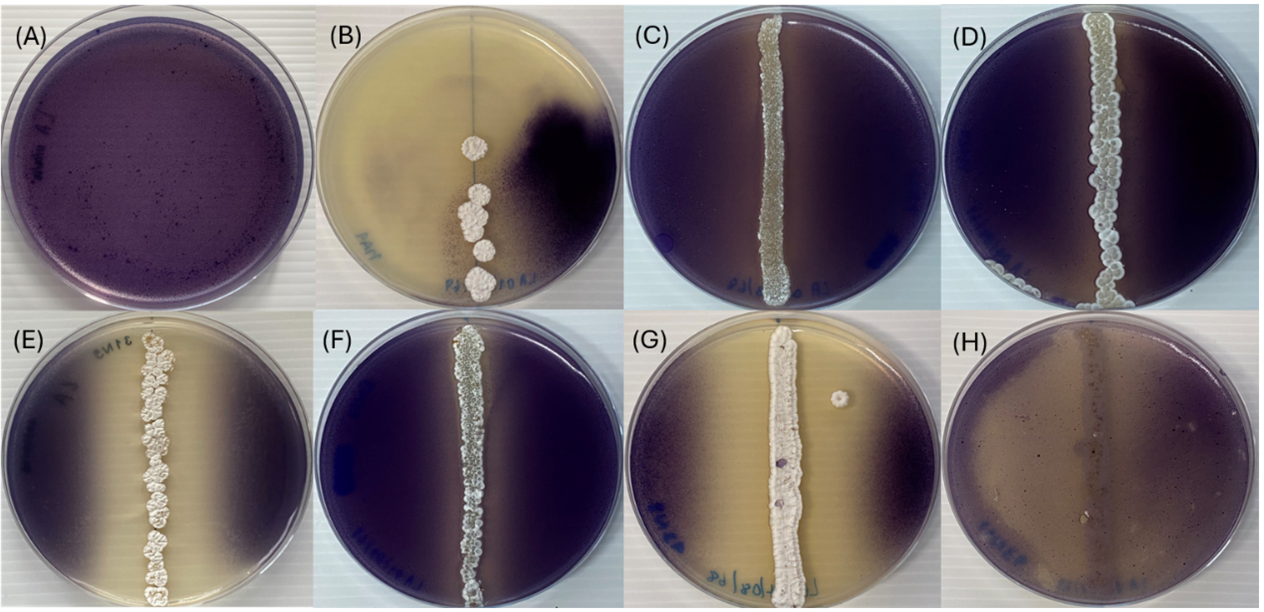
Figure 1. Quorum sensing inhibitory activity of actinobacterial isolates against Chromobacterium violaceum KCTC 2897 as a reporter strain. (A) Control; (B) isolate 11A4; (C) isolate 11N4; (D) isolate 11N7; (E) isolate 31N5; (F) isolate 42N3; (G) isolate 43N8; and (H) isolate 43N11
Evaluation of QSI by actinobacterial crude extracts
The crude ethyl acetate extracts obtained from all seven actinobacterial isolates exhibited QSI activity against C. violaceum KCTC 2897, as evidenced by the suppression of violacein production. Six crude extracts (11A4, 11N4, 11N7, 31N5, 42N3, and 43N8) inhibited violacein production at concentrations ranging from 1.56 to 3.13 mg mL-1, whereas extract 43N11 required a concentration of 6.25 mg mL-1 to produce the same effect (Figure S1).
A comparison of the QSI-active concentrations with the corresponding MIC values revealed that the concentrations required to suppress violacein production by isolates 11A4, 11N4, 11N7, 31N5, 42N3, and 43N8 were lower than the MIC values against C. violaceum (Table 1; Figure 2 and Figure S2). In contrast, the minimum concentration of 43N11 required to inhibit violacein production was identical to the MIC values (6.25 mg mL-1). Therefore, extract 43N11 was excluded from the subsequent experiments.
Table 1. Quorum sensing inhibitory (QSI) activity and minimum inhibitory concentration (MIC) of crude extracts from actinobacterial isolates against the reporter strain Chromobacterium violaceum KCTC 2897
Isolate |
QSI concen. (mg mL-1) |
MIC (mg·mL-1) |
|---|---|---|
11A4 |
1.56 |
6.25 |
11N4 |
3.13 |
6.25 |
11N7 |
1.56 |
6.25 |
31N5 |
3.13 |
6.25 |
42N3 |
1.56 |
6.25 |
43N8 |
3.13 |
6.25 |
43N11 |
6.25 |
6.25 |
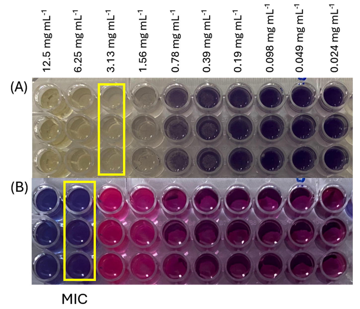
Figure 2. Evaluation of QSI activity (A) and antibacterial activity (B) of crude extracts against C. violaceum KCTC 2897 using two-fold serial dilution assay in 96-well microtiter plates. Yellow boxes indicate the minimum concentrations required to inhibit violacein production (A) and bacterial growth (B)
The yellow boxes indicate the minimum concentrations required to inhibit violacein production (A) and bacterial growth (B).
Minimum inhibitory concentration (MIC) of crude extract
The MICs of the crude extracts of the six selected actinobacterial isolates against P. aeruginosa and S. aureus were determined using a resazurin-based broth microdilution assay. The MIC values ranged from 3.13 to 12.5 mg mL-1 depending on the isolate and test organism (Table 2; Figures S3 and S4). The crude extract of isolate 11A4 exhibited the lowest MIC value (3.13 mg mL-1) against both bacterial strains. The extracts from isolates 11N4, 11N7, and 31N5 showed MIC values of 6.25 mg mL-1 against both P. aeruginosa and S. aureus. For isolate 42N3, the MIC values were 12.5 mg mL-1 against P. aeruginosa and 6.25 mg mL-1 against S. aureus. The crude extract from isolate 43N8 exhibited MIC values of 12.5 mg mL-1 against both bacterial strains. Based on these results, sub-MIC concentrations were selected for subsequent antibiofilm assays.
Table 2. Minimum inhibitory concentrations (MICs) of crude extracts from selected actinobacterial isolates against P. aeruginosa and S. aureus
| Isolate | MIC (mg mL-1) against | |
|---|---|---|
| P. aeruginosa | S. aureus | |
| 11A4 | 3.13 | 3.13 |
| 11N4 | 6.25 | 6.25 |
| 11N7 | 6.25 | 6.25 |
| 31N5 | 6.25 | 6.25 |
| 42N3 | 12.5 | 6.25 |
| 43N8 | 12.5 | 12.5 |
Antibiofilm formation activity
Among the six crude extracts, those from isolates 42N3 and 43N8 inhibited biofilm formation by both P. aeruginosa and S. aureus, whereas extract 11A4 inhibited biofilm formation only in S. aureus. Biofilm reduction, as determined by crystal violet staining and optical density measurements, is shown in Figures S5 and S6.
The crude extract from isolate 43N8 inhibited P. aeruginosa biofilm formation by 58.2, 56.1 and 36.3% at concentrations of 6.25, 3.13 and 1.56 mg mL-1, respectively. These concentrations corresponded to 1/2, 1/4, and 1/8 MIC, respectively. Against S. aureus, biofilm inhibition was 79.7%, 64.3%, and 47.6% at the corresponding concentrations tested (Figures 3 and 4). The crude extract from isolate 42N3 inhibited biofilm formation by both pathogens, with inhibition percentages ranging from 49.9 to 62.8% against S. aureus across the tested sub-MIC concentrations. In P. aeruginosa, biofilm inhibition was detected only at 6.25 mg mL-1 (1/2 MIC), with an inhibition percentage of 15.3%. The crude extract of isolate 11A4 inhibited S. aureus biofilm formation by 18.2% at 1/2 MIC (1.56 mg mL-1), whereas no inhibition was detected at 1/4 or 1/8 MIC (Figure 4).
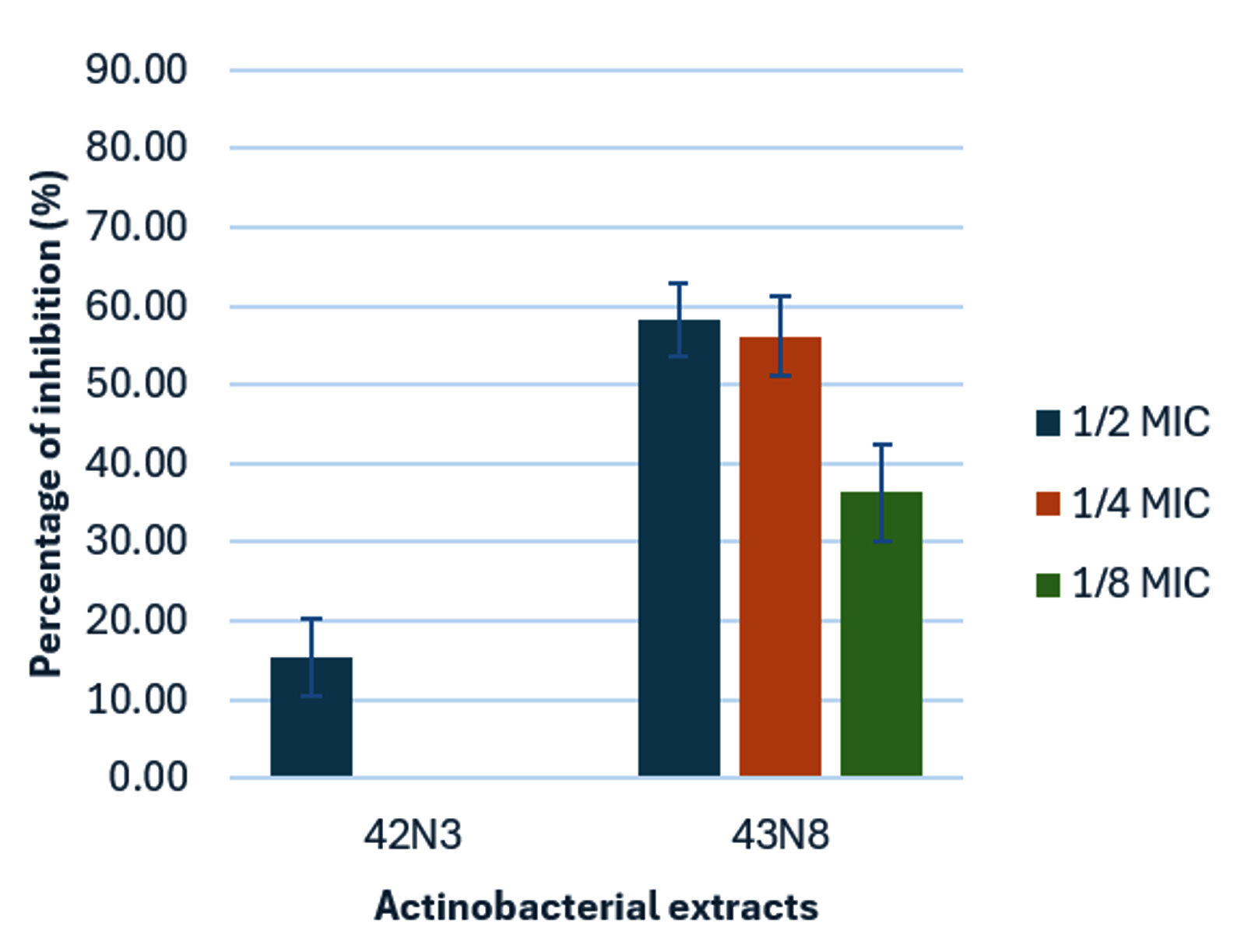
Figure 3. Antibiofilm activity of actinobacterial extracts against Pseudomonas aeruginosa at sub-MIC levels. Bars represent mean ± SD (n = 3)
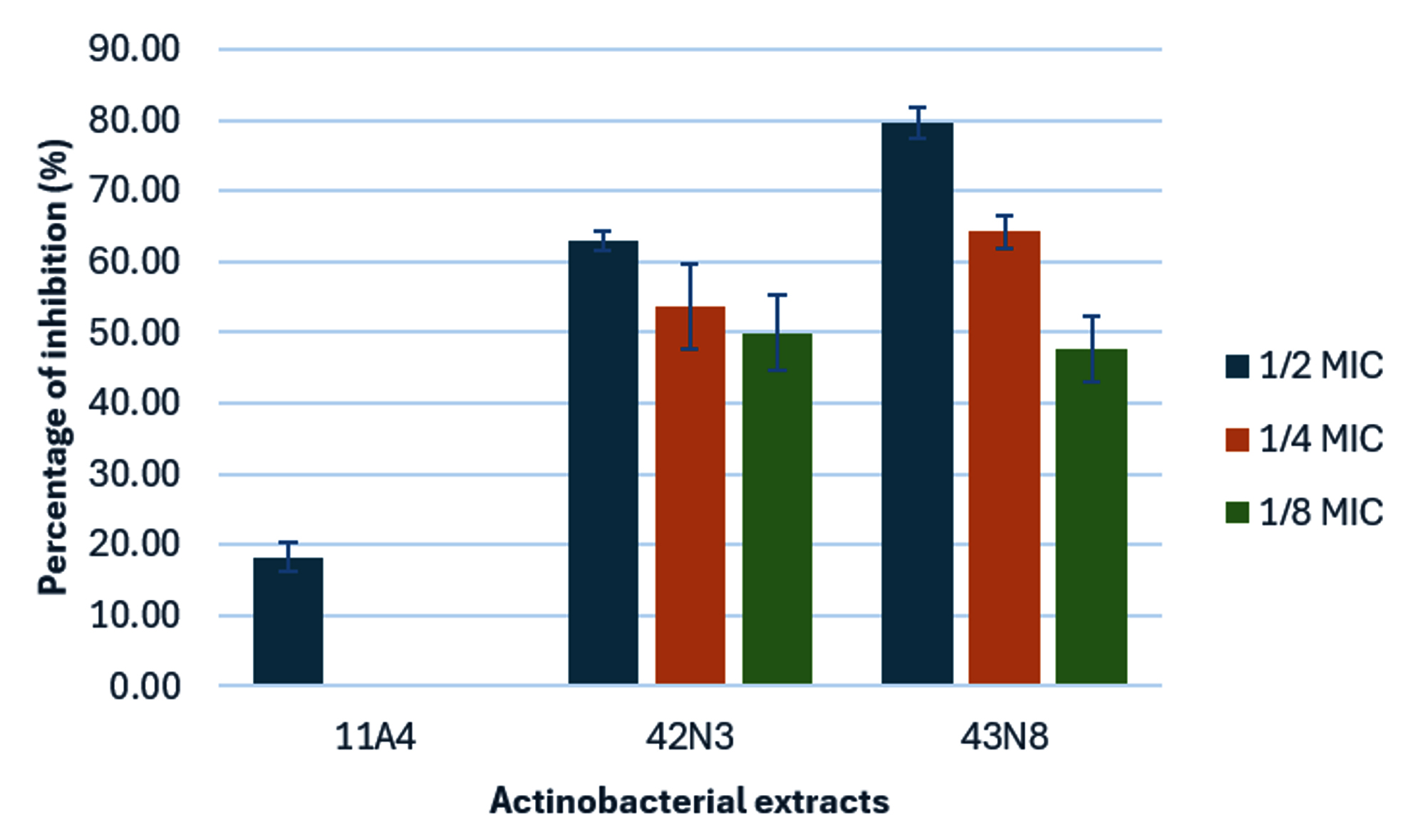
Figure 4. Antibiofilm activity of actinobacterial extracts against Staphylococcus aureus at sub-MIC levels. Bars represent mean ± SD (n = 3)
Microscopic images of biofilm formation on coverslip surfaces showed fewer bacterial cells attached after treatment with crude extract 43N8 (6.25 mg mL⁻¹) compared with the untreated controls (Figures 5A and 6A). Reduced biofilm formation was observed in both S. aureus and P. aeruginosa (Figures 5B and 6B). Similarly, reductions in biofilm formation were observed with crude extract 42N3 (Figures 5C and 6C), although the effect was less than that of crude extract 43N8. These observations are consistent with the percentage inhibition results, further demonstrating the effectiveness of crude 43N8 and 42N3 in inhibiting biofilm formation. In contrast, crude extract 11A4 inhibited biofilm formation by S. aureus but showed no inhibitory effect against P. aeruginosa (Figures 5D and 6D).
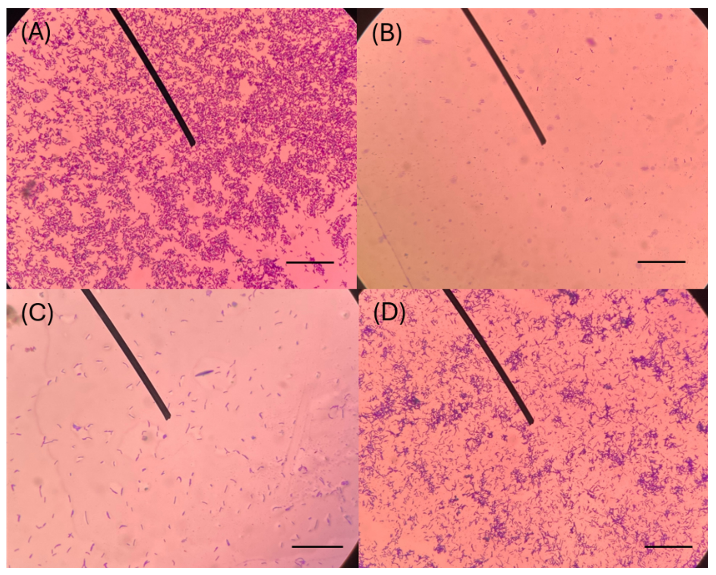
Figure 5. Light microscopic images (x400) of P. aeruginosa biofilms formed on coverslips and treated with crude extract. (A) Untreated control; (B) isolate 43N8; (C) isolate 42N3; and (D) isolate 11A4. Scale bar = 50 µm
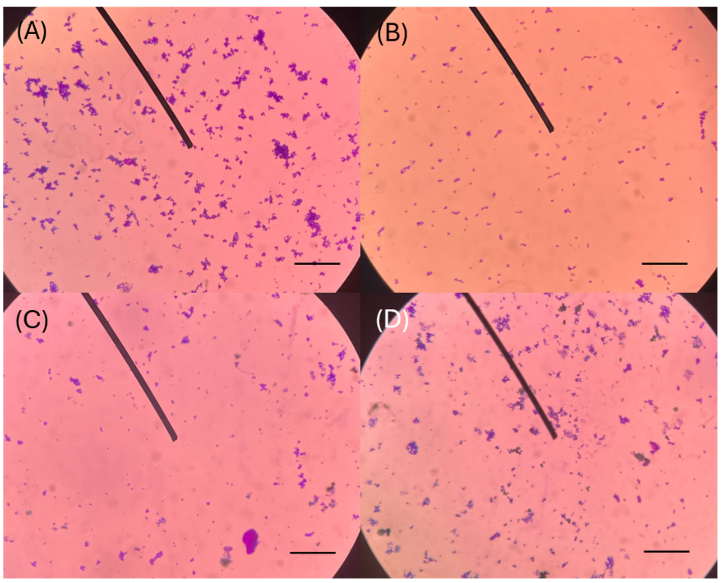
Figure 6. Light microscopic images (x400) of S. aureus biofilms formed on coverslips and treated with crude extract. (A) Untreated control; (B) isolate 43N8; (C) isolate 42N3; and (D) isolate 11A4. Scale bar = 50 µm
Identification of actinobacteria
Seven actinobacterial isolates exhibiting anti-QS activities were identified based on 16S rRNA gene sequence analysis and assigned to the genus Streptomyces (Table 3; Figure 7). Isolates 11A4 (accession number PZ125053) and 43N8 (PZ125058) showed the highest 16S rRNA gene sequence similarity with Streptomyces misionensis NBRC 13063T (99.57%), whereas isolate 42N3 (PZ125057) showed the highest sequence similarity with Streptomyces barringtoniae LMG 32415T (98.03%).
Table 3. Percent similarity analysis of the 16S rRNA gene sequence between the isolates and their closest related strains
Isolate |
Accession number |
Closely related taxa |
Similarity (%) |
Difference/Total nucleotide |
|---|---|---|---|---|
11A4 |
PZ125053 |
Streptomyces misionensis NBRC 13063T (EF178678) |
99.57 |
6/1405 |
11N4 |
PZ125054 |
Streptomyces humi MUSC 119T (KJ632661) |
98.98 |
14/1377 |
11N7 |
PZ125055 |
Streptomyces humi MUSC 119T (KJ632661) |
98.84 |
16/1382 |
31N5 |
PZ125056 |
Streptomyces misionensis NBRC 13063T (EF178678) |
99.43 |
8/1393 |
42N3 |
PZ125057 |
Streptomyces barringtoniae LMG 32415T (LC635741) |
98.03 |
27/1368 |
43N8 |
PZ125058 |
Streptomyces misionensis NBRC 13063T (EF178678) |
99.57 |
6/1388 |
43N11 |
PZ125059 |
Streptomyces sennicomposti RCPT1-4T (OM661191) |
99.27 |
10/1377 |
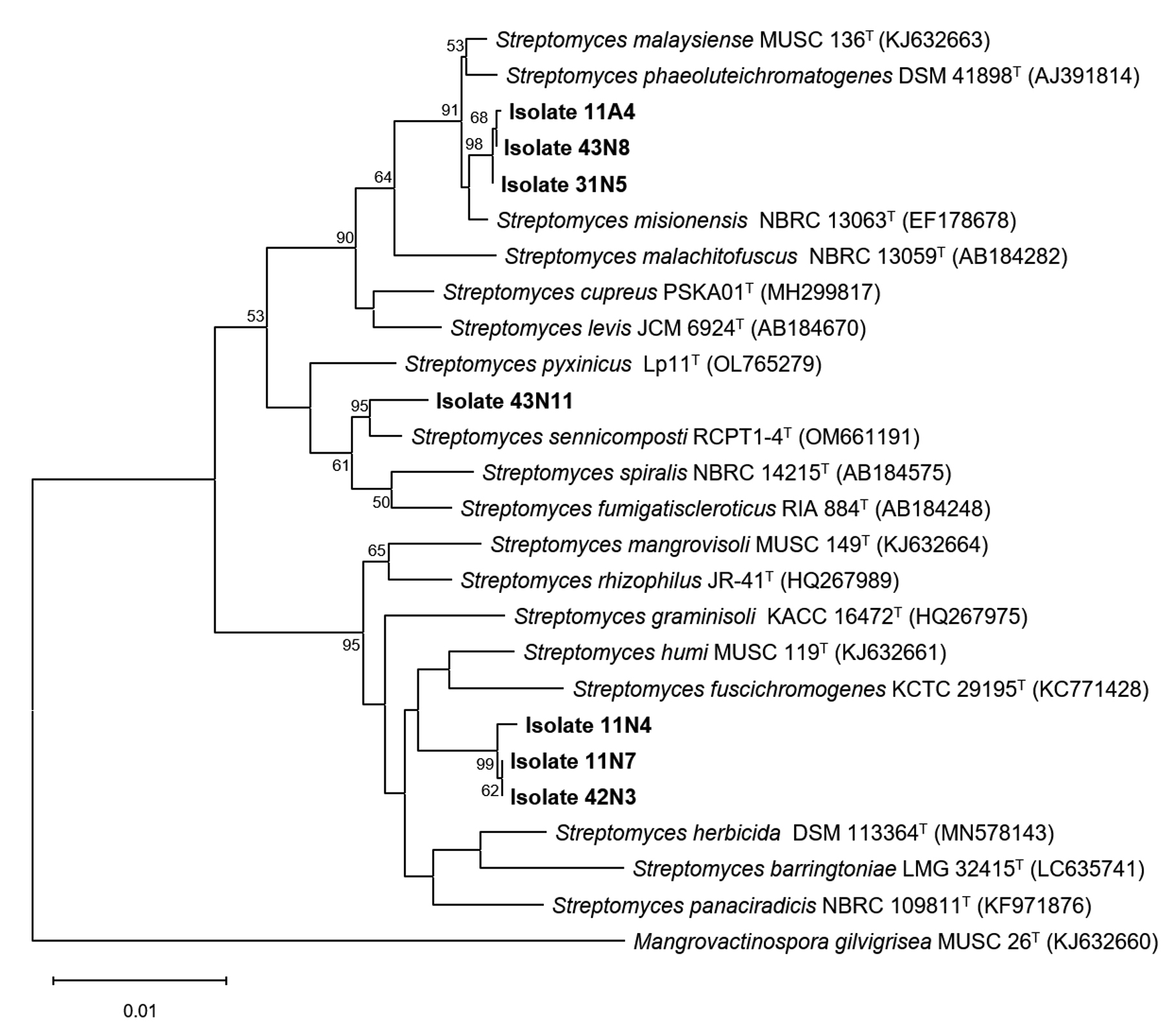
Figure 7. Neighbor-joining (NJ) phylogenetic tree14 constructed via the Tamura–Nei model17 on the basis of 16S rRNA gene sequences, showing the relationships between the active isolates and other members of Streptomyces. Bootstrap values (≥50%) based on 1,000 replications are shown at the branch nodes. Mangrovactinospora gilvigrisea MUSC 26T (KJ632660) was used as an outgroup. Bar, 0.01 nucleotide substitutions per site
Actinobacteria are widely recognized as important sources of bioactive secondary metabolites, including antimicrobial, antibiofilm, and QSI compounds.18 Numerous studies have reported that metabolites produced by actinobacteria can interfere with bacterial QS systems, thereby suppressing the expression of virulence factors and inhibiting biofilm formation without necessarily affecting bacterial growth.19 Such anti-QS activity is considered a promising alternative strategy for controlling pathogenic bacteria while potentially reducing the selective pressure associated with conventional antibiotics. Notably, the actinobacterial isolates investigated in this study originated from peat swamp soils, which are relatively underexplored environments in terms of actinobacterial diversity. The unique ecological conditions of this habitat may favor the production of specialized secondary metabolites, warranting further investigation into its capacity to produce specialized secondary metabolites, including potential QSI compounds.
The selective occurrence of QSI activity observed among the isolates in this study suggests that this bioactive property may be strain-dependent rather than a general characteristic of actinobacteria. This observation is consistent with previous reports showing that only a limited proportion of environmental actinobacterial isolates exhibit QSI activity.19,20 Such strain-specific activity may reflect differences in the biosynthetic capacity of individual actinobacterial strains and their ability to produce secondary metabolites capable of interfering with bacterial signaling pathways. The use of C. violaceum as a reporter strain provides a useful initial screening approach, as violacein production is regulated by an AHL-dependent QS system and can be readily detected by the characteristic purple pigmentation.21 Therefore, inhibition of violacein production in the absence of growth inhibition provides an indication of potential interference with QS rather than a nonspecific antibacterial effect.
To confirm that the observed inhibition of violacein production was due to QS interference rather than antibacterial activity, the crude extracts were evaluated for both QSI and antimicrobial activities. Six of the seven crude extracts inhibited violacein production at concentrations below their respective MICs against C. violaceum, indicating that these extracts interfere with QS-regulated pigment production without suppressing bacterial growth. In contrast, crude extract 43N11 inhibited both violacein production and bacterial growth at the same concentration, suggesting that the apparent QSI activity was likely a consequence of growth inhibition. This distinction is critical because growth suppression can lead to false positive results in QSI assays. Therefore, the exclusion of isolate 43N11 from subsequent analyses ensured that only the extracts exhibiting potential QSI activity were further investigated. However, the absence of a DMSO solvent control is a limitation of this study. Because the crude extracts were dissolved in DMSO, the potential contribution of the solvent to the observed effects could not be excluded. Therefore, future studies should include an appropriate DMSO control to confirm that the observed biological activities are attributable to the crude extract, rather than to the solvent.
Biofilm formation is an important bacterial survival strategy that is frequently regulated by QS systems. Biofilm-associated infections are challenging to treat because biofilm-embedded cells exhibit enhanced tolerance to antimicrobial agents and host immune responses.22 Therefore, compounds capable of interfering with QS may also exhibit antibiofilm activity. In the present study, crude extracts from isolates 42N3 and 43N8 substantially inhibited biofilm formation by both P. aeruginosa and S. aureus at sub-MIC concentrations, indicating that the observed antibiofilm activity was not primarily attributable to growth inhibition. The use of sub-inhibitory concentrations is particularly important for evaluating antibiofilm effects because it minimizes the possibility of false-positive results arising from antimicrobial activity.23 However, the crystal violet assay measures total biofilm biomass and cannot distinguish between viable and non-viable cells. Thus, reduced crystal violet staining alone cannot confirm the inhibition of biofilm development despite the use of sub-MIC concentrations. Complementary viability assays are required to further validate the antibiofilm effects of the extracts. Nevertheless, the observed antibiofilm and QSI activities support the possibility that these extracts interfere with bacterial processes involved in biofilm formation.
Among the tested isolates, crude extract 43N8 exhibited the strongest antibiofilm activity, with inhibition of biofilm formation by both P. aeruginosa and S. aureus. The pronounced activity against both Gram-negative and Gram-positive pathogens suggests that the extract contains bioactive metabolites with broad-spectrum antibiofilm properties. Microscopic observations further supported the quantitative results obtained from the crystal violet assay, which demonstrated a reduction in surface-associated bacterial cells following treatment with the extracts of isolates 43N8 and 42N3. Similar associations between QSI and reduced biofilm formation have been reported for several Streptomyces-derived metabolites, including phenolic compounds, diketopiperazines, and other secondary metabolites capable of interfering with bacterial communication pathways.24
16S rRNA gene sequence analysis identified the active isolates as members of the genus Streptomyces. This finding is consistent with previous reports demonstrating that Streptomyces species are prolific producers of biologically active secondary metabolites, including antimicrobial, antivirulent, and QSI compounds.25 Several Streptomyces-derived metabolites have been reported to interfere with AHL-mediated signaling systems and inhibit biofilm formation by pathogenic bacteria.26,27 Therefore, these findings support the potential involvement of secondary metabolites produced by Streptomyces isolates in QSI and antibiofilm activities.
The results demonstrated that Streptomyces isolates 42N3 and 43N8 are promising sources of bioactive metabolites with potential QSI and antibiofilm properties. The ability of the crude extracts to inhibit biofilm formation at sub-inhibitory concentrations further supports their potential as candidates for the development of alternative approaches to control biofilm-associated infections. Future studies should focus on bioassay-guided fractionation, structural elucidation of active metabolites, and investigation of their mechanisms of action, particularly their effects on bacterial QS pathways.
This study demonstrated that actinobacteria isolated from peat swamp forest soils represent a promising source of QSI and antibiofilm agents. Among the 98 isolates screened, six crude extracts exhibited QSI activity against C. violaceum KCTC 2897 at concentrations below their respective antibacterial MIC values. Of these, isolates 42N3 and 43N8 showed the strongest activity, effectively inhibiting biofilm formation by P. aeruginosa and S. aureus at sub-MIC concentrations, suggesting potential antibiofilm activity without affecting bacterial growth. Phylogenetic analysis identified the active isolates as members of the genus Streptomyces, highlighting their potential to produce QSI and antibiofilm compounds. Further studies should focus on the purification and structural characterization of the active metabolites and elucidation of their mechanisms of action.
Additional file: Figure S1-S6.
ACKNOWLEDGMENTS
The authors gratefully acknowledge the support provided by the Science Classrooms in University-Affiliated School Project, under the supervision of Kasetsart University, Kamphaeng Saen Campus, which contributed to the successful completion of this research
CONFLICT OF INTEREST
The authors declare that there is no conflict of interest.
AUTHORS’ CONTRIBUTION
All authors listed have made a substantial, direct and intellectual contribution to the work and approved it for publication.
FUNDING
None.
DATA AVAILABILITY
All datasets generated or analyzed during this study are included in the manuscript.
ETHICS STATEMENT
This article does not contain any studies on human participants or animals performed by any of the authors.
- Bouyahya A, Chamkhi I, Balahbib A, et al. Mechanisms, anti-quorum-sensing actions, and clinical trials of medicinal plant bioactive compounds against bacteria: A comprehensive review. Molecules. 2022;27(5): 1484.
Crossref - Xu K-Z, Vinothkanna A, Wang D, et al. Bacterial quorum-sensing systems in cell–cell communication. FEMS Microbiol Rev. 2026;50:fuag020.
Crossref - Miller MB, Bassler BL. Quorum sensing in bacteria. Annu Rev Microbiol. 2001;55:165-199.
Crossref - Azeem K, Fatima S, Ali A, Ubaid A, Husain FM, Abid M. Biochemistry of bacterial biofilm: Insights into antibiotic resistance mechanisms and therapeutic intervention. Life. 2025;15(1):49.
Crossref - Zhao A, Sun J, Liu Y. Understanding bacterial biofilms: From definition to treatment strategies. Front Cell Infect Microbiol. 2023;13:1137947.
Crossref - Wijaya M, Delicia D, Waturangi DE. Control of pathogenic bacteria using marine actinobacterial extract with antiquorum sensing and antibiofilm activity. BMC Res Notes. 2023;16(1):305.
Crossref - Miller T, Waturangi DE, Yogiara. Antibiofilm properties of bioactive compounds from Actinomycetes against foodborne and fish pathogens. Sci Rep. 2022;12(1):18614.
Crossref - Polkade AV, Mantri SS, Patwekar UJ, Jangid K. Quorum sensing: An under-explored phenomenon in the Phylum Actinobacteria. Front Microbiol. 2016;7:131.
Crossref - De La Hoz-Romo MC, Diaz L, Gomez-Leon J, et al. Quorum quenching: A key biological activity of marine actinobacteria extracts for acne vulgaris control. Biotechnol Rep. 2025;47:e00903.
Crossref - Tongtoht J, Wongchawalit J, Mingma R. Bioprospecting of cultivable actinobacteria from tropical peat swamp forest soil reveals Streptomyces with potent antimicrobial activity. Antonie van Leeuwenhoek. 2026;119(9):192.
Crossref - Shirling EB, Gottlieb D. Methods for characterization of Streptomyces species. Int J Syst Evol Microbiol. 1966;16(3):313-340.
Crossref - Kataoka M, Ueda K, Kudo T, Seki T, Yoshida T. Application of the variable region in 16S rDNA to create an index for rapid species identification in the genus Streptomyces. FEMS Microbiol Lett. 1997;151(2):249-255.
Crossref - Chalita M, Kim YO, Park S, et al. EzBioCloud: a genome-driven database and platform for microbiome identification and discovery. Int J Syst Evol Microbiol. 2024;74(6):006421.
Crossref - Saitou N, Nei M. The neighbor-joining method: a new method for reconstructing phylogenetic trees. Mol Biol Evol. 1987;4(4):406-425.
- Kumar S, Stecher G, Suleski M, Sanderford M, Sharma S, Tamura K. MEGA12: Molecular evolutionary genetic analysis version 12 for adaptive and green computing. Mol Biol Evol. 2024;41(12):msae263.
Crossref - Felsenstein J. Confidence limits on phylogenies: An approach using the bootstrap. Evolution. 1985;39(4):783-791.
Crossref - Tamura K, Nei M. Estimation of the number of nucleotide substitutions in the control region of mitochondrial DNA in humans and chimpanzees. Mol Biol Evol. 1993;10(3):512-526.
Crossref - Lee LH, Goh BH, Chan KG. Editorial: Actinobacteria: Prolific producers of bioactive metabolites. Front Microbiol. 2020;11:1612.
Crossref - Raissa G, Waturangi DE, Wahjuningrum D. Screening of antibiofilm and anti-quorum sensing activity of actinomycetes isolates extracts against aquaculture pathogenic bacteria. BMC Microbiol. 2020;20(1):343.
Crossref - Mulya E, Waturangi DE. Screening and quantification of anti-quorum sensing and antibiofilm activity of Actinomycetes isolates against food spoilage biofilm-forming bacteria. BMC Microbiol. 2021;21:1.
Crossref - Bassler BL. Small talk: Cell-to-cell communication in bacteria. Cell. 2002;109(4):421-424.
Crossref - Rather MA, Gupta K, Mandal M. Microbial biofilm: formation, architecture, antibiotic resistance, and control strategies. Braz J Microbiol. 2021;52(4):1701-1718.
Crossref - Talla RM, Tamfu AN, Wakeu BNK, et al. Evaluation of anti-quorum sensing and antibiofilm effects of secondary metabolites from Gambeya lacourtiana (De Wild) Aubr. & Pellegr against selected pathogens. BMC Complement Med Ther. 2023;23(1):300.
Crossref - Borges A, Simoes M. Quorum sensing inhibition by marine bacteria. Mar Drugs. 2019;17(7):427.
Crossref - Alam K, Mazumder A, Sikdar S, et al. Streptomyces: The biofactory of secondary metabolites. Front Microbiol. 2022;13:968053.
Crossref - Zhang Z, Sun Y, Yi Y, et al. Screening and identification of a Streptomyces strain with quorum-sensing inhibitory activity and effect of the crude extracts on virulence factors of Pseudomonas aeruginosa. Microorganisms. 2023;11(8):2079.
Crossref - Hassan R, Shaaban MI, Abdel Bar FM, El-Mahdy AM, Shokralla S. Quorum sensing inhibiting activity of Streptomyces coelicoflavus isolated from soil. Front Microbiol. 2016;7:659.
Crossref
© The Author(s) 2026. Open Access. This article is distributed under the terms of the Creative Commons Attribution 4.0 International License which permits unrestricted use, sharing, distribution, and reproduction in any medium, provided you give appropriate credit to the original author(s) and the source, provide a link to the Creative Commons license, and indicate if changes were made.