


ISSN: 0973-7510
E-ISSN: 2581-690X
 , R. Hernández-Gutiérrez,
A. Gutiérrez-Ortega
, M. Martínez-Velázquez, TA. Camacho-Villegas,H. Espinosa-Andrews
, J. Bravo-Madrigal and AH. Alvarez Bovine interferon-gamma (b-IFN-γ) is a key cytokine biomarker, the activity of which increases during chronic animal infection with intracellular pathogens. Disease diagnosis can be performed based on detection of the b-IFN-γ blood levels in infected animals via specific antibodies. The production of specific reagents for b-IFN-γ identification involves the gene cloning of native templates followed by protein expression, and often produces misfolded proteins due to improper translation by heterologous cell platforms. In this study, a computational approach combined with gene engineering expression technology was used for the bacterially production of a recombinant b-IFN-γ yielding 2 µg/ml soluble protein. The rb-IFN-γ was recognized by Western blot with specific monoclonal antibodies and bovine IFN-γ-capture ELISA. The recombinant cytokine demonstrated good in vivo immunogenic properties, raising mouse polyclonal antibodies titers of above 51.2K, and allowing recognition of blood plasma b-IFN-γ. The synthesized bovine cytokine is expected to aid in developing b-IFN-γ specific immunoassays for diagnosing bovine infectious diseases.
Gene Optimization, Bioinformatics, Escherichia coli, Soluble Bovine IFN-γ, Mouse Antiserum, Immunodetection
Interferon-gamma (IFN-γ) is one of the most structurally conserved cytokines in the immune system of vertebrates; thus, the interest in producing and measuring IFN-γ relies on its therapeutic and diagnostic properties as a biomarker of inflammatory responses during pathogen exposure or for effective vaccination assessment.1-5 The in vitro diagnosis of bovine infectious diseases, such as tuberculosis, brucellosis, paratuberculosis, mastitis, and coxiellosis, relies on the specific detection of cellular adaptative immune responses, including IFN-γ production, which follow antigenic stimulus in the blood of peripheral blood mononuclear cells6-11; thus, cytokine standards and specific antibodies are required for devices, such as ELISA assays, bead-arrays, and biosensors.12-16 However, the production of specific antibodies is laborious and may require cross-species sources.17-20 The production of cell mediators via different expression systems from bacteria and yeast to insect and mammal cells with different yields has thus been encouraged in the effort to produce IFN-γ and other chemokines from different species due to the therapeutic and prophylactic properties of these molecules.21-25 Interferons are typically obtained via biotechnological platforms and sophisticated expression systems, such as gene engineering or exogenous gene modification technology.26-32 The b-IFN-γ gene was first cloned from a cDNA library using RNA isolated from concanavalin A-stimulated bovine lymph node cells.33 Obtaining native gene sequences for bacterial cloning is complicated and includes cellular obtention from the spleen or PBMC, gene expression stimulation, and RNA isolation.34-36 Thus, early attempts at manufacturing the cytokine cells for IFN-γ gene cloning relied upon the use of native DNA templates from animal cells, which was often fruitless in terms of yield due to mismatches of rare codons in the heterologous biological mRNA translation system. The heterologous production and purification processes that are used to obtain eukaryotic recombinant proteins are tedious, rendering such products costly on both small-scale and automated production.37,38 Although some researchers have expressed recombinant b-IFN-γ in baculovirus, yeast, and even E. coli, disadvantages such as the formation of non-soluble recombinant proteins within the inclusion bodies, and the acquisition of heterologous proteins renders the process biotechnologically challenging.39,40 With the advent of gene engineering and cloning techniques, several biologically important proteins with high efficiency and solubility for downstream application have been expressed in E. coli.41-43 An efficient and preferred strategy to increase protein production is commercial gene synthesis with computationally designed artificial codon-optimized templates for heterologous expression.44-46 Protein synthesis and folding can be altered by synonymous codons without substituting original amino acids.47,48 We therefore aimed to produce a recombinant bovine IFN-γ (rb-IFN-γ) as a biologic with immunogenic potential and an optimized synthetic gene by codon bias adjustment for the high-level production of a soluble rb-IFN-γ molecule in E. coli. The accomplished molecule was immunogenic in mice, suggesting the production of polyspecific antibodies with diagnostic properties.
Reagents, antibodies, plasmids, and bacterial strains
Restriction enzymes NcoI and XhoI were provided by NEB (New England Biolabs). Anti-His6x tag monoclonal antibody was provided by R&D Systems. Rat anti-b-IFN-γ monoclonal antibody was obtained from R&D Systems Mab, and recombinant bovine IFN-γ (E. coli derived) from R&D Systems. Plasmid pET28a(+) was purchased from Novagen/Sigma-Aldrich (USA). One-Shot TOP10 and BL21 (DE3) E. coli strains were obtained from Invitrogen (USA). Plasmid pUC57 with the rb-IFN-γ gene optimized for codon usage translation in E. coli was manufactured by GenScript (Piscataway, NJ) via the OptimumGene Codon-Optimization platform (https://www.genscript.com/codon-opt.html?src=cft), with the complete amino acid sequence retrieved from the NCBI Protein Bank Database Accession No. NP_776511.1 used to obtain the analog gene template for the efficient E. coli translation of heterologous protein, including the initial signal peptide (amino acids 1-23), to achieve an extended mouse polyspecific IgG humoral response, as the initial protein sequence has an antigenic epitope, as a robust immunoreactive biologic for diagnostic purposes. A commercial in vitro Mycobacterium bovis Gamma IFN-γ microplate ELISA Test kit (BOVIGAMTM, Prionics AG, USA), which is based on bovine tuberculin purified protein derivative (PPDB) and a monoclonal anti-b-IFN-γ as biologics for tuberculous infection diagnosis in cattle, was used for specific cytokine identification. The Bio-Scale Mini Profinity Ni-charged IMAC cartridge for immobilized metal affinity chromatography was from Bio-Rad Laboratories.
In silico analysis of amino acid sequence and antigenicity prediction
Protein sequence in FASTA format was subjected to: (1) secondary and tertiary structure confirmation using the I-TASSER (https://zhanglab.ccmb.med.umich.edu/I-TASSER) and parallel comparison of the 3-D model via the protein structure AI machine learning system AlphaFold (https://alphafold.ebi.ac.uk/), (2) solubility prediction by Protein-sol (https://protein-sol.manchester.ac.uk/), and (3) disulfide bonding and antigenicity predictions by Scratch Protein Predictor (https://scratch.proteomics.ics.uci.edu/). Antigenic peptides were identified using the Kolaskar and Tongaonkar’s approach (http://imed.med.ucm.es/Tools/antigenic.pl). Predictions reflect the occurrence of epitopes that have most probable residues that may elicit an antibody response.
Construction and transformation of recombinant expression plasmid
The purchased plasmid pUC57 (with synthetic rb-IFN-γ gene) was recovered together with the expression vector pET28a(+) from independent E. coli transformant strains. Plasmids were digested individually with the NcoI and XhoI restriction enzymes, and purified from the agarose gel using the MinElute Gel Extraction Kit (QIAGEN) to subclone the gene insert in pET28a(+) with a 5:1 DNA ratio after quantification with a NanoDrop ND-1000 spectrophotometer. Enzyme ligation was performed using T4 DNA ligase at 16 °C for 18 hrs and transformed into E. coli TOP10 for plasmid recovery, after which transformants were obtained on LB agar with selective kanamycin. Plasmid was recovered and confirmed by restriction pattern analysis before final introduction by chemical transformation into the E. coli BL21 (DE3) cells in LB-kanamycin 50 µg/ml agar via clone confirmation.
Culture conditions and protein expression confirmation
Protein induction was performed using a selected BL21 (DE3) clone using standard cell culture conditions described previously.6 Briefly, bacteria were cultured by shake flask incubation in 250 ml of LB medium at 37 °C to an OD600 of 0.7. Isopropyl-β-D-thiogalactopyranoside (IPTG, 1 mM) was then added to induce expression of the recombinant protein over 4 hrs. Cells were pelleted, and the supernatant medium was discarded. Cells were then lysed by ultrasonication in PBS and 0.1% Triton X-100 with a protease inhibitor cocktail. Protein supernatant was obtained by centrifugation, and rb-IFN-γ was confirmed by SDS-PAGE according to Laemmli using a 12% polyacrylamide gel with 5 µg of total protein per lane, Coomassie staining, and Western blot on nitrocellulose sheets using monoclonal mouse anti-His6x and rat anti-b-IFN-γ as primary antibodies.
Recombinant protein purification
Recombinant b-IFN-γ was recovered from the supernatant by His-tag metal affinity chromatography using standard methodology as described previously.6 Bound proteins were then eluted with elution buffer (300 mM KCl, 50 mM KH2PO4, 250 mM imidazole, pH = 8.0) before subjecting fractions to dialysis by membrane tubing (25 mM HEPES, 10% glycerol, 0.15 M NaCl, pH = 7.5) and SDS-PAGE using 12% polyacrylamide gel with Coomassie staining and Western blot as described above.
Thermal denaturation studies
Differential scanning calorimetry (DSC) measurements of the structure were assessed using a DSC2000 TA Instrument. Briefly, 5 mg of freeze-dried recombinant protein was placed in a platinum hermetic pan and heated from 20-50 °C at 10 °C/min in a nitrogen atmosphere.
Immunological confirmation, immunogenicity, and polyvalent serum
In vitro immunologic identity of rb-IFN-γ was confirmed in dialyzed recombinant protein dilutions using the specific in vitro ELISA Test kit BOVIGAMTM in accordance with its technical protocol. Briefly, the initial recombinant protein solution was subjected to serial dilution at 1:100 to 1:6400 before analysis using ELISA anti-b-IFN-γ. Analysis was performed in triplicate, and OD was contrasted to a standard b-IFN-γ curve with a top concentration of 2 µg/ml. For in vivo testing of immunogenicity and polyclonal antibody production, four 8 week-old female Balb/c mice (ENVIGO, Harlan Laboratories) were housed and treated following the ARRIVE guidelines (https://www.nc3rs.org.uk/arrive-guidelines) from the EU Directive 2010/63/EU for animal experiments. The study was approved by the Internal Committee for Care and Use of Laboratory Animals (license number: CICUAL-2018009B). Mice were immunized subcutaneously using 50 µg of recombinant rb-IFN-γ emulsified with Freund’s Incomplete Adjuvant (FIA) on a weekly basis, with three intramuscular boosts of Freund’s Complete Adjuvant (FCA) to a final volume of 200 µl in a 1:1 ratio on days 7, 14, and 21. Mouse tail veins were bled one week after each immunization, and serum aliquots were pooled as a whole polyvalent antiserum in the fifth week. Recognition of mouse antibodies to rb-IFN-γ was then performed by serial dilution of pooled mouse antiserum ranging from 1:200 to 1:51200 on 2 mm thick nitrocellulose foil strips (electroblotted with rb-IFN-γ from a preparative SDS-PAGE gel) by Western blot to form the first antibody. A goat anti-mouse IgG-HRP was used as the secondary antibody. For immuno-testing of mouse antiserum, Dot-Blot analysis was performed on a nitrocellulose sheet with 1 µg/ml of rb-IFN-γ and commercial b-IFN-γ (E. coli generated) at a 1:100 antiserum dilution as primary antibody, while 5 µl of a pooled bovine plasma from 10 whole-blood samples (pre and post-stimuli with the PPDB-Tuberculin reagent), which have a strong positive BOVIGAMTM test result (with an intense optical density), was analyzed by Western blot in parallel with 1 µg/ml of commercial rb-IFN-γ.
Protein yield
The concentration of total protein of the solubilized rb-IFN-γ was detected colorimetrically with two standard curves, bovine serum albumin, and specific bovine IFN-γ, with Bradford assay and the anti-b-IFN-γ ELISA kit BOVIGAMTM, respectively. GraphPad Prism 10.2.0 software was used for graphical analysis.
In silico screening of protein structure, solubility, and antigenicity predictions
Uniprot ID: P07353 was used to elucidate the reposited protein sequence of b-IFN-γ with the NCBI Protein ID NP_776511.1. Secondary structure analysis using the I-TASSER platform showed a predominant α-helix core and good solvent accessibility (Figure 1A), with the predicted scaled solubility of 0.688 obtained using Protein-sol better than that of other average soluble E. coli proteins. A Grand Average of Hydropathicity (GRAVY) index of -0.495 was obtained using the ProtParam in ExPASY tools, indicating the hydrophilic nature of the protein. The solubility of a protein can influence its heterologous overexpression, affecting protein folding, with misfolded proteins degraded as part of the normal protein turnover. However, protein 3-D structures, which are useful parameters to predict protein aggregation, are not always available.49 Early attempts to predict the tertiary structure via an α-helix docking algorithm and crystallographic analysis suggested conserved α-helix folding of the interferons.50,51 The predicted 3-D structure with predominant helical motifs was obtained using I-TASSER and confirmed via the AlphaFold Protein Structure Database (https://alphafold.ebi.ac.uk/entry/P07353, Figures 1B and C), a novel AI system machine learning approach that incorporates physical and biological information about protein structures.52
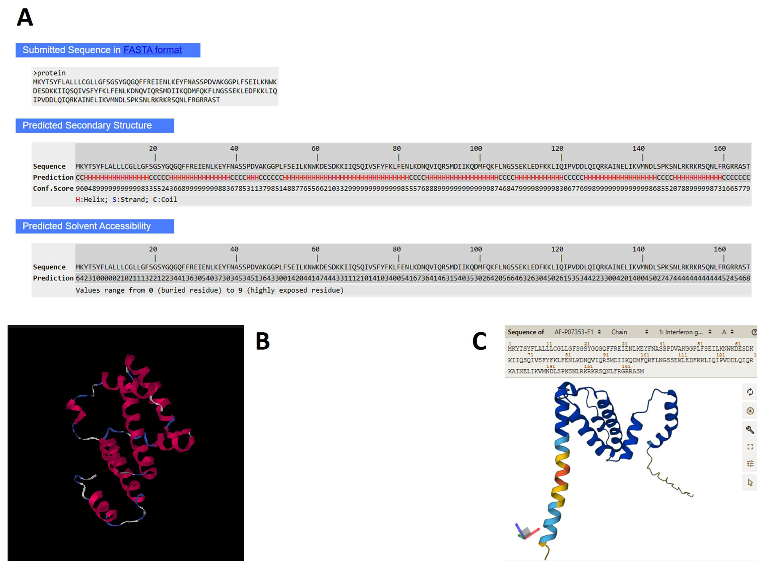
Figure 1. In silico analysis of b-IFN-γ native protein sequence. (A) The complete amino acid sequence was analyzed using individual residues with the predicted secondary structure; (B) related 3-D model from I-TASSER with structural template from the Protein Data Bank and from AlphaFold based on protein structure (C)
The structure has less than two cysteines and probably no disulfide bonding; thus, protein folding does not alter the binding interface with the Major Histocompatibility Complex (MHC) in immune cells.53 According to the Scratch Protein Predictor, the average predicted probability of antigenicity of 0.582472 renders it a good antigen for immunological assessment. Antigenic peptides should be located in solvent accessible regions and contain both hydrophobic and hydrophilic residues. Thus, any modification to the native protein sequence and structure for successful expression of an antigenic recombinant protein in E. coli was elucidated, showing five clear epitopes in the primary protein sequence that influence an antibody response (Figure 2A). However, the expression of a foreign protein in E. coli is influenced by a range of factors, including the host codon usage preference; therefore, we opted to optimize the DNA template of the native gene sequence to produce a precise template for heterologous translation, avoiding mismatches and punctual mutations of the final recombinant molecule (Table). In other similar research investigating the expression of an analog molecule using Pichia pastoris, two codons were removed that code for two cysteines at position 64-69 bp, where they form a disulfide bridge between the structure of IFN-α, possibly leading to inclusion body formation.54 Yin et al.55 synthesized an optimized bovine IFN-α gene by obtaining a 36 kDa recombinant protein from inclusion bodies in E. coli. This protein has six cysteines, with two potential disulfide bonds that could lead to protein aggregation. One hypothesis is that aggregates may affect the immunogenicity of recombinant therapeutic proteins.56 Therefore, when overexpressing foreign genes in E. coli, it is important to consider structural analysis before template optimization to ensure an efficient protein production bioprocess.
Table. Optimized synthetic gene sequence coding for rb-IFN-γ
| aCCATG GGA AAA TAC ACA TCA TAT TTT CTA GCT TTG TTG CTT TGC GGC CTG TTA |
| bM G K Y T S Y F L A L L L C G L L |
| GGT TTT TCC GGT AGC TAC GGC CAA GGT CAG TTT TTC CGT GAA ATT GAG AAT |
| G F S G S Y G Q G Q F F R E I E N |
| TTG AAA GAA TAC TTC AAC GCT AGC TCA CCG GAT GTT GCA AAA GGC GGT CCG |
| L K E Y F N A S S P D V A K G G P |
| CTG TTC TCC GAG ATC CTG AAG AAC TGG AAA GAC GAA AGC GAC AAG AAG ATC |
| L F S E I L K N W K D E S D K K I |
| ATC CAA AGC CAG ATC GTG AGC TTT TAT TTC AAA CTG TTC GAG AAC CTC AAG |
| I Q S Q I V S F Y F K L F E N L K |
| GAC AAC CAG GTT ATT CAA CGC AGC ATG GAT ATT ATC AAG CAG GAT ATG TTT |
| D N Q V I Q R S M D I I K Q D M F |
| CAG AAA TTC CTG AAT GGC TCT TCT GAG AAA CTG GAA GAT TTT AAG AAA CTG |
| Q K F L N G S S E K L E D F K K L |
| ATC CAA ATC CCG GTC GAC GAC CTG CAG ATT CAG CGT AAG GCG ATT AAC GAG |
| I Q I P V D D L Q I Q R K A I N E |
| CTG ATT AAG GTG ATG AAT GAT CTG AGC CCA AAA AGC AAT CTG CGT AAG CGC |
| L I K V M N D L S P K S N L R K R |
| AAA CGT TCG CAA AAC TTG TTC CGC GGT CGT CGT GCG TCC ACC CTCGAG |
| K R S Q N L F R G R R A S T |
a: DNA template optimized for E. coli translation (triplets). Underlined, enzyme restriction sites 5′-NcoI and XhoI-3′. Length 501 bp, total GC%: 40.
b: Single-letter amino acid code of recombinant protein rb-IFN-γ. MW: 19,420.45 Da (167 aa).
Construction and expression of rb-IFN-γ in the bacterial system
The optimized gene sequence of b-IFN-γ was synthesized via a codon adjustment, obtaining an open reading frame of 501 bp in pUC57. The synthetic gene encodes the native and complete mammal protein (Table). The 5′-terminus included the NcoI restriction site in-frame with the initial methionine, and the stop codon was deleted by adding the XhoI restriction site at the 3′-ending. The plasmid pUC57 was first transformed into E. coli TOP10 strain for plasmid multiplication, with confirmation of the plasmid showing the 501 bp insert, which was subcloned into pET28a(+) using the NcoI and XhoI restriction sites before introduction into E. coli BL21 (DE3), as confirmed by sequencing and enzyme restriction analysis of a single clone (Figure 2B). The results showed that the insert had a correct 5′-3′ transcriptional orientation corresponding to the 501 bp insert, giving to pET28a(+)-rb-IFN-γ plasmid.
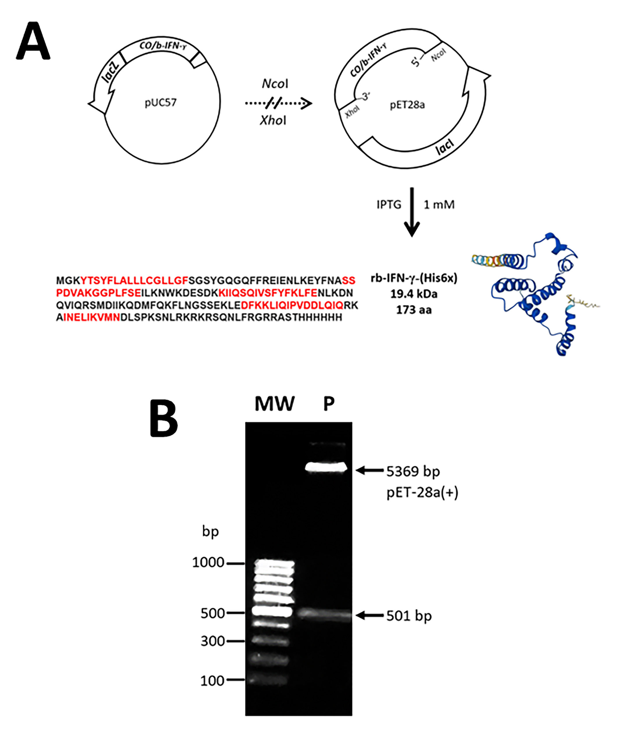
Figure 2. The subcloning strategy for efficient E. coli expression of codon-optimized (CO) b-IFN-γ gene for the obtention of recombinant protein after IPTG-induced culture. (A) Artificial CO/b-IFN-γ gene from pUC57 was subcloned into expression plasmid pET28a(+) by enzyme restriction with NcoI and XhoI. (B) Confirmation of final pET28a(+) with rb-IFN-γ gene from plasmid (P) of a single E. coli BL21 (DE3) clone was obtained by enzymic restriction. Protein 3D model and most probable epitopes (red) of linear amino acid sequence are shown. MW, 100 bp DNA ladder
After cell culture with IPTG addition and cell lysis, Coomassie staining and Western blot were used to search for the recombinant protein in cell fractions, with an expected and overproduced 19 kDa protein in the soluble fraction (Figure 3) after IPTG induction demonstrating successful heterologous production of the recombinant cytokine molecule in E. coli cells identified by a robust protein band at 19 kDa and by using the His6x-tag monoclonal antibody. Although a faint and less abundant protein band was observed in the insoluble fraction (pellet, not shown) following culture lysate, it was considered merely part of unlysed bacteria due to the overexpressed bacterial product that resulted from inefficient lysis.
From the Western blot analysis (Figure 3B, bottom), it is evident that there are truncated fragments of rb-IFN-γ, probably by gene overexpression, however no effects were observed in terms of the protein obtention in the soluble fraction, which was thus used for downstream purposes, with protein purification performed on the soluble fraction via Ni-affinity chromatography.
Figure 3. SDS-PAGE and Western blot analysis of soluble fractions following IPTG induction of E. coli culture. (A) Aliquots of supernatant of IPTG-induced (I) culture were compared with a cell lysate of non-induced (NI) culture and observed by Coomassie staining, and (B), Western blot with mouse anti-His6x tag monoclonal antibody. S, molecular size standards (kDa)
The expressed rb-IFN-γ protein was efficiently obtained from E. coli BL21 (DE3) harboring the pET28a(+)-rb-IFN-γ plasmid with metal affinity His-tag cartridge and recovered with elution buffer containing 250 mM imidazole in almost 95% of pure protein in final elution fractions, and recognition of the produced protein was assessed by a rat anti-b-IFN-γ monoclonal antibody which confirmed its immunological cytokine molecule identity in all the final aliquots of eliution fractions from affinity chromatography evaluated (Figures 4A and B).
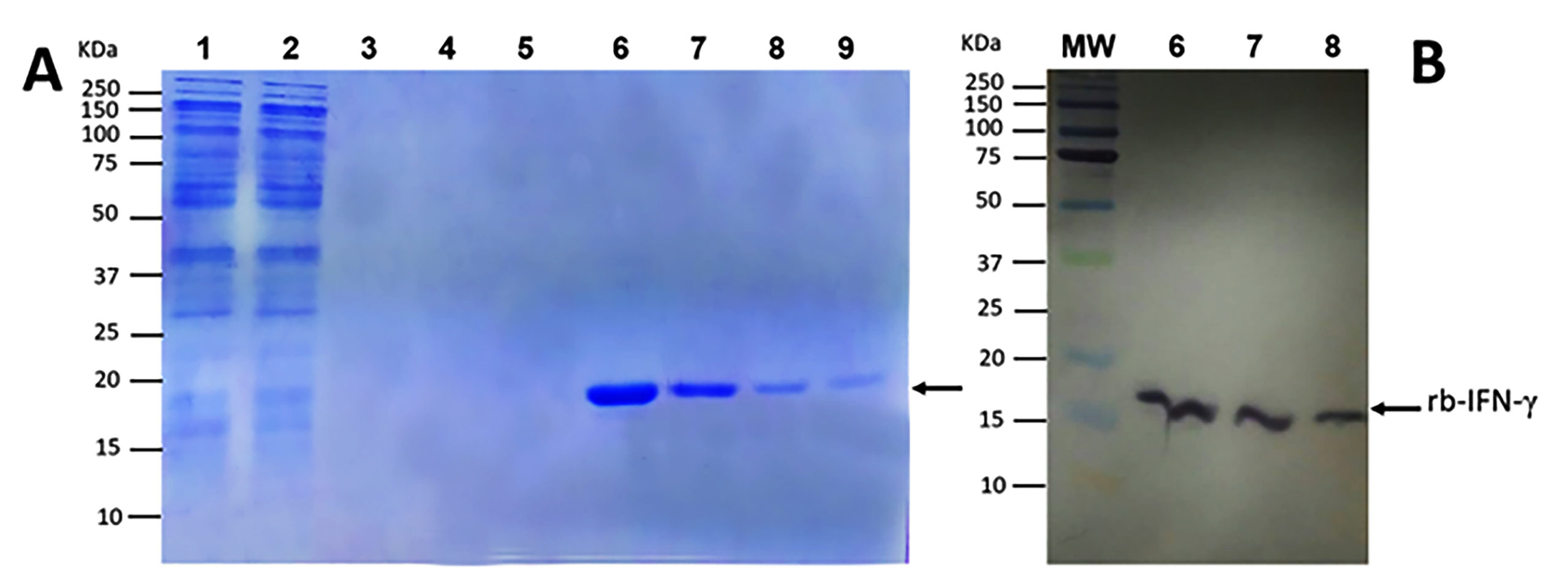
Figure 4. Purification of rb-IFN-γ by Ni-affinity chromatography using the soluble fraction of E. coli BL21 (DE3). (A) SDS-PAGE analysis of fractions, lanes: 1, eluate; 2-5, washes (50-70 mM imidazole); 6-9, elutions (250 mM imidazole). (B) Western blot with rat monoclonal anti-b-IFN-γ for three of the 250 imidazole elutions (lanes 6, 7, and 8). MW, molecular size standards (kDa)
Characterization of rb-IFN-γ by DSC and ELISA
DSC has been used to characterize protein thermal stability, overall conformation, and domain folding integrity by the biopharmaceutical biomolecule industry. The thermal denaturation of proteins is an endothermic process that reflects heat absorption, with conformational changes occurring due to the breaking of intramolecular non-covalent and disulfide bonds. Thus, in this study, DSC was used to search for the heat-denaturation molecular fingerprint of the recombinant protein, with a denaturing temperature (Tm) above 32 °C (Figure 5), which is similar to another recombinant analog IFN-γ molecule from human nature,54 with a relatively low Tm, as has been reported somewhere else with other four-helical-bundle cytokines, with data variations depending on pH stability and the instrumental DSC scanning method.57
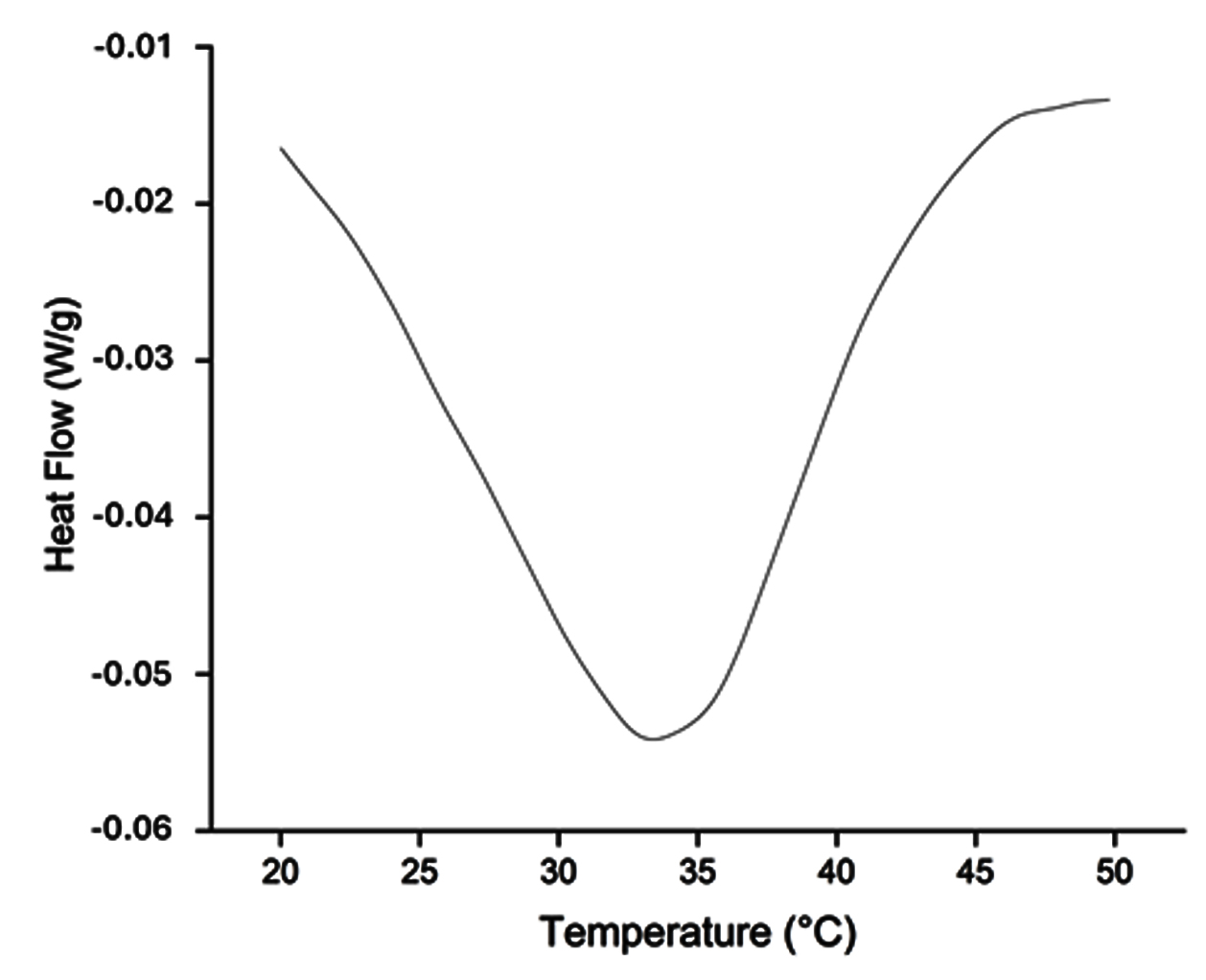
Figure 5. Thermal stability of the rb-IFN-γ. DSC scans were performed from 20-50 °C at a heating rate of 10 °C/min
Specific secondary and tertiary structures directly influence antigenicity through linear and conformational epitopes of the amino acid backbone, with decreasing antigenic levels between 90 and 120 °C. On the other hand, enzyme-linked immunosorbent assay (ELISA) is a common method of measuring epitope binding and antigenicity of a protein source58,59; thus, immunological identity of the purified recombinant protein was confirmed with a specific anti-b-IFN-γ ELISA kit BOVIGAMTM. Additionally, immuno-enzymatic analysis of the recombinant protein solution demonstrated a total protein concentration of 2 µg/ml, which was confirmed colorimetrically by increasing dilutions of 1:100 to 1:6400 with the mentioned ELISA kit, as increasing OD reached the top concentration of 2 µg/ml of the standard control curve in most dilutions (Figure 6A,B).
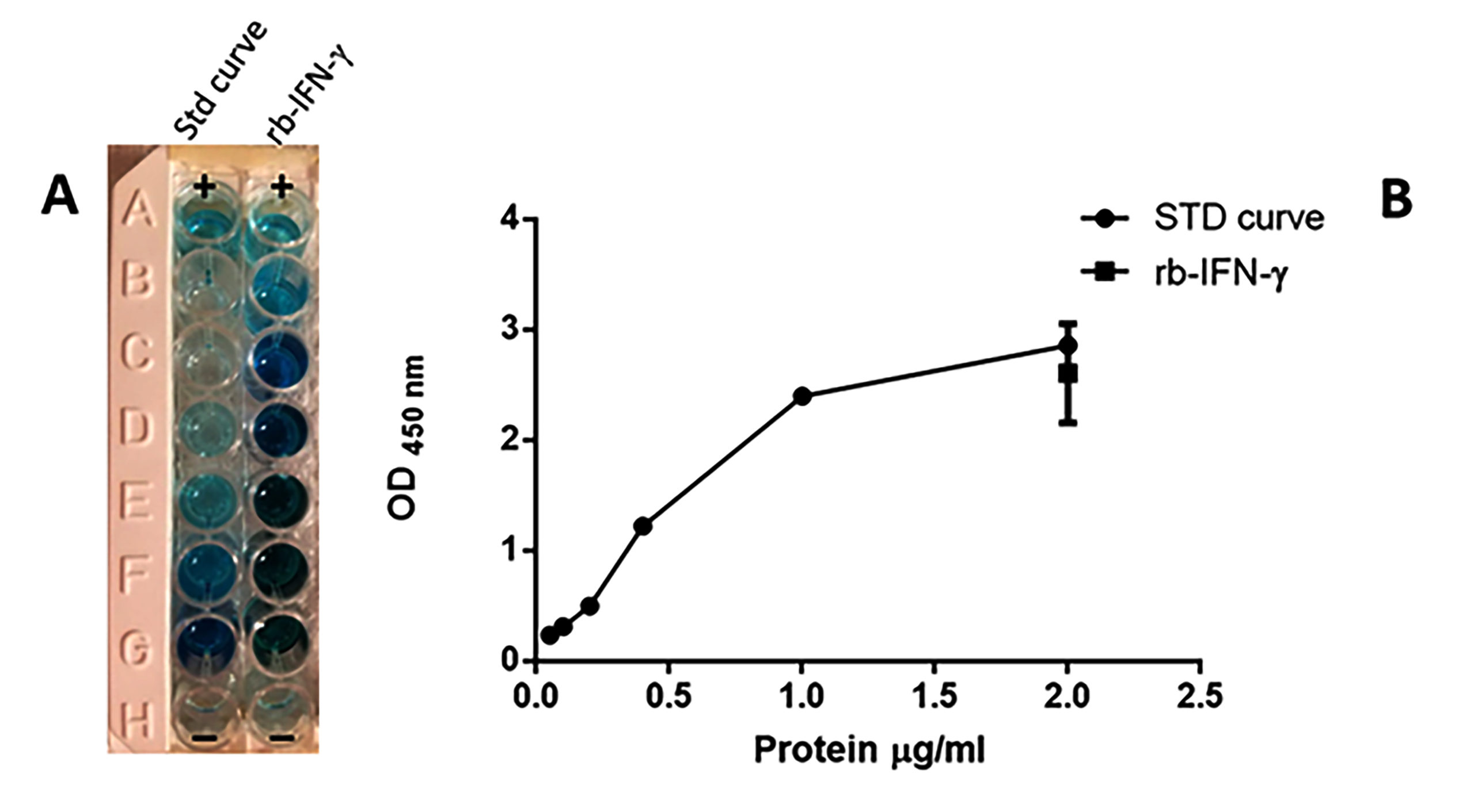
Figure 6. Quantitative immunological evaluation of rb-IFN-γ using a commercial ELISA kit with monoclonal anti-b-IFN-γ. (A) Wells B-G, decreasing dilutions of recombinant molecule (1:6400 to 1:100), and standard commercial b-IFN-γ curve (0.02-2 µg/ml), respectively. (B) Protein standard curve with ODs of protein dilutions. The mean SD of rb-IFN-γ dilutions is shown inside the STD curve. Duplicate positive and negative wells (A and H, respectively) were included as enzyme reaction controls
Another study by Xu et al.32 examined the expression of b-IFN-γ in supernatants obtained from Flp-In™ 293 cells (a stable mammalian expression cell line), with protein yields of 0.33 µg/ml measured with the BOVIGAMTM specific kit. In another eukaryotic biotechnological platform by P. pastoris, expression of human IFN-γ from a gene optimization cassette obtained 200 µg/L of recombinant product.60 Our results confirm that bacterial biosynthesis of heterologous interferon-gamma is also a good choice for stability and reliability for b-IFN-γ obtention, thereby enabling subsequent immunological investigations. The rb-IFN-γ from E. coli lacks the carbohydrate moieties attached to its structure as compared to its natural form. This absence of carbohydrate moieties may reduce the half-life of the cytokine in the bloodstream, affecting its pharmacokinetics, nor the antigenic properties. This considerable difference is important for choosing the heterologous expression system that remains under the downstream application purposes of the biological molecule.
Immunogenic assessment of rb-IFN-γ and mouse polyvalent antiserum
The in vivo immunogenicity of the recombinant cytokine was analyzed via the induction of high total IgG antibody levels in the serum of immunized mice following three consecutive shots of recombinant protein in combination with FCA. Although FCA is not recommended for use in laboratory animals because of associated side effects, researchers still prefer FCA to stimulate a higher humoral response; mouse anti-human IFN-γ polyclonal antiserum was achieved with 25-100 µg of recombinant cytokine from E. coli using FIA and FCA.61 In our work, 50 µg doses of the obtained rb-IFN-γ in FCA successfully triggered an active antibody response as shown by Western blot analysis on thick nitrocellulose foil strips, indicating that practically all the dilutions of the obtained pooled mouse antiserum maintained the capacity to recognize the bacterial biosynthetic protein. High antibody titers, ranging from 0.2-51.2 K, demonstrated the high IgG concentrations resulting from the immunogenic biosynthetic protein stimulus in mice (Figure 7A). However, the specificity of the mouse-generated antiserum was demonstrated by Dot-Blot, with positive recognition of b-IFN-γ by the mouse polyspecific anti-b-IFN-γ antibodies observed on the nitrocellulose sheet. Recombinant and commercial b-IFN-γ were also recognized (Figure 7B). Most importantly, the raised antibodies also reacted with a 19 kDa band in the plasma of a pooled plasma from 10 whole blood bovine samples with previous positive results for M. bovis infection, and first diagnosed with the specific BOVIGAMTM anti-b-IFN-γ in vitro test. The plasma from blood samples were treated for b-IFN-γ induction using PPDB as whole blood cultures according diagnostic test manual, and did not cross-react with a pooled plasma from 10 blood samples without a previous PPDB stimulus (Figure 7C).
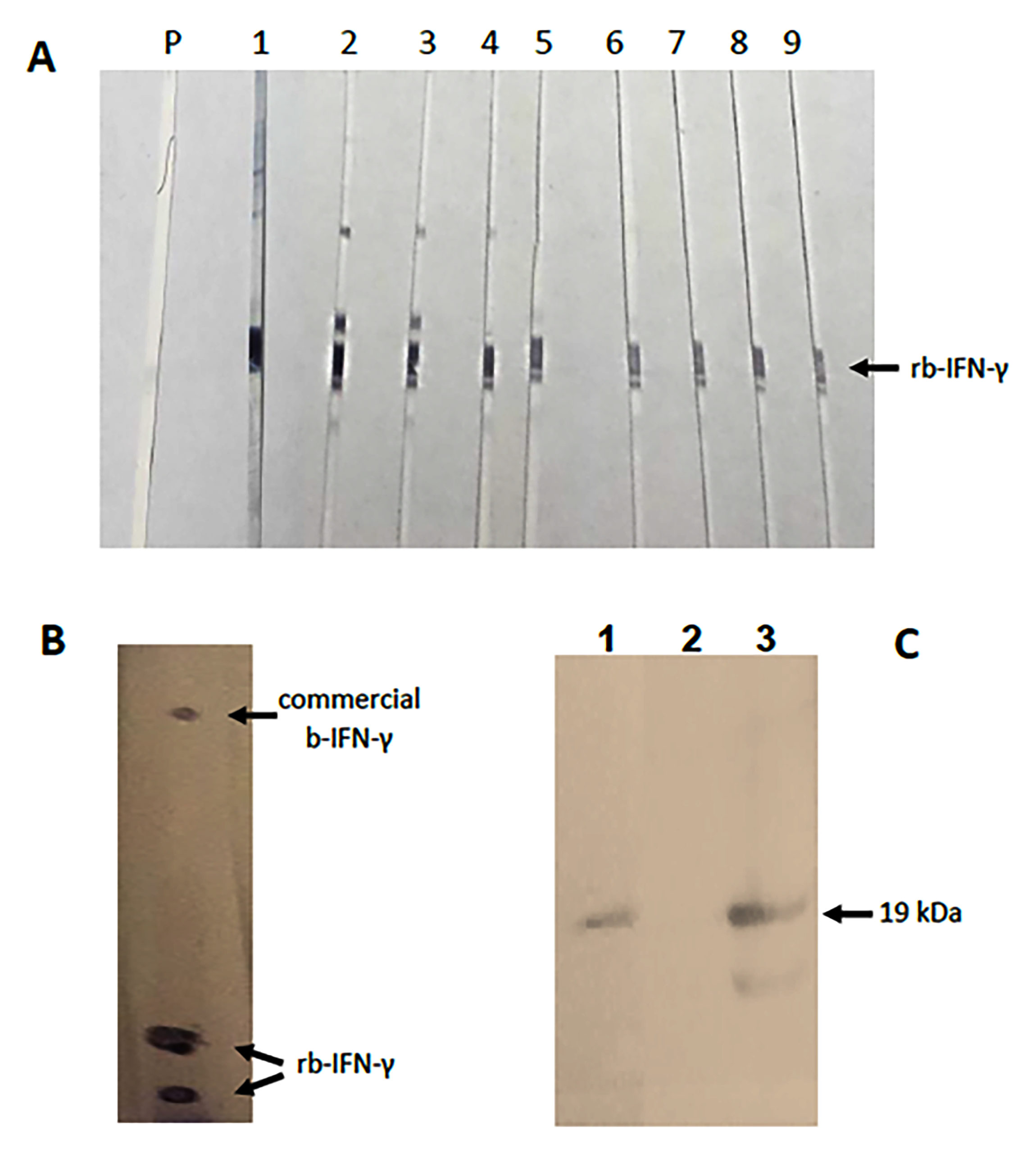
Figure 7. Immuno assessment of mouse anti-rb-IFN-γ antiserum. (A) Western blot of rb-IFN-γ electroblotted in 2 mm thick nitrocellulose foil strips with serial dilution of pooled mouse antiserum:PBS (starting with 1:200 and reaching 1:51,200), lanes: 1-9, respectively; P, pre-immune serum. (B) Dot-Blot analysis of rb-IFN-γ (duplicate) and a commercial b-IFN-γ (E. coli generated) with 1:100 mouse antiserum dilution as primary antibody. (C) Western blot with mouse antiserum; lanes: 1, commercial b-IFN-γ (19 kDa); 2, pooled bovine plasma from non-PPDB induced blood; and 3, pooled bovine plasma from PPDB-induced blood
These results demonstrate the specific recognition of b-IFN-γ in the blood plasma of clinical samples, suggesting the potential for diagnostic purposes. It has been reported that produced monoclonal anti-b-IFN-γ antibodies have recognized recombinant b-IFN-γ overexpressed in the 293 cell line, though not in E. coli.32 However, chicken anti-IFN-γ polyclonal antibodies from egg yolk have been developed with a recombinant cytokine molecule and used as secondary antibodies in prototype capture ELISA formats.17,19 Strong neutralizing antibody responses have been elicited in mice from the achieved protein with E. coli codon optimization bias.62 These observations suggest that polyclonal antibodies may be a better choice to cover a wide spectrum of standard biologics for immunological purposes.
Polyspecific antibodies recognize multiple sites on an antigen and are considered optimal ligands for immunodiagnostics, as almost all protein antigens usually bind to IgG antibodies, with large-scale production lowering costs.63 Besides its technical and economic virtues, the inbred mouse model used for immunogenic development has negligible overall genetic variability, implying high representativeness for the resulting immunity.64 However, the synthesis of bioengineered molecules in E. coli as a heterologous expression host has been performed using various soluble immunological molecules, including fragments and full-length IgGs, without altering their functional conformation.65-67 Due to the lack of post-translational glycosylation and the corresponding influence on the circulatory half-life of the molecule, the bacterial cell factory has a clear disadvantage, as proposed by Gentilomi et al. and Jalali et al.,29,30 who improved biosynthesis of the molecule using eukaryotic cells. It is assumed that the glycosylation of native IFN-γ protects the protein from proteolytic degradation, suggesting a potential role for glycans in therapeutic application.24 Although some contradictory statements assume that glycosylation is not essential for the biological activity of recombinant IFNs,68-70 IFN-γ acts as an adjuvant and enhances the immune response. It would be relevant to analyze whether the synthesized bovine cytokine displays its immunomodulatory activity by promoting gene transcription and lymphocyte recruitment, as indicated in downstream studies of a recombinant b-IFN-γ.71 Even so, optimization of the E. coli gene expression system cassettes is generating a wide range of new recombinant biologics with antigenic and pharmaceutical potential with improved protein yields.72-75 Initial strategies for cytokine synthesis and related antibodies consisted of laborious cell and total mRNA isolation steps that considered the native coding sequences for cDNA template generation and expression.20,36,76,77 Still, the overproduced recombinant proteins were often present in inclusion bodies as non-soluble products or usually could not sustain continuous high-level expression of the protein, needing improved subcloning strategies, adding fusion protein tags or downstream denaturing treatment conditions to solubilize the target molecule, but often making a more complex structure.78-80 Innovations have been continuously implemented to improve the production of these immunological molecules, overcoming their unfavorable yields and aggregative properties after heterologous overexpression.81,82 We used gene retro-engineering for favorable rb-IFN-γ biosynthesis as a biomarker for immunodiagnostic downstream applications with satisfactory protein yields, strategically starting from a rational computational analysis of the protein-retrieved amino acid sequence, overcoming the production challenges inherent in other methods. An IFN-λ molecule was successfully obtained after direct gene sequence retrieval from the NCBI database for gene synthesis with synonymous codon usage bias, with overexpression in E. coli.83 Thus, evidence for preferring gene optimization strategies over standard procedures with native sequences for biotechnological and diagnostic purposes in E. coli is accumulating.84-89 Attempts to express a fully glycoprotein from buffalo in HEK 293F cell line were not successful, opting for the expression of a 124 aa segment of the target protein in E. coli with no glycosylated sites after codon optimization technology.90 Production of recombinant chicken TNF-α from a cDNA cloning protocol and expression in transfected COS-7 cells was unsuccessful, not by a codon optimization strategy for the expression of the extracellular domain of the target protein in E. coli.91 In our work, the complete target bovine cytokine was successfully overproduced with the described gene retro-engineering strategy for bacterial expression. Finally, the achieved rb-IFN-γ may have good immunogenic characteristics of a standard biologic for specific immunoassaying, representing a feasible biotechnological product for accomplishing the immune reagent production for the livestock pharmaceutical industry.
This study indicated the feasible biotechnological synthesis of a soluble immunocompetent b-IFN-γ in an E. coli expression system by codon usage optimization technology, with good protein yields and the generation of high levels of polyclonal antibodies as biologics specific for b-IFN-γ detection. Polyspecific antibodies recognize multiple sites on an antigen and are considered optimal ligands for immunodiagnostics, with the potential for large-scale production lowering costs. This study adds to others favoring the production of such mediators as specific reagents with immunological control potential for infectious diseases in livestock, with secondary application in the production of biologic standards for the biopharmaceutical industry. To our knowledge, this approach could be more suitable for commercial purposes.
ACKNOWLEDGMENTS
The authors are thankful to Mr. Brian Walsh (U.S. Peace Corps) for his assistance with English language editing and Mr. Javier Rodríguez Yáñez (CIATEJ) for his support in the literature compilation.
CONFLICT OF INTEREST
The authors declare that there is no conflict of interest.
AUTHORS’ CONTRIBUTION
AHA conceptualized the study. RH, AHA, AG, MM, HE, TAC, and JB collected resources and supervised the study. RH, AHA, AG, MM, HE, TAC, and JB applied methodology. AC and AOV performed the investigation. AHA performed visualization, project administration, funding acquisition and wrote the original manuscript. RH, AHA, AG, MM, HE, TAC, and JB reviewed and edited the manuscript. All authors read and approved the final manuscript for publication.
FUNDING
This work was partially supported by Consejo Estatal de Ciencia y Tecnología de Jalisco (COECYTJAL) and Secretaría de Ciencia, Humanidades, Tecnología e Innovación (SECIHTI) through grants awarded to AHA (Grant numbers 190426 and CBF-3729).
DATA AVAILABILITY
All datasets generated or analyzed during this study are included in the manuscript.
ETHICS STATEMENT
The study was approved by the Internal Committee for Care and Use of Laboratory Animals (license number: CICUAL-2018009B).
- Casanova JL, MacMicking JD, Nathan CF. Interferon-γ and infectious diseases: Lessons and prospects. Science 2024;384(6693):eadl2016.
Crossref - Holder T, Coad M, Allan G, Hogarth PJ, Vordermeier MH, Jones GJ. Vaccination of calves with Bacillus Calmette-Guerin Danish strain 1331 results in a duration of immunity of at least 52 weeks. Vaccine. 2023;41(48):7290-7296.
Crossref - Kapten K, Orczyk K, Maeser A, Smolewska E. Interferon-γ release assay in the assessment of cellular immunity-A single-centre experience with mRNA SARS-CoV-2 vaccine in patients with juvenile idiopathic arthritis. J Clin Med. 2024;13(9)2523.
Crossref - Kresic N, Philips W, Haegeman A, de Regge N. Evaluation of an interferon-gamma release assay for early detection of lumpy skin disease virus infection and vaccination in cattle. Microbiol Spectr. 2025;13(4):e0293924.
Crossref - Lee YJ, Min J, Myong JP, et al. Incidence of tuberculosis among immigrants in Korea who participated in a latent tuberculosis infection screening program. J Korean Med Sci. 2024;39(28):e207.
Crossref - Alvarez AH, Gutierrez-Ortega A, Gomez-Entzin V, et al. Assessment of antigenic supplementation of bovine purified protein derivative for diagnosis of subclinical infection with Mycobacterium bovis in cattle. Microb Pathog. 2017;108:114-121.
Crossref - Botcher J, Alex M, Danicke S, Gethmann J, Mertens-Scholz K, Janowetz B. Susceptibility, immunity, and persistent infection drive endemic cycles of coxiellosis on dairy farms. Animals 2024;14(7):1056.
Crossref - Bulun H, Bridger PS, Schillinger S, et al. Interferon gamma producing CD4+ T cells quantified by flow cytometry as early markers for Mycobacterium avium ssp. paratuberculosis infection in cattle. Vet Res. 2024;55(69):69.
Crossref - Coad M, Doyle M, Steinbach S, Gormley E, Vordermeier M, Jones G. Simultaneous measurement of antigen-induced CXCL10 and IFN-γ enhances test sensitivity for bovine TB detection in cattle. Vet Microbiol. 2019;230:1-6.
Crossref - Wang YJ, Tong T, Xin XQ, et al. Establishment and evaluation of an interferon-gamma enzyme-linked immunospot method for the detection of Brucella-infected cattle and goats. J Dairy Sci. 2025;108(7):7472-7480.
Crossref - Wenz JR, Fox LK, Muller FJ, Rinaldi M, Zeng R, Bannerman DD. Factors associated with concentrations of select cytokine and acute phase proteins in dairy cows with naturally occurring clinical mastitis. J Dairy Sci. 2010;93(6):2458-2470.
Crossref - Chileshe J, Goosen WJ, Buss PE, et al. A commercial ELISA for detection of interferon gamma in white rhinoceros. J Vet Diagn Invest. 2019;31(4):531-536.
Crossref - Ding S, Brownlee BJ, Parate K, et al. IFN-γ and IL-10 immunosensor with vertically aligned carbon nanotube interdigitated electrodes toward pen-side cattle paratuberculosis monitoring. Glob Chall. 2024;8(9):2400021.
Crossref - Rodrigues V, Baudier JB, Chantal I. Development of a bead-based multiplexed assay for simultaneous quantification of five bovine cytokines by flow cytometry. Cytometry. 2017;91(9):901-907.
Crossref - Sipka A, Mann S, Babasyan S, Freer H, Wagner B. Development of a bead-based multiplex assay to quantify bovine interleukin-10, tumor necrosis factor-a, and interferon-γ concentrations in plasma and cell culture supernatant. JDS Comm. 2022;3(3):207-211.
Crossref - Yerrapragada RM, Mampallil D. Interferon-γ detection in point of care diagnostics: Short review. Talanta 2022;245:123428.
Crossref - Brujeni GN, Jalali SAH, Koohi MK. Development of DNA-designed avian IgY antibodies for quantitative determination of bovine interferon-gamma. Appl Biochem Biotechnol. 2011;163(3):338-345.
Crossref - Mitchell JL, Raper A, Gunn-Moore DA, Hope JC. Recognition of recombinant interferon-gamma from Felidae species by anti-cat antibodies. Vet Immunol Immunophatol. 2021;241:110327.
Crossref - Morar D, Tijhaar E, Negrea A, et al. Cloning, sequencing and expression of white rhinoceros (Ceratotherium simum) interferon-gamma (IFN-γ) and the production of rhinoceros IFN-γ specific antibodies. Vet Immunol Immunopathol. 2007;115(1-2):146-154.
Crossref - Pedra ACK, de Oliveira NR, Maia MAC. Production of recombinant cytokines and polyclonal antibodies for analysis of cellular immune response in golden Syrian hamster. Mol Biol Rep. 2024;51(1):1012.
Crossref - Boyd P, Hudgens E, Loftus JP, et al. Expressed gene sequence and bioactivity of the IFNgamma-response chemokine CXCL11 of swine and cattle. Vet Immunol Immunopathol. 2010;136(1-2):170-175.
Crossref - Gaertner FH, Babiuk LA, Mutwiri G, Armstrong JM, Griebel PJ. Amended recombinant cells (ARCs™) expressing bovine IFN-γ: An economical and highly effective adjuvant system. Vaccine. 2009;27(9):1377-1385.
Crossref - Premraj A, Aleyas AG, Nautiyal B, Rasool TJ. Novel type-I interferons from the dromedary camel: Molecular identification, prokaryotic expression and functional characterization of camelid interferon-delta. Mol Immunol. 2023;153:212-225.
Crossref - Razaghi A, Owens L, Heimann K. Review of the recombinant human interferon gamma as an immunotherapeutic: Impacts of production platforms and glycosylation. J Biotechnol. 2016;240:48-60.
Crossref - Shao J, Cao C, Bao J, Gao M, Wang J. Characterization of the biological activities and physicochemical characteristics of recombinant bovine interferon-a14. Mol Immunol. 2015; 64(1):163-169.
Crossref - An R, Zhang R, Guo Y, et al. Biological activity of optimized codon bovine type III interferon expressed in Pichia pastoris. Viruses 2023;15(5):1101.
Crossref - Barathiraja S, Gangadhara PAV, Umapathi V, Dechamma HJ, Reddy GR. Expression and purification of biologically active bovine Interferon l3 (IL28B) in Pichia pastoris. Protein Expr Purif. 2018;145:14-18.
Crossref - Gao M, Guo Y, Luo X, et al. Design, biological activity and signaling pathway of bovine consensus omega interferon expressed in Pichia pastoris. Mol Immunol. 2019;106:46-52.
Crossref - Gentilomi G, Lelli R, D’Angelo M, et al. Expression of bioactive recombinant bovine interferon-γ using baculovirus. New Microbiol. 2006;29(1):19-24.
- Jalali SAH, Nikbakht-Brujeni G, Tadjbakhsh H, Koohi MK, Gholkar M, Rabbani M. Cloning and high level expression of bovine interferon-gamma gene in eukaryotic cells (COS-7). Iran J Vet Res. 2010;11:125-133.
Crossref - Tu Y, Wang G, Wang Y, et al. Extracellular expression and antiviral activity of a bovine interferon-alpha through codon optimization in Pichia pastoris. Microbiol Res. 2016;191:12-18.
Crossref - Xu Z, Shan F, Meng C, et al. Generation and application of a 293 cell line stably expressing bovine interferon-gamma. Protein Expr Pur. 2014;99:131-132.
Crossref - Cerretti DP, McKereghan K, Larsen A, Cosman D, Gillis S, Baker PE. Cloning, sequence, and expression of bovine interferon-gamma. J Immunol. 1986;136(12):4561-4564.
- Badiee-Kheirabadi SE, Mashayekhi K, Moghadam M, Javad-Mousavi M, Samkian M. Cloning, expression, and purification of recombinant mouse interferon-γ. Res Mol Med. 2021;9:1-10.
Crossref - Das PK, Sahoo A, Veeranki VD. Current status, and the developments of hosts and expression systems for the production of recombinant human cytokines. Biotechnol Adv. 2022;59:107969.
Crossref - Reddy KS, Rao DM, Dechamma HJ, Suryanarayana VVS, Reddy GR. Expression of bovine (Bos indicus) interleukin-18 in Escherichia coli and its biological activity. Microbiol Immunol. 2010;54(9):564-567.
Crossref - Kesidis A, Depping P, Lode A, et al. Expression of eukaryotic membrane proteins in eukaryotic and prokaryotic hosts. Methods. 2020;180:3-18.
Crossref - Saez NJ, Vincentelli R. High-throughput expression screening and purification of recombinant proteins in E. coli. Methods Mol Biol. 2014;1091:33-53.
Crossref - Leimgruber RM, Bishop BF, Levine AD, et al. High level expression and refolding of mouse interleukin 4 synthesized in Escherichia coli. J Biol Chem. 1995;270(13):7445-7452.
Crossref - Li NN, Liu P, Chen SJ, Lin QP, Zhou LF, Zhang SQ. Construction and expression of a novel bioactive IFN-a2b/CM4 fusion protein in Escherichia coli. Microbiol Res. 2010;165(2):116-121.
Crossref - Ariyaratne KANS, Brown R, Dasgupta A, et al. Expression of bovine β-lactoglobulin as a fusion protein in Escherichia coli: a tool for investigating how structure affects function. Int Dairy J. 2002;12(4):311-318.
Crossref - Huo J, Zhang G, Wang L, et al. Combining the advantages of prokaryotic expression and T7 phage display systems to obtain antigens for antibody preparation. Protein Expr Purif. 2021;184:105808.
Crossref - Itkonen JM, Urtti A, Bird LE, Sarkhel S. Codon optimization and factorial screening for enhanced soluble expression of human ciliary neurotrophic factor in Escherichia coli. BMC Biotechnol. 2014;14:92.
Crossref - Konczal J, Gray CH. Streamlining workflow and automation to accelerate laboratory scale protein production. Protein Expr Purif. 2017;133:160-169.
Crossref - Welch M, Govindarajan S, Ness JE, et al. Design parameters to control synthetic gene expression in Escherichia coli. PLoS ONE. 2009;4(9):e7002.
Crossref - Yu K, Ang KS, Lee DY. Synthetic gene design using codon optimization on-line (COOL). Methods Mol Biol. 2017;1472:13-34.
Crossref - Hu S, Wang M, Cai G, He M. Genetic code-guided protein synthesis and folding in Escherichia coli. J Biol Chem. 2013;288(43):30855-30861.
Crossref - Jiang R, Yuan S, Zhou Y, et al. Strategies to overcome the challenges of low or no expression of heterologous proteins in Escherichia coli. Biotechnol Adv. 2024;75:108417.
Crossref - Vihinen M. Solubility of proteins. ADMET DMPK. 2020;8(4):391-399.
Crossref - Samudzi CT, Rubin JR. Structure of recombinant bovine interferon-γ at 3.0 A resolution. Acta Cryst. 1993;D49:513-521.
Crossref - Sternberg MJE, Cohen FE. Prediction of the secondary and tertiary structures of interferon from four homologous amino acid sequences. Int J Biol Macromol. 1982;4(3):137-144.
Crossref - Jumper J, Evans R, Pritzel A, et al. Highly accurate protein structure prediction with AlphaFold. Nature 2021;596(7873):583-589.
Crossref - Sachs A, Moore E, Kosaloglu-Yalcin Z, et al. Impact of cysteine residues on MHC binding predictions and recognition by tumor-reactive T cells. J Immunol. 2020;205(2):539-549.
Crossref - Prabhu AA, Purkayastha A, Mandal B, Kumar JP, Mandal BB, Veeranki VD. A novel reverse micellar purification strategy for histidine tagged human interferon gamma (hIFN-γ) protein from Pichia pastoris. Int J Biol Macromol. 2018;107(Part B):2512-2524.
Crossref - Yin H, Li S, Chai C, et al. Biological activity of recombinant bovine IFN-a and inhibitory effect on BVDV in vitro. Microb Pathog. 2023;181:106155.
Crossref - Hermeling S, Schellekens H, Maas C, Gebbink MFBG, Crommelin DJA, Jiskoot W. Antibody response to aggregated human interferon alpha2b in wild-type and transgenic immune tolerant mice depends on type and level of aggregation. J Pharm Sci. 2006;95(5):1084-1096.
Crossref - Wen J, Arthur K, Chemmalil L, Muzammil S, Gabrielson J, Jiang Y. Applications of differential scanning calorimetry for thermal stability analysis of proteins: Qualification of DSC. J Pharm Sci. 2012;101(3):955-964.
Crossref - Bogahawaththa D, Chandrapala J, Vasiljevic T. Thermal denaturation of bovine β-lactoglobulin in different protein mixtures in relation to antigenicity. Int Dairy J. 2019;91: 89-97.
Crossref - Zuber B, Rudstrom K, Ehrnfelt C, Ahlborg N. Epitope mapping of neutralizing monoclonal antibodies to human interferon-γ using human-bovine interferon-γ chimeras. J Interferon Cytokine Res. 2016;36(9):542-551.
Crossref - Prabhu AA, Veeranki VD, Dsilva SJ. Improving the production of human interferon gamma (hIFN-γ) in Pichia pastoris cell factory: An approach of cell level. Process Biochem. 2016;51:709-718.
Crossref - Uwituze V, Mugasa CM, Namayanja M, et al. Optimization of recombinant human interferon-gamma expression and purification in E. coli, and developing an immunization protocol for the production of optimal humoral response. Biologics. 2026;20:584180.
Crossref - Mansouri M, Mousavy SJ, Ehsaei Z, Nazarian S, Zali MR, Moazzeni SM. The codon-optimization of cfaE gene and evaluating its high expression capacity and conserved immunogenicity in Escherichia coli. Biologicals. 2013;41(3):169-175.
Crossref - Delahaut P. Immunisation – Choice of host, adjuvants and boosting schedules with emphasis on polyclonal antibody production. Methods. 2017;116:4-11.
Crossref - Jiskoot W, Kijanka G, Randolph TW, et al. Mouse models for assessing protein immunogenicity: Lessons and challenges. J Pharm Sci. 2016;105(5):1567-1575.
Crossref - Blake-Hedges J, Groff D, Foo W, et al. Production of antibodies and antibody fragments containing non-natural amino acids in Escherichia coli. MAbs. 2024;16(1):2316872.
Crossref - Leith EM, O´Dell WB, Ke N, et al. Characterization of the internal translation initiation region in monoclonal antibodies expressed in Escherichia coli. J Biol Chem. 2019;294(48):18046-18056.
Crossref - Jung ST, Kang TH, Georgiou G. Efficient expression and purification of human aglycosylated Fcg receptors in Escherichia coli. Biotechnol Bioeng. 2010;107(1):20-31.
Crossref - Chen WS, Villaflores OV, Jinn TR, Chan MT, Chang YC, Wu TY. Expression of recombinant human interferon-γ with antiviral activity in the bi-cistronic baculovirus-insect/larval system. Biosci Biotechnol Biochem. 2011;75(7):1342-1348.
Crossref - Xu Q, Nomura T, Ikeda M, et al. Stability of recombinant bovine interferon-γ antiviral activity in the absence of stabilizing additives. Microbiol Immunol. 2011;55(8):595-598.
Crossref - Holthausen DJ, Bickel KA, Medina GN, et al. Bacterially expressed non-glycosylated recombinant bovine interferon lambda demonstrates antiviral activity against bovine viral diarrhea virus in cell culture. Res Vet Sci. 2026;201:106071.
Crossref - Taubert A, Hermosilla C. Bovine recombinant IFNg induces endothelial cell gene transcription of immunoregulatory molecules and upregulates PMN and PBMC adhesion on bovine endothelial cells. Vet Res Commun. 2008;32(1):35-47.
Crossref - Pal D, Tripathy RK, Teja MS, Kumar M, Banerjee UC, Pande AH. Antibiotic free expression system for the production of human interferon beta protein. 3 Biotech. 2018;8:36.
Crossref - Pandey R, Kumar N, Monteiro GA, Veeranki VD, Prazeres DMF. Re-engineering of an Escherichia coli K-12 strain for the efficient production of recombinant human Interferon Gamma. Enzyme Microb Technol. 2018;117:23-31.
Crossref - Sun H, Li J, Liu G, et al. Expression of codon-optimized TgMIC16 in three Escherichia coli strains. 3 Biotech. 2017;7(4):270.
Crossref - Wu W, Gupta W, Sagan SA, Moseley CE, Zamvil SS, Pak JE. High-yield production of recombinant human myelin oligodendrocyte glycoprotein in SHuffle bacteria without a refolding step. J Immunol Methods. 2024;534:113764.
Crossref - Chen C, Herzig CTA, Baldwin CL. Expressed gene sequence of bovine IL23A and IL23R. Vet Immunol Immunopathol. 2009;128(4):425-430.
Crossref - Kashima T, Morishita A, Iwata H, Maeda K, Inoue T. Expression of bovine cytokines in Escherichia coli. J Vet Med Sci. 1999;61(2):171-173.
- Yang F, Pan Y, Chen Y, et al. Expression and purification of Canis interferon a in Escherichia coli using different tags. Protein Expr Purif. 2015;115:76-82.
Crossref - Yasamut U, Thongheang K, Weechan A, Sornsuwan K, Juntit O, Tayapiwatana C. Evaluating the ability of different chaperones in improving soluble expression of a triple-mutated human interferon gamma in Escherichia coli. J Biosci Bioeng. 2024;138(3):232-238.
Crossref - Zhao J, Yu HY, Zhao Y, et al. Soluble expression, rapid purification, biological identification of chicken interferon-alpha using a thioredoxin fusion system in E. coli and its antiviral effects to H9N2 avian influenza virus. Prep Biochem Biotechnol. 2019;49(2):192-201.
Crossref - Mobasher MA, Ghasemi Y, Montazeri-Najafabady N, et al. Two step production of optimized interferon beta 1b; a way to overcome its toxicity. J Pure Appl Microbiol. 2013;7(4):2867-2871.
- Shaldzhyan A, Zabrodskaya Y, Yolshin N, et al. Clean and folded: Production of active, high quality recombinant human interferon-λ1. Process Biochem. 2021;111:32-39.
Crossref - Akhtar H, Akhtar S, Jan SU, et al. Over expression of a synthetic gene encoding interferon lambda using relative synonymous codon usage bias in Escherichia coli. Pak J Pharm Sci. 2013;26(6):1181-1188.
- Ayyar BV, Arora S, Ravi SS. Optimizing antibody expression: The nuts and bolts. Methods. 2017;116:51-62.
Crossref - Kaur J, Kumar A, Kaur J. Strategies for optimization of heterologous protein expression in E. coli: Roadblocks and reinforcements. Int J Biol Macromol. 2018;106:803-822.
Crossref - Lu X, Wang J, Jin X, Zhu J. High-level expression of a novel liver-targeting fusion interferon with preferred Escherichia coli codon preference and its anti-hepatitis B virus activity in vivo. BMC Biotechnol. 2015;15:54.
Crossref - Mehta D, Chirmade T, Tungekar AA, Gani K, Bhambure R. Cloning and expression of antibody fragment (Fab) I: Effect of expression construct and induction strategies on light and heavy chain gene expression. Biochem Eng J. 2021;176:108189.
Crossref - Montazeri-Najafabady N, Ghasemi Y, Mobasher MA, Gashemian A, Rasoul-Amini S, Ebrahimi S. Codon optimization, cloning and expression of interleukin 11 in two different E. coli systems. J Pure Appl Microbiol. 2013;7(4):2717-2722.
- Nasr SM, Samir S, Okasha S. Interdisciplinary gene manipulation, molecular cloning, and recombinant expression of modified human growth hormone isoform-1 in E. coli system. Int J Biol Macromol. 2024;257(Part 1):128637.
Crossref - Srinivasan SV, Ghosh J, Nazar SB, Roy KS. Buffalo early pregnancy biomarker coding sequence cloning and partial length expression in E. coli after codon optimization. Heliyon. 2019;5(11):e02863.
Crossref - Yang Y, Meng Y, Chen L, et al. Development of specific monoclonal antibodies for the detection of natural chicken tumor necrosis factor-alpha. Heliyon. 2022;8(12):e12446.
Crossref
© The Author(s) 2026. Open Access. This article is distributed under the terms of the Creative Commons Attribution 4.0 International License which permits unrestricted use, sharing, distribution, and reproduction in any medium, provided you give appropriate credit to the original author(s) and the source, provide a link to the Creative Commons license, and indicate if changes were made.