

ISSN: 0973-7510
E-ISSN: 2581-690X
 , Chandan Dharmashekar2, H.S. Pruthvi2 and
Krishnaveni Ramakrishna1
Hospital-generated biomedical waste is a critical, yet understudied, reservoir for multidrug-resistant pathogens, particularly methicillin-resistant Staphylococcus aureus (MRSA). This study investigated the prevalence, characteristics, and virulence potential of MRSA in biomedical waste from healthcare facilities across three districts in Karnataka, India. Sixty biomedical waste samples were collected and subjected to systematic microbiological analyses, including culture-based isolation, biochemical characterization, molecular identification through 16S rRNA sequencing, and comprehensive antimicrobial susceptibility testing. Advanced bioinformatics tools including, PICRUSt2 were employed to assess the virulence profiles and functional potential. The results revealed that 63.3% of the samples showed bacterial growth, with 36.8% testing positive for presumptive S. aureus on mannitol salt agar. Biochemical and molecular characterization confirmed the presence of multidrug-resistant MRSA with extensive resistance to multiple antibiotic classes including β-lactams, fluoroquinolones, aminoglycosides, and macrolides. The isolated strains demonstrated significant virulence characteristics, including robust hemolytic activity and diverse metabolic capabilities that enhance environmental persistence. Phylogenetic analysis revealed close relationships with clinically significant strains, suggesting potential bidirectional transmission between clinical and environmental reservoirs. Geographic variations in the prevalence across districts indicate disparities in waste management practices and infection control protocols. Computational analyses revealed genes associated with nitrate reduction, environmental persistence, adhesion proteins, and immune evasion. These findings establish biomedical waste as an active ecological niche for enhanced pathogenic traits, posing substantial risks for healthcare-associated infections and environmental contamination. This study highlights the urgent need for enhanced waste surveillance, improved treatment technologies, and targeted management interventions to prevent the dissemination of MDR pathogens and protect public health.
Biomedical Waste, Multidrug-resistant bacteria, Methicillin-resistant Staphylococcus aureus, Virulence Factors, Antibiotic Resistance, Hospital Waste Management, Public Health
Methicillin-resistant Staphylococcus aureus (MRSA) remains a major global healthcare problem, associated with significant morbidity, mortality, and increased healthcare costs.1 Recent surveillance data indicate that MRSA was directly responsible for 1.27 million deaths globally in 2019, with an additional 4.9 million associated deaths, making drug-resistant infections more deadly than HIV/AIDS or malaria.2 The ability of the pathogen to acquire resistance to multiple antibiotic classes through horizontal gene transfer, combined with its extensive virulence arsenal, makes it particularly problematic in healthcare environments.3 Contemporary surveillance shows that MRSA bloodstream infections continue to pose substantial clinical challenges, with incidence rates varying significantly across regions but maintaining persistent burden in hospital settings.4
Hospital biomedical waste represents a complex microenvironment that serves as a significant reservoir for antimicrobial-resistant bacteria and resistance genes.5 Recent studies demonstrate that medicinal activities in hospitals lead to high consumption of antibiotics, resulting in substantial excretion rates of antibiotic residues that are not fully absorbed by the body and are subsequently discharged into the environment through biological waste.6 This creates conditions characterized by high organic load, diverse microbial populations, and selective pressure from residual pharmaceuticals that favor the emergence and maintenance of multidrug resistance.7 Advanced metagenomic analyses have revealed that hospital wastewater contains remarkably diverse resistomes, with the potential for extensive horizontal gene transfer between bacterial species.8
The biomedical waste environment facilitates rapid evolution of antibiotic resistance through multiple interconnected mechanisms, including enhanced transformation, conjugation, and transduction mediated by high cell density and environmental stress.9 Recent research demonstrates that hospital effluents serve as hotspots for the dissemination of antibiotic molecules and multidrug-resistant bacteria in various ecosystems, with resistance genes persisting in environmental matrices long after initial contamination.10 The selective pressure imposed by sub-therapeutic concentrations of antibiotics commonly found in medical waste creates optimal conditions for co-selection and maintenance of resistance traits.11,12
Despite technological advances, current biomedical waste management practices show significant limitations in controlling multidrug-resistant pathogen dissemination, with recent studies revealing that existing treatment systems primarily target conventional pollutants and lack specific protocols for antimicrobial resistance mitigation.13 The COVID-19 pandemic has further highlighted critical gaps in waste management infrastructure, with increased biomedical waste volumes overwhelming existing systems and potentially facilitating pathogen transmission.14 Surveillance of antimicrobial-resistant pathogens in healthcare waste remains inadequate globally, with less than 10% of hospitals conducting routine waste surveillance despite WHO recommendations.15 The complex and heterogeneous nature of biomedical waste presents unique challenges for microbiological sampling and analysis, including interference from organic matrices and the presence of inhibitory substances that complicate detection efforts.16
The One Health approach has gained prominence in understanding antimicrobial resistance transmission, recognizing the interconnections between human, animal, and environmental health in the context of resistance development and dissemination.17 Recent evidence demonstrates direct links between hospital waste management practices and community-acquired resistant infections, with environmental contamination affecting water bodies near healthcare facilities in up to 65% of surveyed locations.18 The Quadripartite collaboration between WHO, FAO, WOAH, and UNEP has emphasized the critical need for integrated surveillance systems that address antimicrobial resistance across all sectors, including environmental monitoring of healthcare waste streams.19 Advanced molecular techniques, including shotgun metagenomics and AI-driven pathogen analysis, now enable real-time assessment of resistance and virulence profiles with unprecedented accuracy, facilitating comprehensive characterization of microbial communities in clinical waste environments.20
Given the mounting evidence that biomedical waste is a critical reservoir for multidrug-resistant pathogens and the urgent need for evidence-based intervention strategies, systematic surveillance studies employing advanced molecular characterization are essential. The growing recognition of environmental transmission routes for antimicrobial-resistant organisms, particularly following the COVID-19 pandemic, has increased the importance of understanding waste-associated microbial communities and their public health implications.21 This study addresses critical knowledge gaps by employing state-of-the-art molecular techniques to systematically isolate and characterize MRSA populations in hospital biomedical waste, providing essential data for evidence-based infection control strategies and informing next-generation waste management protocols to safeguard public health.
Site selection
MRSA samples were collected from three districts of Karnataka—Bidar, Kalaburagi, and Yadgiri, between April 2022 and September 2023. These sites were selected based on their healthcare density and waste-generation potential.
Geographic Information System (GIS) mapping
The spatial distribution of MRSA across the selected regions was analyzed using the Earth Explorer database (https://earthexplorer.usgs.gov/). The final three-dimensional vector maps were generated using ArcGIS 10.3 software. Sample coordinates were manually uploaded to the system to visualize the spatial distribution and determine the microbial diversity and severity across regions.22
Sample collection
Biomedical waste samples were collected using appropriate personal protective equipment, including gloves, lab coat, and face masks, to ensure safety and prevent contamination. The waste was segregated on-site into specific categories: sharps, infectious waste, pathological waste, pharmaceutical waste, and non-hazardous waste, using color-coded containers in accordance with biomedical waste management guidelines. Red bags were designated for contaminated materials, such as swabs, needles, syringes, scalpel blades, and items saturated with blood or other potentially infectious fluids. Yellow bags are used for chemical, pharmaceutical, or cytotoxic residues, such as medication vials, intravenous tubing, or chemotherapy waste. Black bags were used for non-infectious materials, such as food waste and paper products, as shown in Figure 1. Swabs were collected from each waste category and submerged in separate flasks containing Luria-Bertani (LB) broth. The inoculated broths were incubated at 37 °C for 24 hrs to allow bacterial proliferation before further analysis.23
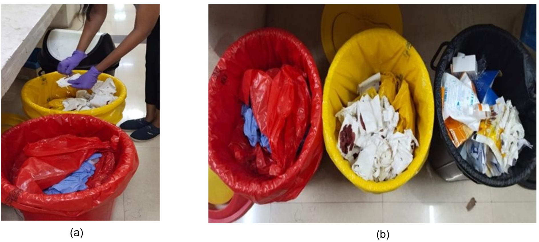
Figure 1. (a) Sample collection from Hospitals; (b) Different types of samples like sharps, infectious waste, pathological waste, pharmaceutical waste, and non-hazardous waste are collected in different bins as per Pollution Control Board regulation
Isolation of bacterial samples using serial dilution method
To isolate bacterial colonies, LB broth cultures containing biomedical waste samples were serially diluted. A 5 mL aliquot of the broth was transferred into a series of eight sterile test tubes, followed by serial dilution using 500 µL transfers at each step to achieve decreasing bacterial concentrations. Each dilution was thoroughly mixed to ensure uniform distribution. This method allowed for the quantitative estimation of the bacterial load and facilitated the isolation of distinct colonies upon plating. Nutrient agar media were prepared, poured into sterile Petri dishes under aseptic conditions, and allowed to solidify. Using a sterile inoculation loop, bacterial samples were collected from the diluted broth, and streak plating was performed in a zigzag pattern to ensure even distribution and isolation of individual colonies. The plates were incubated in an inverted position at 37 °C for 24 hrs to promote bacterial growth. Visible colonies were observed and sub-cultured for further studies.24
S. aureus Screening on Mannitol Salt Agar (MSA)
Distinct colonies were selected based on morphological characteristics such as size, shape, and pigmentation. Selected colonies were screened on MSA (HiMedia, India) by spot inoculation and incubated at 37 °C for 24-48 hrs. Colonies exhibiting yellow coloration with surrounding yellow zones were considered presumptive S. aureus.25
Microbial morphology profiling
Gram staining was performed to differentiate bacterial isolates based on their cell wall characteristics. Bacterial colony smears were prepared on clean glass slides and heat-fixed. Slides were sequentially stained with crystal violet (primary stain), iodine solution (mordant), ethanol (decolorizer), and safranin (counterstain). After air-drying, the slides were examined under a compound microscope at 10× and 40× magnifications to determine Gram staining. Gram-positive bacteria appeared purple, whereas Gram-negative bacteria appeared pink.26
Biochemical characterization
Methyl Red (MR) test
MR-VP broth (HiMedia, India) was prepared as per manufacturer’s instructions, dispensed in 6 mL aliquots into sterile test tubes, and autoclaved at 121 °C for 15 min. Each tube was inoculated with two loops of test culture and incubated at 37 °C for 48 hrs. After incubation, 5-6 drops of methyl red indicator (pH range 4.4-6.2) were added. The development of a stable bright red color indicated a positive MR test result, confirming mixed-acid fermentation. Yellow color was recorded as negative.27
Voges-Proskauer (VP) test
The inoculated MR-VP broth cultures were incubated at 37 °C for 48 hrs. To 1 mL of culture, 0.6 mL of Barritt’s reagent A (5% ±-naphthol in absolute ethanol) and 0.2 mL of reagent B (40% potassium hydroxide) were added sequentially. The tubes were shaken gently and allowed to stand at room temperature for 10-15 minutes. The appearance of a pink to red color indicated acetoin production (positive VP test).28
Starch hydrolysis test
Starch agar plates were prepared using 1% (w/v) soluble starch (HiMedia) in a Nutrient Agar base. After autoclaving and solidification, the test strains were streaked linearly and incubated at 37 °C for 72 hrs. The plates were flooded with Gram’s iodine solution and observed after 5-10 min. A clear halo around the growth indicated starch hydrolysis (amylase activity), whereas a dark blue color indicated no hydrolysis.29
Hemolytic activity assessment
Hemolytic activity was quantified on 5% defibrinated sheep blood agar plates prepared and poured under sterile conditions: isolates were stab-inoculated or streaked in a single line and incubated at 37 °C for 18-24 hrs in ambient air (or 5% CO2 for fastidious organisms). Plates were visually inspected against a black background for areas surrounding colonies: β-hemolysis was noted as a clear, translucent area of total red blood cell destruction; α-hemolysis as a greenish-brown hemolysis or met-hemoglobinization of agar below and around colonies; and γ-hemolysis as no visible change. Where hemolysis was weak or dubious, plates were re-examined at 24-48 hrs and incubated under other atmospheric conditions if clinically warranted; photographic records and measurement of hemolytic zone diameter were conducted for comparative studies, and sterility and media controls were added to validate blood agar quality.30
Gram staining
Gram staining is a microbiological method used to distinguish various types of bacterial cells based on their cell wall composition. A small sample was taken from each of the sub-cultured colonies of randomly collected from three different regions: samples 1, 2, and 3. The samples were spread onto separate slides and heat-fixed to allow adherence to the bacterial cells. The slides were then flooded with crystal violet stain and left to sit for the designated time. The excess stain was then rinsed with water. An iodine solution was applied as a mordant to enhance staining. The slides were then briefly treated with a decolorizing agent such as ethanol or acetone to differentiate between Gram-positive and Gram-negative bacteria. The slides were then rinsed again with water. A counterstain, typically safranin, was applied to color the Gram-negative bacteria. The slides were then rinsed once with water, excess moisture was blotted off, and the slides were allowed to air dry. The stained slides were observed under a microscope at 10× and 40× magnificent to determine the Gram staining of the bacteria.31
Molecular characterization
The bacterial culture was resuspended in 1 mL of DNAiso Reagent (Takara Bio) and mixed thoroughly by pipetting to ensure complete homogenization. The samples were incubated at room temperature for 5 min and centrifuged at 10,000 rpm for 10 min. The supernatant was transferred to a fresh tube, and DNA precipitation was performed by adding absolute ethanol (0.5 mL), followed by gentle inversion. After incubation for 3 min at room temperature, the mixture was centrifuged at 10,000 rpm for 10 min to obtain a DNA pellet. The pellet was washed twice with 75% ethanol, air-dried for 20-30 min, and dissolved in 30 µL Milli-Q water. The quality of the extracted DNA was assessed using 1% agarose gel electrophoresis, and the DNA was stored at -20 °C until further use. PCR amplification of the 16S rRNA gene was performed in a total reaction volume of 20 µL, containing 1 µL of bacterial DNA (10-50 ng), 1 µL each of forward and reverse primers, 10 µL EmeraldAmp GT PCR Master Mix (Takara Bio), and nuclease-free water to make up the volume. The universal primers 27F (5′ -AGAGTTTGATC(AC)TGGCTCAG-3′) and 1492R (5′-GGTTACCTTGTTACGACTT-3′) were used for amplification. The PCR cycling conditions consisted of initial denaturation at 94 °C for 2 min, followed by 30 cycles of denaturation at 94 °C for 30 sec, annealing at 55 °C for 30 sec, extension at 72 °C for 1.5 min, and a final extension at 72 °C for 10 min. The amplified PCR products were resolved on a 1% agarose gel containing ethidium bromide in 1× TAE buffer at 120 V for 45 min, along with a 100 bp DNA ladder, to confirm amplification. The PCR products were sequenced using the BDT v3.1 Cycle Sequencing Kit on an ABI 3730xl Genetic Analyzer. The forward and reverse sequences obtained were assembled to generate a consensus sequence using the Aligner software. The consensus sequence was subjected to BLAST analysis against the NCBI GenBank database (“nr” database). Based on the maximum identity scores, closely related sequences were selected and aligned using ClustalW in MEGA 11, and a phylogenetic tree was constructed using the Neighbor-Joining method to determine the taxonomic position of the isolate.32
Virulent factor characterization
Nitrate reductase test
Reduction of nitrate was determined in nitrate broth with 0.1-0.2% KNO3: the culture was inoculated into 5-10 mL of sterile nitrate broth and incubated aerobically for 24-48 hrs at 37 °C (extended incubation up to 72 hrs may be applied for slow reducers), using uninoculated broth as the negative control and a positive control being a known nitrate-reducing organism. Following incubation, 0.5 mL quantities were taken into sterile tubes (or directly added reagents to culture) and sequentially added reagents A (sulfanilic acid) and B (α-naphthylamine) (usually 0.5 mL each); red/pink color development immediately is a sign of nitrite production (nitrate → nitrite). If no color is seen, a pinch (10 mg) of zinc dust is added: appearance of red after zinc suggests presence of unreduced nitrate (i.e., organism did not reduce nitrate), while the absence of any color following addition of zinc suggests complete reduction of nitrate to beyond nitrite (to N2 or other nitrogenous gases), taken as a positive reduction; results are noted and matched with gas production (where Durham tubes have been used), incubation period, and repeat testing if required for weak reactions.30
Antimicrobial Activity-Drug Resistance Test (Agar Disc Diffusion/Kirby-Bauer)
Antimicrobial susceptibility was conducted by the standardized Kirby-Bauer disc diffusion technique on Mueller-Hinton agar (MHA) following CLSI recommendations: log phase cultures of bacteria were inoculated to a 0.5 McFarland turbidity standard (~1.5 × 10⁸ CFU/mL) in sterile saline; within 15 min of inoculation, a sterile cotton swab was wetted with the suspension, excess liquid removed by pressing against the tube wall, and the entire MHA plate surface inoculated by streaking in three directions to achieve an even lawn and permit the plate surface to dry (5-10 min) prior to application of antibiotic discs. Commercial antibiotic discs (lot and manufacturer documented) were gently applied on the inoculated agar using sterile forceps or an automatic disc dispenser with at least 24 mm center-to-center spacing; plates were inverted and incubated for 18-24 hrs at 37 °C. Following incubation, zone diameters of inhibition were measured to the nearest millimeter with a caliper or ruler, tabulated in mm, and interpreted according to the most current CLSI breakpoints as Susceptible, Intermediate, or Resistant. Quality control organisms were run concurrently to confirm the procedure, and all testing was repeated for any questionable zones or confluent growth on plates or other technical problems.33
Computational analysis
PICRUSt2: Functional profiling
Functional profiling of the identified bacterial isolate was performed using PICRUSt2 (Phylogenetic Investigation of Communities by Reconstruction of Unobserved States). The 16S rRNA gene sequence, along with the corresponding OTU table, was used as the input for the PICRUSt2 full pipeline executed on the Galaxy Europe platform. The workflow involved phylogenetic placement of the input sequence into a reference tree, followed by the prediction of gene family abundance using evolutionary modeling. Based on this, functional annotations were assigned in terms of KEGG Orthologs (KO) and Enzyme Commission (EC) numbers, which were further mapped to metabolic pathways. The Nearest Sequenced Taxon Index (NSTI) value was used to assess the reliability of the predictions, where lower NSTI values indicated closer similarity to reference genomes and higher confidence in the inferred functions. Predicted pathway abundances were used to evaluate the metabolic and functional potential of the isolates. Notably, PICRUSt2 provides predictive functional profiling based on 16S rRNA data, and does not represent the presence or expression of experimentally validated genes.34
GIS mapping and sample collection
MRSA samples were collected from hospital waste across the Bidar, Kalaburagi, and Yadgiri districts of Karnataka. The Figure 2 shows the mapped locations of the targeted sampling zones based on hospital density and waste generation. This spatial representation highlights the regional variation in the potential sources of MRSA contamination for further microbiological assessment. The number of samples collected from the different regions of Bidar, Kalaburagi, and Yadgir and samples showing growth under laboratory conditions are listed in Table 1.
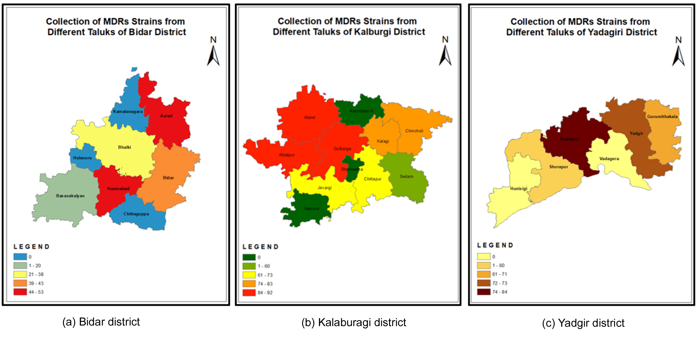
Figure 2. The ArcGIS map depicts the geographical distribution and selected sampling locations for the collection of MRSA samples from hospital waste throughout Bidar, Kalaburagi, and Yadgiri Districts of Karnataka. The displayed sampling areas may have varying degrees of potential exposure to MRSA as indicated by each area’s location on the map. (a) Bidar district; (b) Kalaburagi district; (c) Yadgir district (Source: ArcGIS mapping software)
Table 1. Distribution of collected samples and obtained positive isolates across districts
District |
Collected Samples |
Obtained Results |
|---|---|---|
Yadagiri District |
20 Samples |
12 Samples |
Bidar District |
20 Samples |
16 Samples |
Kalburgi District |
20 Samples |
10 Samples |
Total |
60 |
38 |
A total of 60 samples were collected from three districts, out of which 38 samples yielded positive results for bacterial isolation
Sample collection and processing
Sixty biomedical waste samples were collected from healthcare facilities across three districts of Karnataka, India. The samples were distributed equally with 20 samples each from the Yadagiri, Bidar, and Kalburgi districts. Following microbiological processing and screening, 38 samples (63.3%) showed bacterial growth suitable for further analysis (Figure 3). The recovery rates varied across districts, with Bidar district showing the highest recovery rate of 80% (16/20), followed by Yadagiri district at 60% (12/20) and Kalburgi district at 50% (10/20). These variations in recovery rates may reflect differences in waste management practices, storage conditions, and microbial loads across the different healthcare facilities in each district.
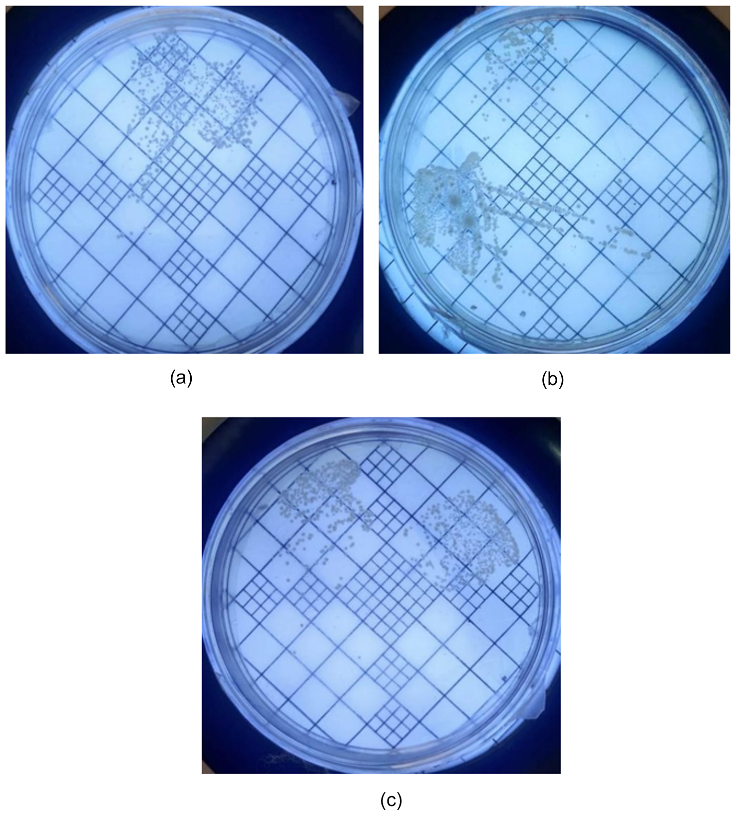
Figure 3. Colonies of isolated bacterial culture from medical waste on NA media. (a) Bidar; (b) Yadgir; (c) Kalaburagi
Microbial morphology profiling
Microscopic examination of the bacterial cultures (Figure 4) revealed progressive growth characteristics with increasing colony density and uniform distribution across the agar surface over the incubation period. The observed growth patterns indicated that the optimal environmental conditions were conducive to bacterial proliferation, with colonies exhibiting consistent morphological features throughout the cultivation process. This progressive development reflects successful bacterial adaptation to the culture medium and maintenance of viable cell populations. Uniform colony distribution and sustained growth dynamics confirmed the establishment of healthy bacterial cultures suitable for subsequent identification and characterization procedures.
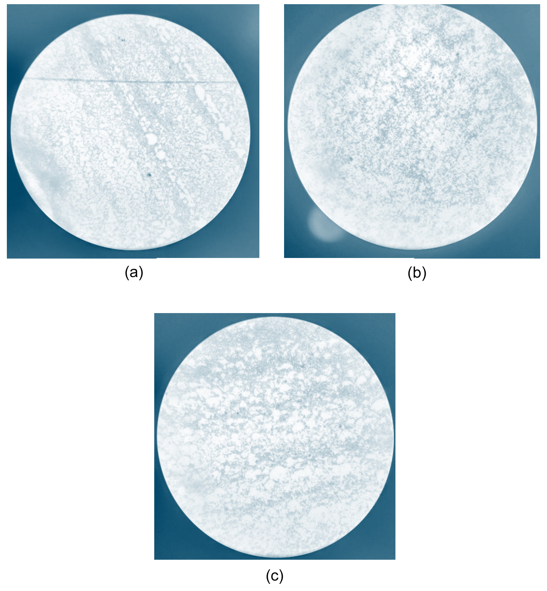
Figure 4. Microscopic morphological characterization of bacterial isolates collected from different districts. (a) Bidar; (b) Yadgir; (c) Kalaburagi
MSA screening
Following screening on MSA, 14 of 38 samples (36.8%) were positive for presumptive S. aureus identification. The positive isolates exhibited a characteristic yellow coloration with distinct yellow zones surrounding the colonies, indicating mannitol fermentation. The district-wise distribution of MSA-positive samples showed 6 samples from Bidar district (37.5% of district samples), 5 samples from Yadagiri district (41.7% of district samples), and 3 samples from Kalburgi district (30.0% of district samples). All 14 MSA-positive isolates were subjected to further confirmatory testing and molecular characterization as mentioned in Table 2.
Table 2. Screening of Samples on Mannitol Salt Agar (MSA) for Presumptive Staphylococcus aureus
District |
Total Samples (n) |
MSA-Positive Samples (n) |
Percentage Positive (%) |
|---|---|---|---|
Bidar |
16 |
6 |
37.5 |
Yadagiri |
12 |
5 |
41.7 |
Kalburgi |
10 |
3 |
30.0 |
Total |
38 |
14 |
36.8 |
MR test
Of the 14 MSA-positive isolates, 3 (21.4%) demonstrated positive results for the MR test. The positive isolates exhibited a distinct red coloration after the addition of the methyl red indicator, indicating the production of stable acid end products from glucose fermentation with a resultant pH of 4.4 or below. The remaining 11 isolates (78.6%) showed negative results, with yellow coloration indicating pH > 4.4. The threeMR-positive isolates were considered for further biochemical characterization and confirmatory testing, as shown in Figure 5.
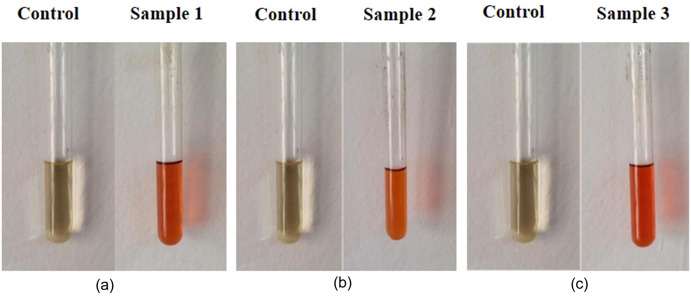
Figure 5. Methyl Red analysis; Red test positive results with change in colour to red. A red colour indicates a positive MR test, meaning the organism has produced a stable acid end product from glucose fermentation, lowering the pH of the medium to 4.4 or below. (a) Sample 1; (b) Sample 2; (c) Sample 3
VP test
Of the 14 MSA-positive isolates, 3 (21.4%) were subjected to the VP test, and all showed negative results, as shown in Figure 6. The tested isolates exhibited a yellow-to-copper-brown coloration following the addition of VP reagents, indicating the absence of acetoin production or insufficient acetoin levels for detection. No red coloration was observed in any of the tested isolates, confirming negative VP test results for all three isolates examined.
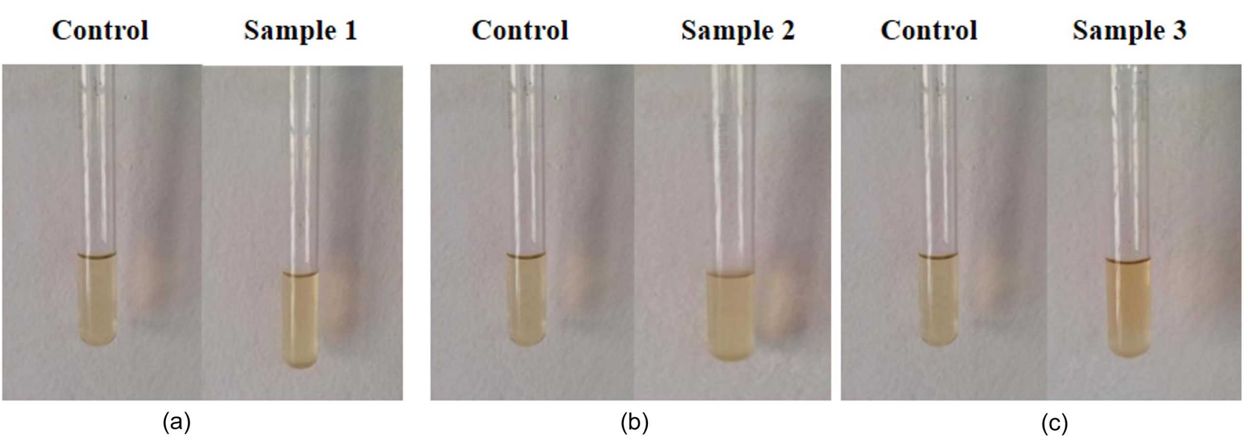
Figure 6. Voges-Proskauer test; Negative results with no change in colour of the isolated bacteria. A yellow or copper-brown color indicates a negative VP test, meaning the organism has not produced acetoin or the amount produced is insufficient to be detected by the reagents. (a) Sample 1; (b) Sample 2; (c) Sample 3
Starch hydrolysis test
Starch hydrolysis testing was conducted on all 14 MSA-positive isolates on starch agar plates. Following inoculation and incubation, the plates were flooded with an iodine solution to detect starch degradation. Three isolates (21.4%) exhibited clear zones around their colonies, indicating starch hydrolysis and amylase production (Figure 7). The remaining 11 isolates (78.6%) showed no clear zones, indicating negative starch hydrolysis.
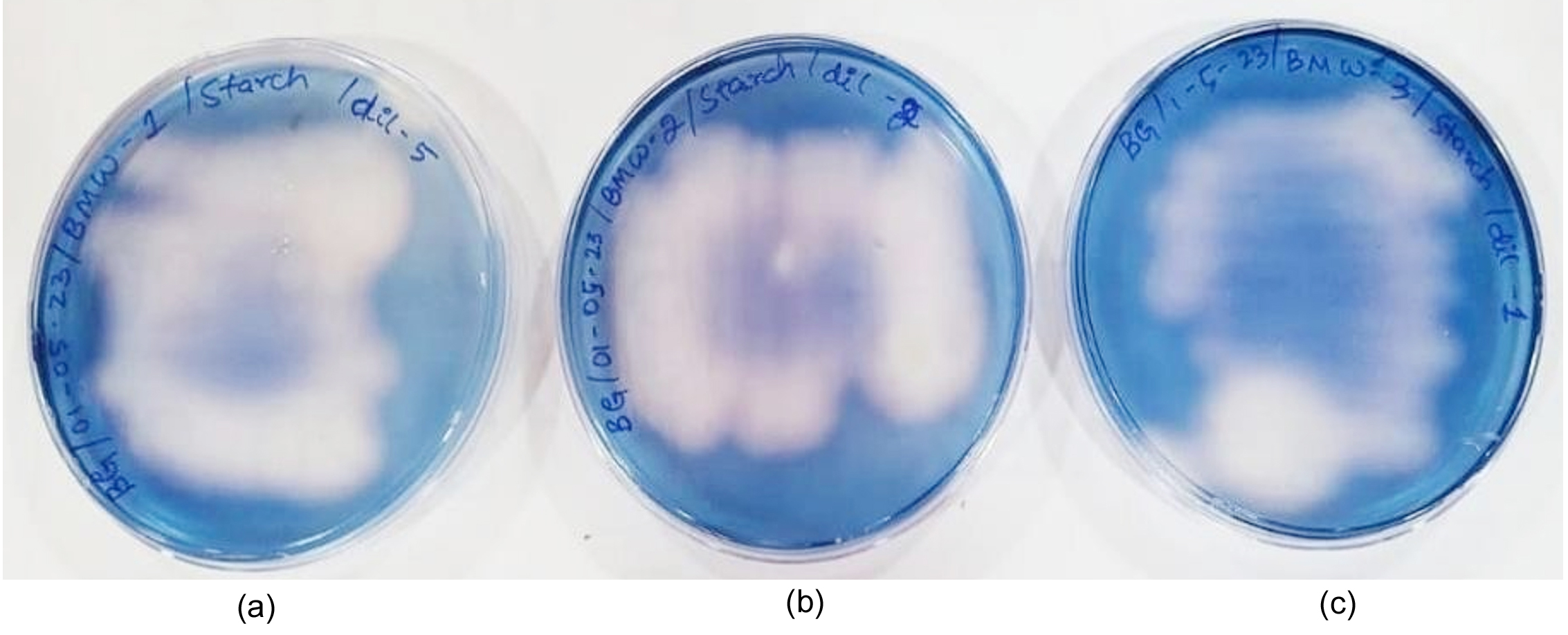
Figure 7. Starch hydrolysis test; Positive result with clean zone around the addition of iodine. The presence of a clear zone indicates that the bacterium hydrolyzes the starch, and thus amylase was produced. (a) Sample 1; (b) Sample 2; (c) Sample 3
Hemolytic activity assessment
Following biochemical screening, three isolates were selected for evaluation of hemolytic activity on 5% sheep blood agar plates. Upon incubation at 37 °C for 18-24 hrs, hemolytic patterns were examined under transmitted light. Figure 8 shows isolate demonstrated complete hemolysis (β-hemolysis), characterized by clear, colorless zones surrounding the bacterial colonies where red blood cells were completely lysed. The remaining 2 isolates showed no hemolytic activity (γ-hemolysis), with no visible clearing zones around the colonies. The β-hemolytic isolate was considered the most probable S. aureus candidate and was selected for molecular confirmation and further characterization studies.
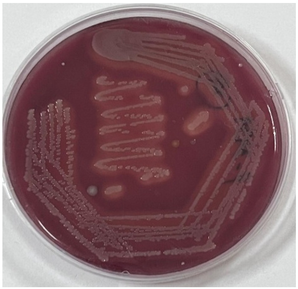
Figure 8. Isolate exhibited β-hemolysis with clear zones indicating complete lysis of red blood cells, while the remaining isolates showed γ-hemolysis with no visible clearing after incubation at 37 °C for 18-24 hrs
Gram staining
Microscopic examination of the bacterial isolates revealed Gram-positive cocci appearing as purple spherical cells arranged in characteristic grape-like clusters as shown in Figure 9. Retention of the crystal violet-iodine complex following alcohol decolorization confirmed the presence of a thick peptidoglycan layer typical of Gram-positive bacteria. The observed cellular morphologies and clustering patterns were consistent with the identification of Staphylococcus species. These findings support preliminary identification based on MSA screening and provide crucial morphological evidence for further biochemical characterization.
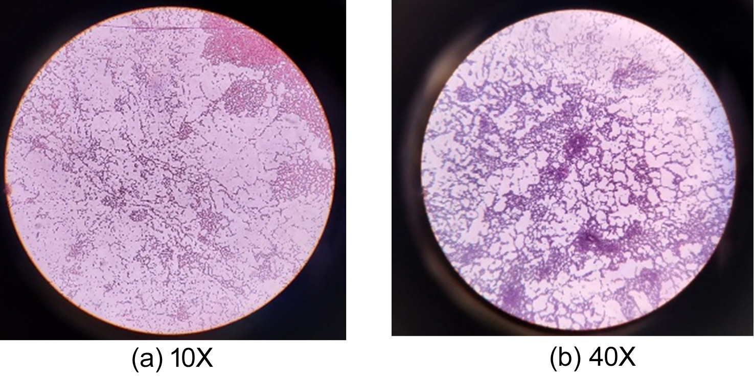
Figure 9. Microscopic view of Gram-stained bacteria at different magnifications. (a) 10x; (b) 40x
Molecular characterization
The quality of the extracted genomic DNA was confirmed by agarose gel electrophoresis, which showed a distinct high molecular weight band. PCR amplification of the 16S rRNA gene yielded a single discrete band of approximately 1500 bp, confirming successful amplification of the target gene (Figure 10). A phylogenetic tree was constructed by the neighbor-joining method using 16S rRNA gene sequences, and MEGA 11 software was used for phylogenetic analysis. Isolate P_3462 showed the highest sequence similarity with S. aureus strains, exhibiting 100% query coverage and upto 100% sequence identity with multiple reference sequences in the NCBI GenBank database. The isolate showed close similarity to S. aureus strains (e.g., MN565965.1, MN567481.1), with identity values ranging from 99.80%-100%. BLAST analysis confirmed that the isolate was closely related to S. aureus which was deposited in GenBank under the accession number PX451269.1 and the selected sequences were used for phylogenetic reconstruction. The phylogenetic tree shown in Figure 11 demonstrates that the isolate clustered tightly with S. aureus reference strains, indicating strong evolutionary relatedness. Furthermore, genetic distance analysis revealed negligible variation between the isolate and the reference sequences, confirming high sequence conservation.
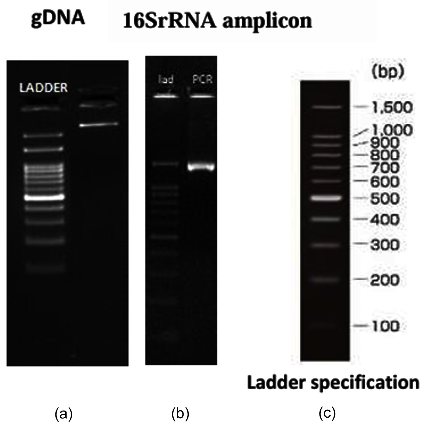
Figure 10. (a) Agarose gel electrophoresis of gDNA and a distinct band is observed; (b) 1500 bp of 16S rRNA amplification; (c) DNA ladder (100-1500 bp)
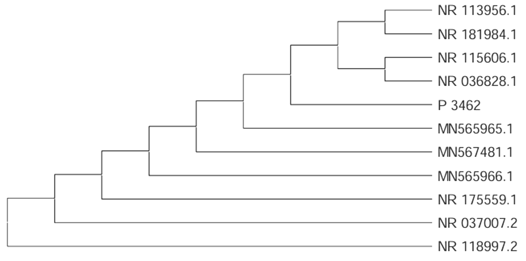
Figure 11. Phylogenetic analysis of the bacterial isolate based on 16S rRNA gene sequences
The neighbour-joining tree shows that isolate P_3462 clusters closely with Staphylococcus aureus reference strains, confirming its taxonomic identity
Virulent factor characterizations
Nitrate reductase test
The nitrate reductase test was positive for the isolate as shown in Figure 12. A positive result suggests that the isolate can reduce nitrate to nitrite, a trait commonly found in facultative anaerobes such as S. aureus. This ability supports bacterial survival in low-oxygen environments such as infected tissues or biofilms, which are common in hospital-acquired infections. Nitrate reduction is also linked to enhanced persistence of medical devices and contributes to biofilm formation. These biofilms protect the bacteria from antibiotics and aid in antibiotic resistance. In contrast, a negative result may indicate limited adaptability to hostile conditions, suggesting a lower virulence and resistance potential.
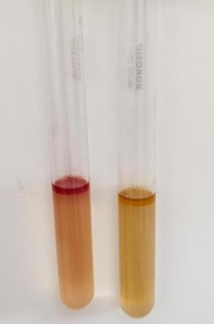
Figure 12. Nitrate reduction test showing positive and negative reactions
Antimicrobial activity-drug resistance test (Agar disc diffusion/Kirby-Bauer)
A bacterial isolate obtained from the swab sample was identified as MRSA using the automated VITEK 2 identification system with a 99% probability of identification. The isolate demonstrated resistance to several β-lactam antibiotics including benzylpenicillin, oxacillin, cefuroxime, cefpodoxime, and cefotaxime, confirming methicillin-resistance. The strain was also resistant to macrolides and fluoroquinolones such as erythromycin, clarithromycin, ciprofloxacin, and levofloxacin. However, the isolate was susceptible to linezolid, vancomycin, teicoplanin, daptomycin, tetracycline, tigecycline, nitrofurantoin, and rifampicin, indicating potential efficacy of these antibiotics. The antimicrobial susceptibility profiles are presented in Table 3.
Table 3. The MRSA isolate demonstrated extensive multidrug resistance to 14 antibiotics across β-lactam, fluoroquinolone, macrolide, and lincosamide classes, with sensitivity retained to glycopeptides, linezolid, daptomycin, tetracycline, tigecycline, nitrofurantoin, and rifampicin
Antibiotic |
Result |
|---|---|
Cefoxitin |
Resistant |
Benzylpenicillin |
Resistant |
Oxacillin |
Resistant |
Amoxicillin/Clavulanic acid |
Resistant |
Cefuroxime |
Resistant |
Cefpodoxime |
Resistant |
Cefotaxime |
Resistant |
Ciprofloxacin |
Resistant |
Levofloxacin |
Resistant |
Erythromycin |
Resistant |
Clarithromycin |
Resistant |
Clindamycin |
Resistant |
Azithromycin |
Resistant |
Cotrimoxazole |
Resistant |
Gentamicin |
Intermediate |
Linezolid |
Sensitive |
Vancomycin |
Sensitive |
Teicoplanin |
Sensitive |
Daptomycin |
Sensitive |
Tetracycline |
Sensitive |
Tigecycline |
Sensitive |
Nitrofurantoin |
Sensitive |
Rifampicin |
Sensitive |
Computational analysis
The input data were processed using the PICRUSt2 full pipeline with the default parameters to infer the functional potential of the identified isolate. The NSTI value obtained was 0.0194, indicating high similarity to reference genomes and reliable functional predictions. Phylogenetic placement analysis performed using the PICRUSt2 workflow revealed that the 16S rRNA sequence was accurately positioned within a reference phylogenetic tree containing diverse bacterial genomes. The resulting tree, visualized in a radial layout (Figure 13), showed that the query sequence clustered within the Staphylococcus aureus group alongside closely related reference strains. Dense clustering and short branch lengths indicated minimal evolutionary divergence, thereby supporting the taxonomic identification established by BLAST and phylogenetic analysis.
Functional pathway prediction in the Table 4, revealed a wide range of metabolic and biosynthetic pathways associated with the isolate. Core energy metabolism pathways, including glycolysis, anaerobic glycolysis, the tricarboxylic acid cycle, and central carbon metabolism, were identified, indicating active energy production mechanisms. Additionally, multiple amino acid biosynthesis pathways, such as valine, isoleucine, tryptophan, arginine, histidine, and threonine metabolism pathways, were observed, reflecting the metabolic versatility of the organism. Lipid metabolic pathways, including fatty acid biosynthesis and elongation, were also predicted. Notably, the pathways involved in cell wall biosynthesis, including peptidoglycan, teichoic acid, and lipoteichoic acid synthesis, were highly represented. These pathways are characteristic features of S. aureus and play important roles in maintaining the cell structure and contributing to its pathogenic potential.
Table 4. Predicted functional pathways identified using PICRUSt2 analysis
Pathway ID |
Predicted Function |
Relative Abundance |
|---|---|---|
ANAEROFRUCAT-PWY |
Anaerobic fructose catabolism |
0.619 |
FASYN-ELONG-PWY |
Fatty acid elongation (unsaturated) |
0.571 |
REDCITCYC |
Reductive citric acid cycle |
0.636 |
GLYCOLYSIS |
Glycolytic pathway |
0.581 |
HISDEG-PWY |
Histidine degradation |
0.500 |
HISTSYN-PWY |
Histidine biosynthesis |
0.538 |
THRESYN-PWY |
Threonine biosynthesis |
0.545 |
ILEUSYN-PWY |
Isoleucine biosynthesis |
0.571 |
SER-GLYSYN-PWY |
Serine and glycine synthesis |
0.500 |
ARO-PWY |
Aromatic amino acid biosynthesis |
0.500 |
COMPLETE-ARO-PWY |
Complete aromatic amino acid biosynthesis |
0.545 |
PWY-7222 |
Amino acid biosynthesis |
0.600 |
PWY-7208 |
Cell wall glycan biosynthesis |
0.667 |
PWY-5667 |
Coenzyme A biosynthesis |
0.571 |
PWY-6386 |
Central carbon metabolism |
0.545 |
PWY-6121 |
Nucleotide metabolism |
0.577 |
PWY-6126 |
Pyrimidine metabolism |
0.583 |
PWY-6147 |
Amino sugar metabolism |
0.571 |
PWY-5897 |
Fatty acid degradation |
0.500 |
PWY-5837 |
Purine metabolism |
0.500 |
Metagenome prediction based on the EC classification identified 565 predicted enzymes involved in key metabolic processes such as fermentation, redox balance, and energy generation. Representative enzymes, including alcohol dehydrogenase, lactate dehydrogenase, pyruvate kinase, and fumarase, indicate active central metabolic pathways. KO prediction identified 1,267 functional orthologs involved in diverse biological processes. The key functional genes included those associated with glycolysis, peptidoglycan biosynthesis, teichoic acid synthesis, and regulatory systems involved in environmental adaptation and signaling. Gene copy number analysis indicated that most core metabolic genes were single-copy genes, whereas certain regulatory and virulence-associated genes exhibited slightly elevated copy numbers, suggesting the potential enrichment of adaptive and pathogenic traits.
Overall, the PICRUSt2 analysis provided comprehensive insights into the predicted functional capabilities of the isolate, highlighting its metabolic adaptability and potential biological functions. However, these findings represent computational predictions based on reference genomes and do not correspond to experimentally validated gene presence or expression as shown in Figure 13.
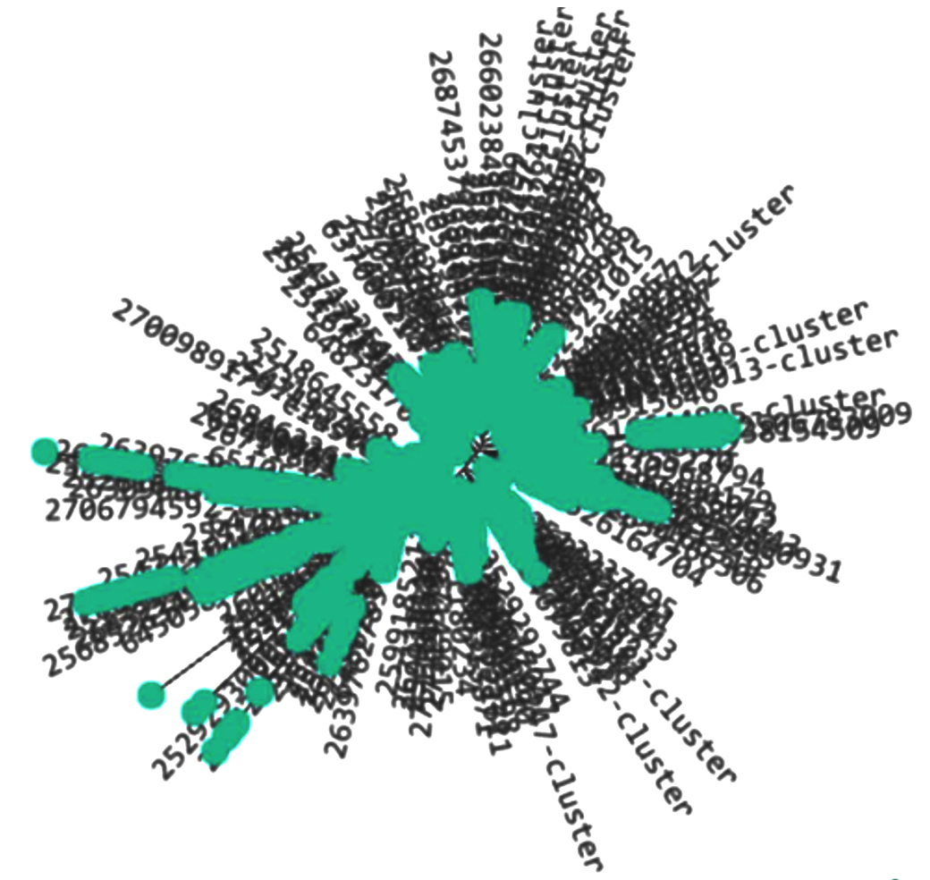
Figure 13. Phylogenetic placement of the 16S rRNA sequence using PICRUSt2
The radial phylogenetic tree represents the placement of the query 16S rRNA sequence within a reference database of bacterial genomes generated by the PICRUSt2 pipeline. Each branch corresponds to related reference sequences, while the turquoise-highlighted node indicates the position of the query sequence.
The identification of multidrug-resistant MRSA in hospital biomedical waste fundamentally challenges the traditional view of waste as a passive byproduct, revealing it as an active ecological niche that selects for enhanced pathogenic traits.35 The selective pressures inherent in these environments, including sub-therapeutic antibiotic concentrations, diverse nutrients, and competitive microbial communities, create evolutionary conditions that may produce pathogens with greater virulence and resistance than their clinical counterparts.36 This paradigm shift necessitates reconceptualizing biomedical waste management from simple disposal to active pathogen surveillance and control, with implications extending far beyond immediate healthcare settings.
The observed biochemical and molecular characteristics suggest significant evolutionary adaptations that enhance bacterial survival in harsh waste environments while simultaneously increasing pathogenic potential. The metabolic versatility demonstrated by isolates reflects selection for competitive advantages in nutrient-limited, chemically stressed conditions typical of biomedical waste.37 More concerning is the evidence for ongoing horizontal gene transfer and resistance accumulation, creating genetic reservoirs that may outpace clinical surveillance efforts. The close phylogenetic relationship between waste-derived and clinically significant strains indicates bidirectional transmission pathways that challenge traditional epidemiological models that focus solely on patient-to-patient spread.8
Geographic variations in MRSA prevalence reveal systemic vulnerabilities in the healthcare infrastructure that extend beyond resource availability to encompass complex interactions between antimicrobial prescription practices, waste handling protocols, and regulatory compliance. These disparities suggest that standardized approaches may be insufficient without considering regional adaptation and implementation capacity.5 The higher prevalence in certain districts indicates potential hotspots for resistance development that could serve as sources for broader dissemination, emphasizing the need for targeted surveillance and intervention strategies that address local contexts rather than uniform national policies.
The presence of virulent, multidrug-resistant MRSA in biomedical waste has profound implications for the One Health framework, demonstrating how healthcare waste serves as critical nodes in resistance transmission networks that extend into environmental and community settings. In a comparable study, Volvoikar et al. collected samples from various tertiary care hospitals across India. Their findings revealed that 44.8% of the isolates were resistant to MRSA, showing the highest resistance to ciprofloxacin and gentamicin, along with other antibiotics. These observations are consistent with the results of our findings.38
The documented environmental contamination surrounding hospitals creates exposure pathways that affect not only healthcare workers but also broader populations through water, soil, and air contamination.18 This interconnectedness suggests that effective resistance control requires coordinated interventions across multiple sectors, with waste management representing a critical but underappreciated component of antimicrobial resistance mitigation strategies.19
The current regulatory frameworks governing biomedical waste management are inadequate for addressing multidrug-resistant organisms with enhanced survival capabilities. Existing guidelines focus on traditional sterilization approaches that may be ineffective against resistant pathogens embedded in biofilms or possessing enhanced environmental persistence.13 The absence of mandatory surveillance requirements for resistant bacteria in waste streams creates critical blind spots in pathogen monitoring, while technological limitations in rapid detection methods hamper real-time response capabilities. The integration of advanced molecular approaches with routine surveillance remains constrained by resource requirements and technical complexity, particularly in low-resource settings where enhanced monitoring may be required.16
The economic implications of resistant pathogen emergence in biomedical waste extend far beyond immediate healthcare costs and encompass broader societal impacts, including environmental remediation, outbreak response, and compromised antimicrobial effectiveness. The potential for environmental reservoirs to serve as sources of novel resistance mechanisms represents a significant threat to global health security, particularly given the rapid international spread of resistance genes through multiple pathways.2 Investment in enhanced waste management infrastructure and surveillance systems must be evaluated against the long-term costs of uncontrolled resistance dissemination. Economic modeling suggests that proactive approaches may be cost-effective compared to reactive outbreak responses.21
Future research must prioritize longitudinal studies tracking the evolution of resistance in waste environments, development of rapid field-deployable detection methods, and evaluation of novel treatment technologies against multidrug-resistant organisms. The integration of artificial intelligence and machine learning with traditional microbiological approaches offers promise for predictive surveillance systems that can transform waste monitoring from reactive to preventive strategies.39 Critical knowledge gaps remain regarding the effectiveness of current treatment processes against biofilm-embedded resistant bacteria and the potential for waste-derived pathogens to acquire enhanced virulence through environmental stress responses.
The identification of biomedical waste as a reservoir of enhanced pathogens necessitates the immediate revision of waste management protocols, worker protection standards, and environmental monitoring requirements. Healthcare facilities should implement routine surveillance of waste streams as early warning systems for emerging resistance threats, while policy makers must update regulations to specifically address multidrug-resistant organisms and require enhanced treatment protocols.40 The development of international cooperation frameworks for waste-associated resistance surveillance could provide critical early detection capabilities for global health security. Ultimately, addressing the challenge of waste-associated antimicrobial resistance requires fundamental shifts in the conceptualization, regulation, and management of the environmental dimensions of healthcare delivery.
The functional pathways and virulence-associated traits identified in this study were based on PICRUSt2 predictive analysis and therefore represent computational inferences rather than experimentally validated functions. Further genomic and experimental validation studies are required to confirm the predicted biological activities and pathogenic traits.
This study highlights the potential role of hospital biomedical waste as a critical reservoir for multidrug-resistant MRSA with enhanced virulence profiles, highlighting concerns regarding the traditional concept of waste as merely disposal. The identification of extensively drug-resistant MRSA isolates exhibiting robust nitrate reduction, diverse metabolic capabilities, and multiple virulence factors demonstrated that environmental selection pressures in waste streams may contribute to the persistence and potential pathogenicity of organisms in biomedical waste environments. The geographic variation in prevalence across districts reveals systemic vulnerabilities in the healthcare infrastructure that require targeted, region-specific interventions rather than uniform national approaches.
These findings highlight the potential relevance of biomedical waste within the One Health framework and demonstrate how inadequately managed biomedical waste creates interconnected transmission networks that extend far beyond healthcare facilities and affect community and environmental health. The observed resistance patterns and virulence characteristics suggest that current waste management protocols are inadequate for controlling multidrug-resistant organisms, necessitating immediate regulatory reform and technological innovation. Economic implications extend beyond immediate healthcare costs to broader societal impacts, including environmental contamination, occupational health risks, and compromised antimicrobial effectiveness that threaten global health security.
Further investigations and improved monitoring strategies may help address the identified threats through enhanced waste surveillance systems, improved treatment technologies, and strengthened worker protection protocols. Healthcare facilities must implement routine monitoring of waste streams as an early warning system for emerging resistance threats, and policymakers should mandate enhanced treatment protocols specifically targeting multidrug-resistant organisms. Future research should focus on developing rapid detection methods, evaluating novel treatment approaches, and establishing international cooperative frameworks for surveillance of waste-associated resistance. The integration of advanced molecular techniques with artificial intelligence is promising for transforming waste monitoring from a reactive to a predictive strategy.
In conclusion, this study established biomedical waste as an active ecological niche that selects and maintains highly adapted, virulent bacterial populations, with significant implications for healthcare-associated infections and the dissemination of antimicrobial resistance. These findings underscore the urgent need for paradigm shifts in the conceptualization, regulation, and management of the environmental dimensions of healthcare delivery. Addressing the challenge of waste-associated antimicrobial resistance requires coordinated efforts across the clinical, environmental, and policy domains to prevent these hidden reservoirs from undermining global efforts to combat antimicrobial resistance and protect public health.
ACKNOWLEDGMENTS
The authors sincerely acknowledge the District Government Hospital Gulbarga, Bidar and Yadgir district for helping to collect Biomedical waste of hospitals for the work and also like to thank the Department of Microbiology, VSK University, Ballari (Jnana Sagara Campus, Karnataka, India), and the JSS Academy of Higher Education & Research, Mysuru (Karnataka, India), for providing the necessary laboratory facilities, infrastructure, and support to carry out this research work successfully.
CONFLICT OF INTEREST
The authors declare that there is no conflict of interest.
AUTHORS’ CONTRIBUTION
MM and KR conceptualized the study and applied methodology. CD performed data curation, formal analysis and visualization. MM and KR wrote the original draft. HSP performed supervision, validation, wrote, reviewed and edited the manuscript. All authors read and approved the final manuscript for publication.
FUNDING
None.
DATA AVAILABILITY
All datasets generated or analyzed during this study are included in the manuscript.
ETHICS STATEMENT
This article does not contain any studies on human participants or animals performed by any of the authors.
- Hassoun A, Linden PK, Friedman B. Incidence, prevalence, and management of MRSA bacteremia across patient populations—a review of recent developments in MRSA management and treatment. Crit Care. 2017;21(1):211.
Crossref - Antimicrobial Resistance Collaborators. Global Burden of Bacterial Antimicrobial Resistance in 2019: a Systematic Analysis. Lancet. 2022;399(10325):629-655.
Crossref - Lakhundi S, Zhang K. Methicillin-Resistant Staphylococcus aureus: Molecular Characterization, Evolution, and Epidemiology. Clin Microbiol Rev. 2018;31(4):E00020-18.
Crossref - Brinkwirth S, Feig M, Noll I, et al. Changing dynamics of bloodstream infections due to methicillin-resistant Staphylococcus aureus and vancomycin-resistant Enterococcus faecium in Germany, 2017–2023: A continued burden of disease approach. Antimicrob Resist Infect Control. 2025;14(1):4.
Crossref - Hounmanou YMG, Houefonde A, I-CRECT Consortium Nguyen TT, Dalsgaard A. Mitigating antimicrobial resistance through effective hospital wastewater management in low-and middle-income countries. Front Public Health. 2025;12:1525873.
Crossref - Chandja WBE, Onanga R, Nguema PPP, et al. Emergence of Antibiotic Residues and Antibiotic-Resistant Bacteria in Hospital Wastewater: A Potential Route of Spread to African Streams and Rivers, a Review. Water. 2024;16(22):3179.
Crossref - Abosse JS, Megersa B, Zewge F, Eregno FE. Healthcare waste management and antimicrobial resistance: a critical review. J Water Health. 2024;22(11):2076-2093.
- CDC Weekly C. Reported Cases and Deaths of National Notifiable Infectious Diseases — China, September 2023. China CDC Weekly. 2023;5(46):1045-1046.
Crossref - von Wintersdorff CJH, Penders J, van Niekerk JM, et al. Dissemination of Antimicrobial Resistance in Microbial Ecosystems through Horizontal Gene Transfer. Front Microbiol. 2016;7(173).
Crossref - Chowdhury AMMA, Uddin KN. Analysis of the Occurrence of Antibiotic Resistant Bacteria in the Hospital’s Effluent and its Receiving Environment. Microbiol Insights. 2022;15:117863612210782.
Crossref - Gullberg E, Cao S, Berg OG, et al. Selection of Resistant Bacteria at Very Low Antibiotic Concentrations. PLoS Pathogens. 2011;7(7):e1002158.
Crossref - Stewart PS, William Costerton J. Antibiotic resistance of bacteria in biofilms. Lancet. 2001;358(9276):135-138.
Crossref - Perez-Bou L, Gonzalez-Martinez A, Jesus-Gonzalez Lopez, Correa-Galeote D. Promising bioprocesses for the efficient removal of antibiotics and antibiotic-resistance genes from urban and hospital wastewaters: Potentialities of aerobic granular systems. Environ Poll. 2023;342:123115.
Crossref - Parida VK, Sikarwar D, Majumder A, Gupta AK. An assessment of hospital wastewater and biomedical waste generation, existing legislations, risk assessment, treatment processes, and scenario during COVID-19. J Environ Manag. 2022;308:114609.
Crossref - Tosas AO, Dean A, Van KE, Bertagnolio S. Global antibiotic resistance surveillance report 2025. World Health Organization; 2025. https://ora.ox.ac.uk/objects/uuid:bdc10423-c80c-42e8-8de3-5b370bca9178
- Shuai X, Zhou Z, Zhu L, et al. Ranking the risk of antibiotic resistance genes by metagenomic and multifactorial analysis in hospital wastewater systems. J Hazard Mater. 2024;468:133790-133790.
Crossref - McEwen SA, Collignon PJ. Antimicrobial Resistance: a One Health Perspective. Microbiol Spectr. 2018;6(2).
Crossref - Liu K, Gan C, Peng Y, et al. Occurrence and source identification of antibiotics and antibiotic resistance genes in groundwater surrounding urban hospitals. J Hazard Mater. 2023;465:133368-133368.
Crossref - World Health Organization (WHO). A one health priority research agenda for antimicrobial resistance;2023. https://www.who.int/publications/i/item/9789240075924
- Franklin AM, Weller DL, Durso LM, et al. A one health approach for monitoring antimicrobial resistance: developing a national freshwater pilot effort. Front Water. 2024;6.
Crossref - Endalamaw K, Tadesse S, Asmare Z, Kebede D, Erkihun M, Abera B. Antimicrobial resistance profile of bacteria from hospital wastewater at two specialized hospitals in Bahir Dar city, Ethiopia. BMC Microbiol.2024;24(1):525.
Crossref - Nakamura-Silva R, Dias LL, Sousa RC, Fujimoto RY, Pitondo-Silva A. Multidrug-resistant and potentially pathogenic Enterobacteriaceae found in a tertiary hospital sewage in southeastern Brazil. Environ Monit Assess. 2022;194(10):782.
Crossref - Hussein YA, AL Jabbar WAA, Al-Aubaidy H. Identification and Characterization of Antibiotic-Resistant Bacterial Strains in Hospital Wastewater. J Biomed Biochem.2015;4(2):21-41.
Crossref - Adiba RA, Ferdous RM. Characterization of Pseudomonas aeruginosa from hospital sewage water and nearby community water based on their multi-drug-resistant gene [Doctoral Dissertation], Brac University. 2023
- Thakur P, Nayyar C, Tak V, Saigal K. Mannitol-fermenting and tube coagulase-negative staphylococcal isolates: unraveling the diagnostic dilemma. J Lab Physicians. 2017;9(1):065-066.
Crossref - Hassan MA, Abd El-Aziz S, Elbadry HM, El-Aassar SA, Tamer TM. Prevalence, antimicrobial resistance profile, and characterization of multi-drug resistant bacteria from various infected wounds in North Egypt. Saudi J Biol Sci.2022;29(4):2978-2988.
Crossref - Manik RK, Mahmud Z, Mishu ID, Hossen MS, Howlader ZH, Nabi AHMN. Multidrug Resistance Profiles and Resistance Mechanisms to β-Lactams and Fluoroquinolones in Bacterial Isolates from Hospital Wastewater in Bangladesh. Curr Issues Mol Biol. 2023;45(8):6485-6502.
Crossref - Oluwatayo Makinde. Prevalence of multidrug resistant and extended spectrum beta-lactamase producing Pseudomonas aeruginosa in the soil of hospital waste dumps. Microbes and Infectious Diseases. 2023.
Crossref - Das T, Das A, Das N, et al. Genetic Diversity and Phylogenetic Analysis Among Multidrug-Resistant Pseudomonas spp. Isolated from Solid Waste Dump Sites and Dairy Farms. Acta Microbiol Hell. 2025;70(3):30.
Crossref - Barnwal SK, Saleh AM. Antibiotic resistance patterns of environmental bacteria from sewage water in Vellore, India: isolation, virulence analysis, and characterization. Front Microbiol. 2025;16:1640369.
Crossref - Kunarisasi S, Yuliandari P, Pramono AK, et al. Genomic characterization of multidrug-resistant Escherichia coli isolates from hospital wastewater in Jakarta, Indonesia. Mol Biol Rep. 2025;52(1):960.
Crossref - Gotkowska-Plachta A. The Prevalence of Virulent and Multidrug-Resistant Enterococci in River Water and in Treated and Untreated Municipal and Hospital Wastewater. Int J Environ Res Public Health. 2021;18(2):563.
Crossref - Mahmud MS, Hosen MA, Hossion MI, et al. Isolation, identification, and characterization of resistant bacteria to antibiotics from pharmaceutical effluent and study of their antibiotic resistance. Front Microbiol. 2023;14:1307291.
Crossref - Nawaz A, Zafar S, Alessa AH, et al. Characterization of ES10 lytic bacteriophage isolated from hospital waste against multidrug-resistant uropathogenic E coli.Front Microbiol. 2024;15:1320974.
Crossref - Berendonk TU, Manaia CM, Merlin C, et al. Tackling antibiotic resistance: the environmental framework. Nat Rev Microbiol. 2015;13(5):310-317.
Crossref - Andersson DI, Hughes D. Microbiological effects of sublethal levels of antibiotics. Nat Rev Microbiol. 2014;12(7):465-478.
Crossref - Manoharan RK, Srinivasan S, Shanmugam G, Ahn YH. Shotgun metagenomic analysis reveals the prevalence of antibiotic resistance genes and mobile genetic elements in full scale hospital wastewater treatment plants. J Environ Manag. 2021;296:113270.
Crossref - Volvoikar H, Preethy R, Shatriyaa C, et al. Multidrug-resistance Profile of Staphylococcus aureus in a Tertiary Care Centre. J Pure Appl Microbiol. 2026;20(1):157-166.
Crossref - Briki A, Alkhatib N, Aloba B, et al. Molecular epidemiology of methicillin-resistant Staphylococcus aureus in Gulf Cooperation Council countries (2010–2025): a scoping review. Front Microbiol. 2025;16:1714252.
Crossref - Lan L, Wang Y, Chen Y, Wang T, Zhang J, Tan B. A Review on the Prevalence and Treatment of Antibiotic Resistance Genes in Hospital Wastewater. Toxics. 2025;13(4):263-263.
Crossref
© The Author(s) 2026. Open Access. This article is distributed under the terms of the Creative Commons Attribution 4.0 International License which permits unrestricted use, sharing, distribution, and reproduction in any medium, provided you give appropriate credit to the original author(s) and the source, provide a link to the Creative Commons license, and indicate if changes were made.