


ISSN: 0973-7510
E-ISSN: 2581-690X
 , Sidhartha Singh3 and Shashank Garg1
 Microalgae have gained considerable attention as sustainable sources of biomass and valuable metabolites, including lipids. The unicellular green alga Parachlorella kessleri has shown potential for lipid production. In this study, an algae species was isolated from the Yamuna River in Delhi. The organism was cultured and characterized using morphological, biochemical, and molecular approaches, with species identification primarily supported by morphological and taxonomic characterization. This study was performed to examine the influence of media pH, incubation temperature, photoperiod, and CO2 sparging duration on the growth characteristics of P. kessleri. One-factor-at-a-time (OFAT) approach was used to assess the effect of parameters on cell growth. The lipid content of the organism was also determined. The findings indicated that the maximum biomass production occurred in a 16 day-old culture under the following conditions: pH 8, a temperature of 25 °C, a photoperiod of 16:8 (light:dark), and CO2 sparging for 2 min twice daily. This species exhibited significant potential for lipid production under normal culture conditions.
Parachlorella kessleri, Lipid Production, Biomass, Optimization, Algae, Biofuel
Microalgae have become a focal point of research due to their potential to produce valuable bioproducts such as biodiesel, bioplastics, and bioenergy.1-3 This growing interest is driven by the global demand for sustainable alternatives to fossil fuels and conventional plastics, both of which pose significant environmental challenges.4 The growth dynamics of microalgae are highly strain-specific and strongly influenced by environmental factors such as pH, light intensity, photoperiod, temperature, and CO2 availability.5,6 These parameters are critical in determining microalgal growth rates, adaptability, and biomass productivity, which are closely linked to photosynthetic efficiency.7,8 Optimizing these parameters is therefore essential to maximize yield, lower production costs, and enhance overall efficiency.9,10
Among various microalgal strains, Parachlorella kessleri has gained significant attention for its industrial potential due to its ability to produce both starch and lipids, making it a valuable resource for a wide range of bioproducts.11-13 Notable for its high lipid content, rapid growth rate, and substantial biomass productivity, P. kessleri ensures a reliable and abundant raw material supply.14,15 Additionally, its adaptability to diverse cultivation media, including brewery wastewater,16 chicken processing wastewater,17 agro-industrial byproducts, and wastewater, underscores its potential for resource recovery and environmental sustainability.18
Despite its promising applications, previous research has primarily focused on optimizing individual growth parameters such as pH,19 photoperiod,20 and temperature,21 often overlooking the intricate interactions among multiple factors. This narrow approach limits the scalability of P. kessleri for industrial applications. To address this gap, the present study systematically evaluated the effects of pH, temperature, photoperiod, and CO2 sparging duration on the growth dynamics and biomass productivity of P. kessleri. By integrating these factors, this research aims to provide a comprehensive understanding of optimal cultivation strategies, facilitating large-scale production and enhancing its potential for industrial applications such as bioplastics.
The present study was conducted to investigate the effects of different environmental parameters, including pH, temperature, photoperiod, and CO2 sparging duration on the growth and biomass productivity of P. kessleri. By adopting an integrated optimization approach, this study aims to identify ideal cultivation conditions that enhance biomass yield and overall efficiency.
Chemicals
Analytical-grade chemicals such as Bold’s Basal Medium, ampicillin, Lugol’s solution, Nile red, CTAB buffer, and PCR reagents were sourced from Sigma-Aldrich. The equipment used included a Magnus MLXi Plus microscope, REMI C852 centrifuge, and Lasany Instruments UV-VIS spectrophotometer (Model No. LI 295).
Sample collection, isolation and growth conditions
A microalgal sample was isolated from the stream of Yamuna River at Okhla, Delhi (latitude 28.52° N, longitude 77.28° E). Water samples were collected using the grab sampling method in a sterile white bottle. To reduce the microbial load and facilitate isolation, the sample was diluted with sterile distilled water and transported to the laboratory, where it was processed on the same day. The streak plate method was used to culture the algae in Bold Basal Medium (BBM) in Petri plates. Successive rounds of streaking were performed on BBM plates supplemented with ampicillin to eliminate bacterial contaminants and obtain a purified (axenic) culture. Ampicillin, a β-lactam antibiotic, selectively inhibits bacterial cell wall synthesis without affecting algal cells, thereby aiding in the purification of the algal culture. The resulting axenic culture was maintained in BBM and incubated at 25 °C under continuous illumination (100 lux) with constant shaking at 150 rpm on an orbital shaker.15
Characterization of P. kessleri
Light microscopy
The algal sample was transferred onto a clean glass slide using a pipette, followed by addition of Lugol’s solution to enhance visibility. A coverslip was gently placed over each sample to prepare a wet mount. The algal cells were examined under a bright-field microscope at 100x magnification, and the features and characteristics of the microalgal isolate PDP_01 were documented. The isolate was morphologically identified as P. kessleri based on its observed characteristics and comparison with the taxonomic descriptions available in AlgaeBase.22,23 The morphological identification was further confirmed by Dr. U. Elaya Perumal, Scientist, Annakkili Amma Research Institute (AARI), Chennai, Tamil Nadu, India.
Fluorescent microscopy
Intracellular lipid droplets were identified using Nile Red (9-(diethylamino)-5H-benzo[α]phenoxazine-5-one) fluorescence staining. Microalgae biomass was first collected by centrifugation at 250 g for 10 minutes and resuspended in 200 µL of distilled water by vortex mixing. The cell suspension was then treated with a 1% (w/v) Nile Red solution and incubated for 10 min under dark conditions. Excess dye was removed by washing, and lipid accumulation was visualized using fluorescence microscopy within an excitation range of 535-650 nm.24
Molecular identification
DNA extraction
Genomic DNA was isolated from algal samples using a modified CTAB-based method adapted from Doyle.25 The samples were suspended in CTAB extraction buffer and incubated at 60 °C for 1 hrs with intermittent mixing. After cooling to ambient temperature, chloroform:isoamyl alcohol (24:1, v/v) was added, and the mixture was gently mixed for 15 min, followed by centrifugation at room temperature at approximately 16,000 g for 10 min. The aqueous phase was carefully transferred to a new tube and combined with an equal volume of chilled isopropanol to precipitate the DNA, then incubated at -20 °C for 20-30 min. The DNA was pelleted by centrifugation at 4 °C for 10 min, washed with 70% ethanol, and resuspended in TE buffer. DNA integrity and quality were assessed using 0.8% agarose gel electrophoresis prior to PCR amplification.19
PCR amplification
Amplification of partial 18S rRNA gene sequences was carried out using a thermal cycler. PCR was performed with the forward primer A (5′-AACCTGGTTGATCCTGCCAG-3′) and the reverse primer SSU-inR1 (5′-CACCAGACTTGCCCTCCA-3′), which target conserved regions of the 18S rDNA. Each 20 µL PCR reaction contained 1 µL of genomic DNA template along with the forward and reverse primers. The amplification program consisted of an initial denaturation step at 94 °C for 30 sec, followed by annealing at 55 °C for 30 sec and extension at 72 °C for 1 min, with a final extension at 72 °C for 7 min.22,26
Sequence alignment and phylogenetic analysis
The generated partial 18S rRNA gene sequence was initially examined and manually refined using BioEdit software (version 7.2.5) to eliminate ambiguous regions at the 5′ and 3′ termini. The refined sequence was then compared with reference sequences from the National Center for Biotechnology Information (NCBI) database using the BLAST (Basic Local Alignment Search Tool) algorithm.22,27 For phylogenetic analysis, the sequence of isolate PDP_01 (GenBank accession OR364517) was aligned with multiple P. kessleri reference sequences and closely related taxa obtained from GenBank. Phylogenetic analysis was performed in MEGA 12 using the Maximum Likelihood method based on the Kimura 2-parameter model with gamma-distributed rates and invariant sites (K2P+G+I), as determined by model selection according to the lowest Bayesian Information Criterion (BIC). Bootstrap analysis was conducted with 1,000 replicates, and sites with less than 95% coverage were excluded by partial deletion. Auxenochlorella protothecoides (MK191970.1) served as the outgroup.
Effect of cultivation factor on the growth behavior of P. kessleri
This study aimed to identify the key factors that influence microalgae growth under controlled conditions. A one-factor-at-a-time experiment was conducted in triplicate, focusing on pH, temperature, photoperiod, and CO2 sparging duration. The pH levels were varied at 6, 7, 8, 9, and 10, while temperature conditions were set at 25 °C, 30 °C, 35 °C, 40 °C, and 45 °C. The photoperiod was adjusted to different light/dark cycles, including 8:16, 10:14, 12:12, 14:10, and 16:8 hrs. The effect of CO₂ supplementation was evaluated by sparging CO₂ directly from a CO₂ cylinder into the cultures for 1, 2, 3, 4, and 5 min. CO₂ was supplied intermittently twice daily, in the morning and evening, throughout the cultivation period. The CO₂ flow was manually regulated to maintain a low, steady bubbling rate sufficient to ensure uniform gas dispersion without excessive foaming. A flow meter was not used; therefore, the gas flow rate was not quantitatively recorded. All experiments were maintained at a consistent light intensity of 100 lux (Orbit) throughout the study. The cultivation process was carried out in 250 mL Erlenmeyer flasks, each containing 100 mL of Bold’s Basal Medium as the working volume. The experiment lasted 16 days, during which growth was monitored periodically to assess the impact of different parameters on microalgal proliferation.
Growth kinetics
The growth of P. kessleri was tracked by recording the optical density (OD) at 670 nm every three days using a UV spectrophotometer. For biomass quantification, 100 mL culture samples were collected on the sixteenth day and centrifuged at 5000 rpm for 10 minutes to separate the algal biomass. After weighing, the wet biomass was dried at 40 °C in a hot air oven until it reached a stable weight, allowing calculation of dry biomass yield (g/L-1). The data obtained were used to analyze specific growth rates (µ) and biomass productivity (P) as per equations 1 and 2 respectively. One-factor-at-a-time (OFAT) approach was utilized to determine the optimum conditions for maximum biomass amount.
Specific growth rate (µ) = [In x2 – In x1 / t2 – t1] ….1
Where x1 and x2 denote biomass concentrations (g/L–¹) measured at time t1 and t2.
Biomass productivity: P = [ xf – xi / t ] …2
Where “xf” and “xi” represent final and initial biomass concentrations (g/L-1), respectively and “t” represents the cultivation period in days
Validation of growth at optimized condition
After determining the optimum levels for all four input variables, a comparative growth experiment was conducted under optimized and non-optimized conditions. All experiments were performed in triplicate using 250 mL Erlenmeyer flasks with a 100 mL working volume. For the preparation of experimental culture, 1 mL of actively growing P. kessleri inoculum (exponential phase) was aseptically transferred into 99 mL of sterile Bold’s Basal Medium, yielding a total working volume of 100 mL. The optimized conditions were set at pH 8, 25 °C temperature, a 16:8 hrs photoperiod (light-dark), and CO2 sparging for 2 min twice daily. Optical density (OD) was recorded at three-day intervals, and the dry cell weight (DCW) of the biomass was measured on day 16 to evaluate growth differences between the optimized and non-optimized treatments.
Extraction and estimation of lipid
The extraction and estimation of total lipids were performed from 16 day-old dried algal biomass using the Bligh and Dyer method. The algal culture was centrifuged at 5000 rpm for 10 minutes, after which the resulting pellet was freeze-dried. Following lyophilization, 0.1 g of the dried algal powder was precisely weighed and placed into a 5 mL glass vial. The empty glass bottle was weighed in advance, after which 2 mL of methanol and 1 mL of chloroform were added, and the mixture was refrigerated for 12 hours to facilitate lipid extraction. After incubation, the mixture was centrifuged at 5000 rpm for 10 min. After centrifugation, the lower (bottom) organic phase containing chloroform and extracted lipids was carefully collected. The lipid content was quantified by calculating the ratio of extracted lipid weight to the total biomass weight, expressed as a percentage of dry cell weight using equation 3:
Lipid %= WI / WB × 100 …3
where Wl represents the weight of the extracted lipid, and WB is the total weight of the dry biomass.28
Statistical analysis
All experiments were performed in triplicate, and the results are presented as mean values. Differences between groups were assessed using one-way and two-way analysis of variance (ANOVA) in GraphPad PRISM version 8.0, with a P-value of < 0.05 considered statistically significant.
Analysis and characterization of P. kessleri
The morphological features of the algal sample were consistent with those of P. kessleri. The individual cells of the colonies were in the range of 10 µm. Cells were dark green, spherical, and unicellular in shape, as shown in Figure 1A. Furthermore, Nile Red staining of lipid granules shown in Figure 1B confirmed the presence of intracellular lipid accumulation in PDP_01.
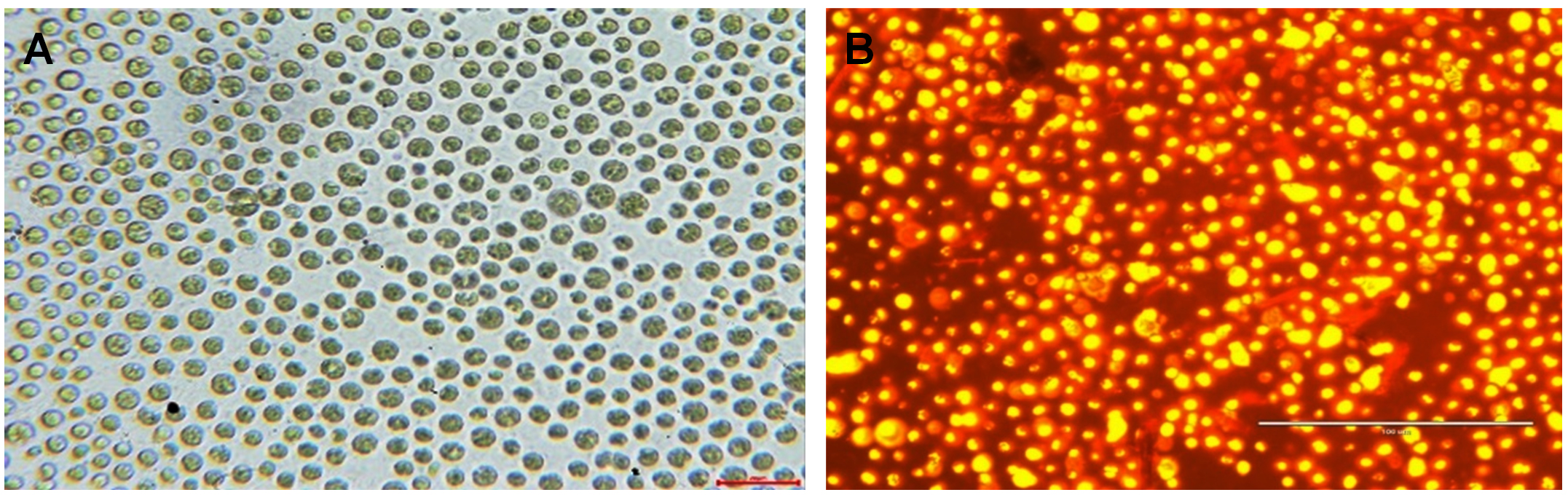
Figure 1. (A) Bright-field microscopic image of P. kessleri, illustrating its cellular morphology. (B) Fluorescent microscopic image of P. kessleri stained with Nile Red, highlighting intracellular lipid granules, which indicate lipid accumulation within the cells
Identification of microalgae species
The isolate PDP_01 was identified as P. kessleri through morphological assessment, comparison with AlgaeBase taxonomic data, and expert confirmation. Molecular analysis of the partial 18S rRNA gene provided further phylogenetic context. Maximum Likelihood phylogenetic analysis placed PDP_01 (GenBank accession OR364517) in a strongly supported clade with Dictyosphaerium sp. YN8 (MF664512.1), supported by a bootstrap value of 98%. However, because the initial BLAST analysis showed relatively low sequence identity, the partial 18S rDNA sequence was insufficient for definitive species-level molecular identification. Consequently, the assignment of PDP_01 as P. kessleri relied primarily on morphological and taxonomic evidence, while the molecular data provided supporting information regarding its phylogenetic relationship with closely related taxa (Figure 2).
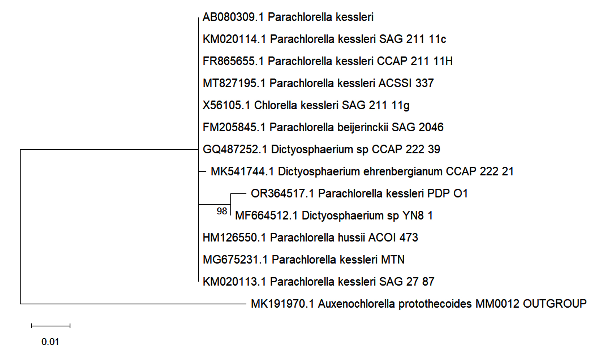
Figure 2. Maximum Likelihood phylogenetic tree showing the placement of isolate PDP_01 (OR364517.1) based on partial 18S rDNA sequences. The tree was constructed using the Kimura 2-parameter model with a gamma distribution and invariant sites (K2P+G+I). Bootstrap analysis was performed with 1,000 replicates, and bootstrap values ≥50% are shown at the nodes. Auxenochlorella protothecoides (MK191970.1) was used as the outgroup. Isolate PDP_01 clustered with Dictyosphaerium sp. YN8 (MF664512.1) with 98% bootstrap support
Effect of pH on growth of P. kessleri
The effect of pH on the cell number and mass of P. kessleri is depicted in Figure 3A and 3B. The data indicate that growth was highest at pH 8, with the lowest growth observed at pH 6. On the 16th day, the optical density of algal biomass reached 0.70 ± 0.021, and the maximum dry weight was recorded at 0.52 ± 0.036 g. Similarly, a previous study investigated the growth of P. kessleri and inorganic carbon (IC) concentration across various nitrogen levels and found that maintaining the pH at 8 enhanced algal growth and achieved a carbon removal efficiency of up to 95% over 34 days, yielding 3.4 g of dry biomass.29 Additionally, another researcher also investigated algal biomass and extracellular polysaccharide production in both indoor and outdoor settings, observing that under indoor conditions, the highest biomass was produced at pH 8 in combination with 2% CO2, outperforming two other treatments.30 These findings are consistent with existing literature, which demonstrates that pH 8 provides favorable conditions for enhanced growth rates and effective nitrogen removal from wastewater. Furthermore, this supports the strain’s adaptability to slightly alkaline conditions, although it can also grow across a broader pH range.
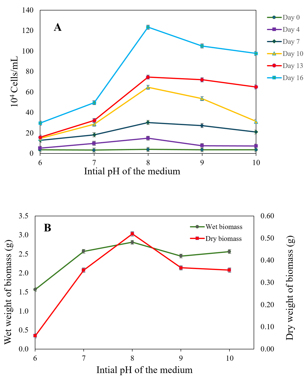
Figure 3. (A) Represents the growth curve of P. kessleri under different pH conditions, showing variations in growth over time. (B) Wet and dry biomass yields obtained at different initial pH levels, with initial pH represented on the x-axis and wet and dry biomass represented on the respective y-axis (n = 3)
Effect of temperature on growth of P. kessleri
Temperature is a critical determinant of microalgal physiology, and P. kessleri exhibited its highest biomass productivity at 25 °C. At this temperature, the culture reached a maximum optical density of 0.693 ± 0.006 and a dry biomass yield of 0.234 ± 0.076 g. Cell growth was completely inhibited at 40°C and 45°C from day 10 onward (Figure 4A). The dry biomass yield decreased to 0.128 ± 0.013 g at 30°C and 0.094 ± 0.006 g at 35°C (Figure 4B). The superior growth at 25 °C is likely attributable to two key factors: (1) enhanced photosynthetic and metabolic efficiency, as core enzymes such as RuBisCO and components of the electron transport chain function optimally at moderate temperatures; and (2) minimal thermal stress, since temperatures above ~28-30 °C are known to induce ROS accumulation, membrane destabilization, and photoinhibition, thereby suppressing biomass formation. These results are consistent with a previous study,31 which also identified 25 °C as the optimal growth temperature, and with Lv et al.,32 who reported that P. kessleri achieved the highest biomass concentrations (2750-2765 mg L-1) among five algal species after seven days of cultivation at this temperature.
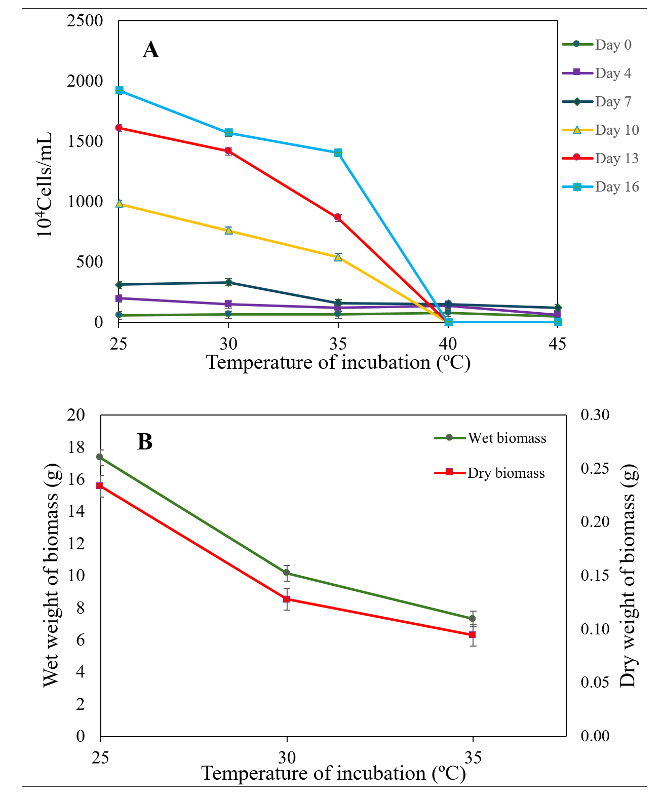
Figure 4. (A) Represents the growth curve of P. kessleri under different temperature conditions, illustrating how temperature influences its growth over time. (B) Wet and dry biomass yields obtained at different incubation temperatures, with temperature represented on the x-axis and wet and dry biomass represented on the respective y-axis (n = 3)
Effect of photoperiod on growth of P. kessleri
The highest algal growth was observed under a 16:8 hour light-dark cycle, achieving the maximum growth rate of 1.22 ± 0.025 and biomass productivity of 0.067 ± 0.004. The effects of photoperiod on cell number and biomass are shown in Figures 5A and 5B, respectively. Similarly, another researcher examined the effects of different photoperiod treatments on algal biomass growth and found no significant difference between the 12:12 and 16:8 cycles.31 Hidasi et al. studied triglyceride production under a 16:8 light-dark regime for P. kessleri in heterotrophic and autotrophic conditions, reporting that P. kessleri exhibited significantly greater triglyceride accumulation in heterotrophic conditions.15 In contrast, Taleb et al. observed a 26% reduction in triacylglycerol (TAG) concentration with a day/night cycle compared to continuous illumination.33 Magierek et al. also compared photoperiodic and continuous illumination and reported that continuous illumination supported higher algal growth.34
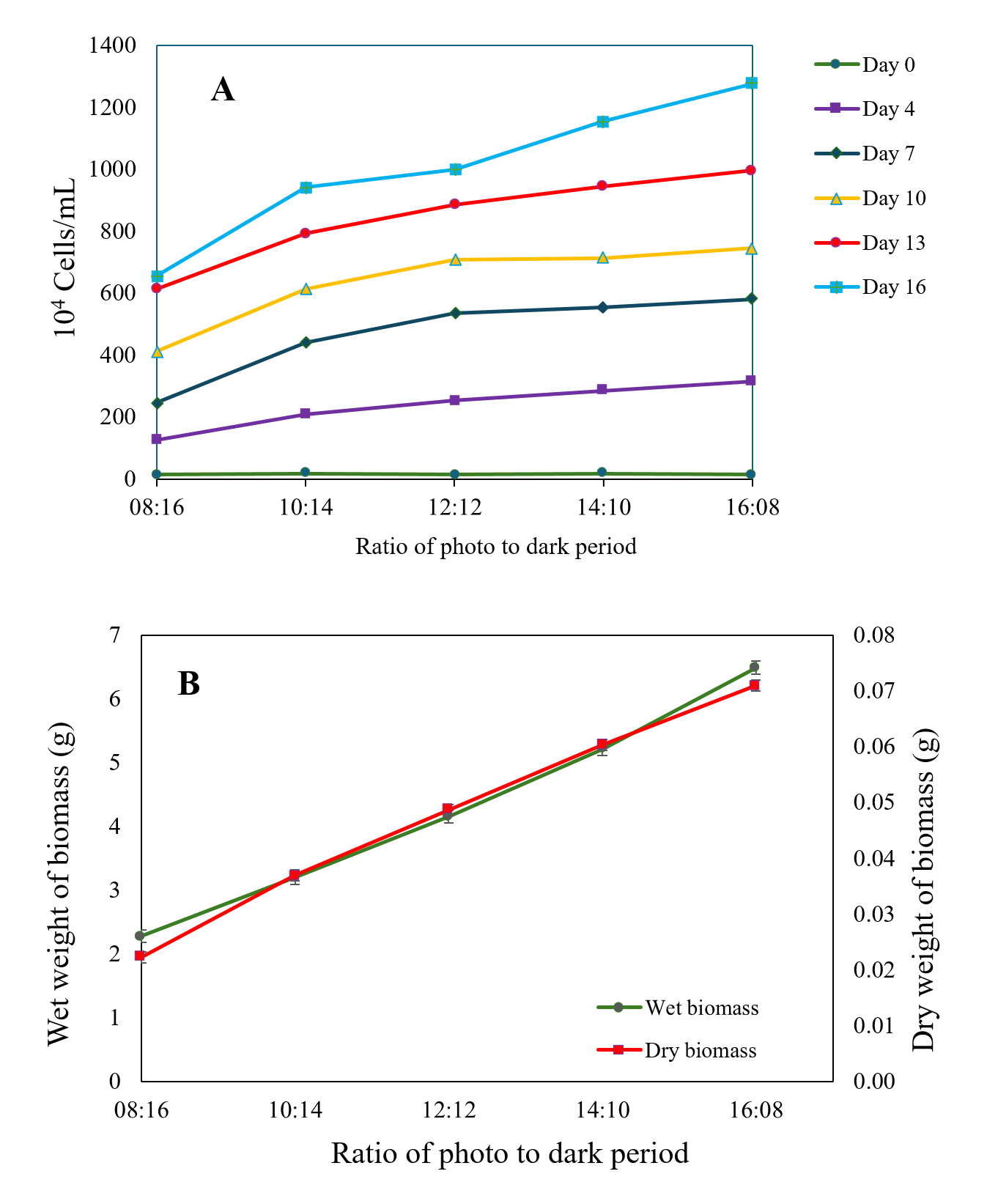
Figure 5. (A) Represents the growth profile of P. kessleri under varying light-dark cycles, demonstrating the impact of photoperiod on growth over time. (B) Depicts the wet and dry biomass yields obtained under different light-dark regimes, with the light-to-dark ratio on the x-axis, wet biomass (g) on the left y-axis, and dry biomass (g) on the right y-axis, highlighting the effect of light exposure on biomass accumulation (n = 3)
Effect of CO2 sparging duration on growth of P. kessleri
The changes in cell numbers and biomass during algal growth under different CO2 sparging durations are depicted in Figures 6A and 6B, respectively. In comparison to the other treatments, the 1 minute and 3 minute groups initially exhibited the highest growth from day 7. By day 10, there was no statistically significant difference in growth between the 1, 2, and 3 minute treatments compared to the 4 and 5 minute treatments. However, on day 13, the 4 minute treatment showed a statistically significant difference, followed by the 1, 3, 2, and 5 minute treatments. By day 16, the growth rates for the 2, 1, and 3 minute treatments were significantly higher than those of the 4 and 5 minute treatments, although no significant differences were observed between the 2, 1, and 3 minute groups. Similar to our study, another researcher investigated algal biomass and extracellular polysaccharide (EPS) production under three different CO2 concentrations (0.04%, 2%, and 5%) and found that algal biomass productivity and EPS production were highest at 2% CO2 compared to the other two treatments.30
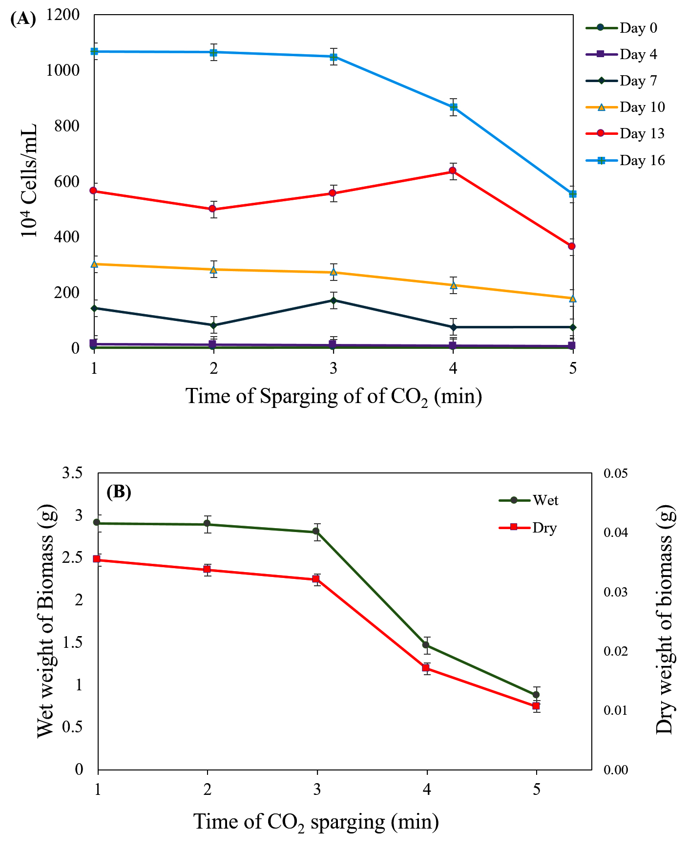
Figure 6. (A) Growth curve of P. kessleri under different CO₂ sparging durations (1-5 min), illustrating the impact of varying CO₂ sparging durations on growth over time. (B) Shows the wet and dry biomass yields obtained under different CO₂ sparging durations, with sparging duration on the x-axis, wet biomass (g) on the left y-axis, and dry biomass (g) on the right y-axis (n = 3)
Growth of algae under optimized conditions
The results indicated that under optimized conditions, the microalgal cell number reached approximately 1035 × 104 cells/mL, compared with 160 × 104 cells/mL under non-optimized conditions on day 16 (Figure 7A). Furthermore, the biomass under optimized conditions was 0.295 ± 0.075 g, compared with 0.091 ± 0.001 g under non-optimized conditions (Figure 7B).
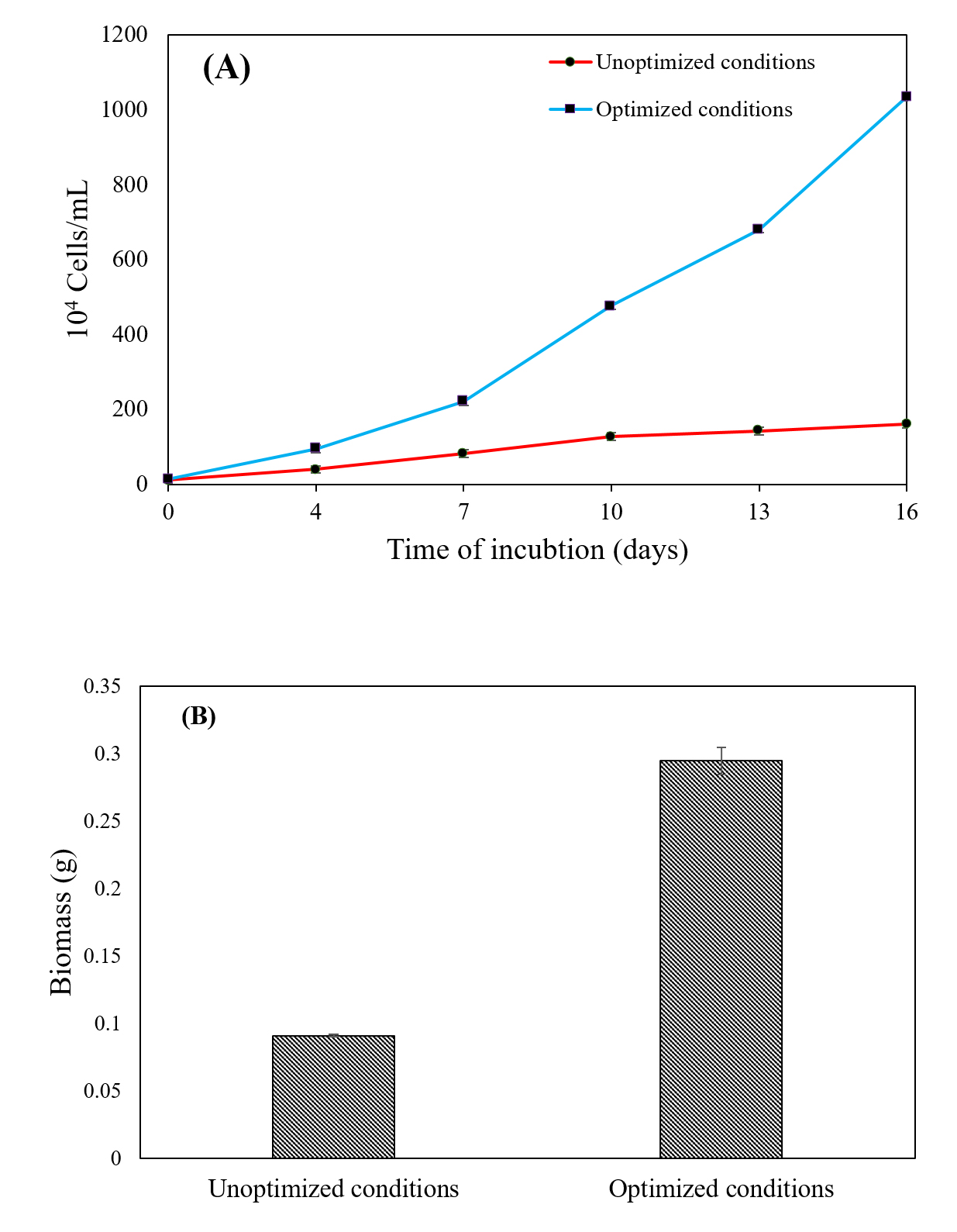
Figure 7. (A) shows the growth of P. kessleri under optimized and non-optimized conditions, while (B) illustrates the biomass growth after 16 days under optimized and non-optimized conditions
Lipid content of microalgae
The biochemical composition of the 16 day-old stationary-phase culture of P. kessleri showed a lipid content of 17%. Beigbeder and Lavoie reported that P. kessleri cultivated in Bold Basal Medium with NaHCO3 at pH 8 exhibited a similar lipid content of 17%.20 Gao et al. observed that lipid content in P. kessleri TY02 varied with culture duration and was more pronounced under nitrogen stress (N-) than in nitrogen-supplemented conditions (N+), ranging from 11.2% to 33.2% under N+ and 12.5%-45.5% under N-.22 Pribyl et al. evaluated biomass productivity and lipid content in ten different strains over seven days, finding that P. kessleri achieved high biomass productivity (1.291 ± 0.032) with lipid contents of 51.06 ± 1.27% under nutrient-depleted conditions.28 Nitrogen stress has been widely reported to enhance lipid accumulation in microalgae; for example, Li et al. found that nitrogen deficiency can induce lipid accumulation of up to 60% in five days.14 Similarly, Fernandes et al. observed up to 28% lipid accumulation in seven days across three different media: fresh medium, 0.2 medium, and 0.1 medium.35
Understanding the response of microalgae to different environmental conditions is important for optimizing biomass and metabolite production. Investigating the interactions between these factors and growth parameters helps to determine the optimal conditions for large-scale production. The results indicated that the highest cell density and biomass yield were achieved at pH 8, 25 °C, a 16:8 hrs light-dark cycle, and CO2 sparging for 2 min twice daily. The isolate collected from the Yamuna River in Okhla was identified as P. kessleri primarily based on classical morphological and taxonomic characterization, while partial 18S rDNA analysis was used to assess its phylogenetic relationship with closely related taxa. Neutral lipids were confirmed through Nile red staining, with lipid quantification showing a 17% accumulation after 16 days of growth. The isolate also responded to different durations of intermittent CO2 supplementation and a wide pH range, and it grew under continuous illumination. These findings demonstrate the isolate’s adaptability to different cultivation conditions and its capacity for lipid accumulation. However, this study did not investigate PHA production; further studies involving PHA extraction, quantification, and structural characterisation are needed to evaluate its potential for PHA-based bioplastic production.
The study identifies the key factors—pH, photoperiod, temperature and CO2 sparging duration—that govern the growth rate of PDP_01. The results suggest that maintaining a pH of 8, a temperature of 25 °C, a photoperiod of 16:8, and CO2 sparging for 2 min twice daily can enhance the growth of PDP_01 for large-scale cultivation in photobioreactors. Biochemical analysis revealed a lipid content of 17%, demonstrating the lipid-accumulating capacity of PDP_01 under the tested cultivation conditions. These findings provide a basis for further investigation of PDP_01 for value-added applications, including the future evaluation of its potential for PHA-based bioplastic production.
ACKNOWLEDGMENTS
The authors thank Lovely Professional University for providing the necessary infrastructure and support during the research.
CONFLICT OF INTEREST
The authors declare that there is no conflict of interest.
AUTHORS’ CONTRIBUTION
JS conceptualized the study and developed the experimental protocols. SG designed the experiments. PR performed the experiments. SG and SS performed data analysis. PR and SG drafted, reviewed, and revised the manuscript. SG communicated the manuscript. All authors read and approved the final manuscript for publication.
FUNDING
None.
DATA AVAILABILITY
The datasets generated and/or analysed during the current study are available from the corresponding author upon reasonable request.
ETHICS STATEMENT
This article does not contain any studies on human participants or animals performed by any of the authors.
- Dolganyuk V, Belova D, Babich O, et al. Microalgae: a promising source of valuable bioproducts. Biomolecules. 2020;10(8):1153.
Crossref - Ferreira De Oliveira AP, Bragotto APA. Microalgae-based products: Food and public health. Future Foods. 2022;6:100157.
Crossref - Wang X, Zhang Y, Xia C, Alqahtani A, Sharma A, Pugazhendhi A. A review on optimistic biorefinery products: Biofuel and bioproducts from algae biomass. Fuel. 2023;338:127378.
Crossref - Iroegbu AOC, Ray SS, Mbarane V, Bordado JC, Sardinha JP. Plastic pollution: a perspective on matters arising: challenges and opportunities. ACS Omega. 2021;6(30):19343-19355.
Crossref - Josephine A, Kumar TS, Surendran B, Rajakumar S, Kirubagaran R, Dharani G. Evaluating the effect of various environmental factors on the growth of the marine microalgae, Chlorella vulgaris. Front Mar Sci. 2022;9:954622.
Crossref - Chowdury KH, Nahar N, Deb UK. The growth factors involved in microalgae cultivation for biofuel production: a review. CWEEE. 2020;9(4):185-215.
Crossref - Gani P, Sunar NM, Matias-Peralta HM, Apandi N. An overview of environmental factor’s effect on the growth of microalgae. J Appl Chem Nat Resour. 2019;1(2):1-5
- Elisabeth B, Rayen F, Behnam T. Microalgae culture quality indicators: a review. Crit Rev Biotechnol. 2021;41(4):457-473.
Crossref - Subramanian S, Sayre RT. The right stuff; realizing the potential for enhanced biomass production in microalgae. Front Energy Res. 2022;10:979747.
Crossref - Yu BS, Pyo S, Lee J, Han K. Microalgae: a multifaceted catalyst for sustainable solutions in renewable energy, food security, and environmental management. Microb Cell Fact. 2024;23(1):308.
Crossref - Balouch H, Zayadan BK, Sadvakasova AK, et al. Prospecting the biofuel potential of new microalgae isolates. Int J Hydrogen Energy. 2023;48(50):19060-19073.
Crossref - Singh AK, Nawkarkar P, Bhatnagar VS, Tripathi S, Mock T, Kumar S. Assessing the potential of a genetically modified Parachlorella kessleri-I with low CO2 inducible proteins for enhanced biomass and biofuel productivity. J Environ Chem Eng. 2024;12(5):113795.
Crossref - Ota S, Oshima K, Yamazaki T, et al. Highly efficient lipid production in the green alga Parachlorella kessleri: draft genome and transcriptome endorsed by whole-cell 3D ultrastructure. Biotechnol Biofuels. 2016;9(1):13.
Crossref - Li X, Pribyl P, Bisova K, et al. The microalga Parachlorella kessleri—a novel highly efficient lipid producer. Biotechnol Bioeng. 2013;110(1):97-107.
Crossref - Hidasi N, Badary A, Jenkins HD, Fields FJ, Mayfield SP, Ferrari S. Selection and characterization of a Parachlorella kessleri microalgal strain able to assimilate lactose and grow on dairy waste. Biomass Bioenergy. 2024;188:107344.
Crossref - O’Rourke R, Gaffney M, Murphy R. The effects of Parachlorella kessleri cultivation on brewery wastewater. Water Sci Technol. 2016;73(6):1401-1408.
Crossref - Primo TARDC, Vargas LB, Alves RD, De Farias Neves F, Skoronski E. New insights into chicken processing wastewater treatment: the role of the microalgae Parachlorella kessleri on nitrogen removal. Environ Technol. 2025;46(8):1229-1241.
Crossref - Da Silva GDA, Joao JJ, Sales RDOJ, Becker D, Skoronski E, Neves FDF. Utilizing Parachlorella microalgae and Arthrospira cyanobacteria for tertiary wastewater treatment and biomass valorization as raw material for biopolymer production. Clean Techn Environ Policy. 2025;27(9):3985-3998.
Crossref - Song X, Liu BF, Kong F, Song Q, Ren NQ, Ren HY. Lipid accumulation by a novel microalga Parachlorella kessleri R-3 with wide pH tolerance for promising biodiesel production. Algal Res. 2023;69:102925.
Crossref - Beigbeder JB, Lavoie JM. Effect of photoperiods and CO2 concentrations on the cultivation of carbohydrate-rich P. kessleri microalgae for the sustainable production of bioethanol. J CO2 Util. 2022;58:101934.
Crossref - Zachleder V, Kselikova V, Ivanov IN, et al. Supra-optimal temperature: an efficient approach for overaccumulation of starch in the green alga Parachlorella kessleri. Cells. 2021;10(7):1806.
Crossref - Gao Y, Lv J, Feng J, Liu Q, Xie S. Morphology, phylogeny and lipid components of an oil-rich microalgal strain. J Appl Bot Food Qual. 2017;90:298-305.
Crossref - Guiry MD. AlgaeBase. World-wide electronic publication, National University of Ireland, Galway. 2010. Accessed January 11, 2026. https://www.algaebase.org
- Huy M, Kumar G, Kim HW, Kim SH. Photoautotrophic cultivation of mixed microalgae consortia using various organic waste streams towards remediation and resource recovery. Bioresour Technol. 2018;247:576-581.
Crossref - Doyle JJ, Doyle JL. A rapid DNA isolation procedure for small quantities of fresh leaf tissue. Phytochem Bull. 1987;19:11-15.
- Haddad R, Alemzadeh E, Ahmadi AR, Hosseini R, Moezzi M. Identification of Chlorophyceae based on 18S rDNA sequences from Persian Gulf. Iran J Microbiol. 2014;6(6):437-442.
- Sureshkumar P, Thomas J, Bhakta S. Development of DNA barcode for freshwater microalgae species revealing it’s evolutionary insights on diversity in Noyyal river of Western ghats, India. [Preprint] Research Square. 2022.
Crossref - Pribyl P, Cepak V, Zachleder V. Production of lipids in 10 strains of Chlorella and Parachlorella, and enhanced lipid productivity in Chlorella vulgaris. Appl Microbiol Biotechnol. 2012;94(2):549-561.
Crossref - Beigbeder JB, Sanglier M, De Medeiros Dantas JM, Lavoie JM. CO2 capture and inorganic carbon assimilation of gaseous fermentation effluents using Parachlorella kessleri microalgae. J CO2 Util. 2021;50:101581.
Crossref - Takagi A, Nagao M, Uejima Y, Sasaki D, Asayama M. Efficient pH and dissolved CO2 conditions for indoor and outdoor cultures of green alga Parachlorella. Front Bioeng Biotechnol. 2023;11:1233944.
Crossref - Ziarati A, Shadi AR. Effect of light and temperature factors in optimizing the growth of Parachlorella microalgae. First International Conference on Marine and Atmospheric Sciences: Environment, Renewable Energy. 2017. Accessed August 17, 2026. https://civilica.com/doc/680747
- Lv J, Wang X, Feng J, et al. Comparison of growth characteristics and nitrogen removal capacity of five species of green algae. J Appl Phycol. 2019;31(1):409-421.
Crossref - Taleb A, Legrand J, Takache H, Taha S, Pruvost J. Investigation of lipid production by nitrogen-starved Parachlorella kessleri under continuous illumination and day/night cycles for biodiesel application. J Appl Phycol. 2018;30(2):761-772.
Crossref - Magierek E, Krzemiסska I, Tys J. Stimulatory effect of indole-3-acetic acid and continuous illumination on the growth of Parachlorella kessleri. Int Agrophys. 2017;31(4):483-489.
Crossref - Fernandes B, Teixeira J, Dragone G, et al. Relationship between starch and lipid accumulation induced by nutrient depletion and replenishment in the microalga Parachlorella kessleri. Bioresour Technol. 2013;144:268-274.
Crossref
© The Author(s) 2026. Open Access. This article is distributed under the terms of the Creative Commons Attribution 4.0 International License which permits unrestricted use, sharing, distribution, and reproduction in any medium, provided you give appropriate credit to the original author(s) and the source, provide a link to the Creative Commons license, and indicate if changes were made.