ISSN: 0973-7510
E-ISSN: 2581-690X
Neonatal sepsis is a blood-stream infection that affects newborns under the age of 28 days. Sepsis is common in NICUs and has a high prevalence of Klebsiella species. As a result, the study aims to find the antibiotic resistance profile, virulence factors, and the prognosis of K. pneumoniae-infected neonates. A prospective study was conducted which included 140 neonates with clinical sepsis. Characterization of Klebsiella pneumonia isolates was done by conventional methods. Drug resistance and virulence factors were detected by phenotypic methods. Genotypic methods included 16s rRNA amplification and sequencing. Detection of multidrug-resistant genes by PCR was performed. K. pneumoniae (26.9%) was the most common pathogen isolated. A high prevalence of ESBL was detected (58.8%). The prevalence of CRKP and MβL was about 29.4%, and 23.5% respectively. Two strains were Strong biofilm producers and nine isolates showed Beta hemolysis.7 strains were positive for the string test. Four strains were positive for the wcaG gene. 3 positive for magA (K1) and 2 were for gene wzy (K2). Three isolates carried blaCTX–M, four isolates harbored blaVIM, two for IMP, and one for NDM and KPC gene. K. pneumoniae isolates in the NICU increased in frequency and antibiotic resistance. It is a serious hazard to the healthcare system, and it necessitates strict infection control methods in healthcare settings, as well as antibiotic stewardship to prevent the overuse of antibiotics in neonatal sepsis.
Neonatal Sepsis, Hypervirulent Klebsiella pneumonia, Drug Resistance, Virulence Factors, Genotypic Methods
Neonatal sepsis (NS) is a blood-stream infection that affects newborns under the age of 28 days. NS is a primary cause of fatality and morbidity.1 The impact of newborn death is disproportionately spread worldwide, with developing nations facing the burden of it. India is one of the five countries responsible for half of all newborn fatalities.2 The diversity of organisms that cause sepsis differs geographically, and in the same location, it changes with time.3
NS is characterized as early-onset sepsis (EOS) or late-onset sepsis (LOS) depending on the timing of symptoms. The EOS is sepsis that occurs within 72 hours of birth and is primarily caused by organisms acquired before and during delivery. LOS, on the other hand, occurs after the first three days of life and most organisms are acquired after delivery.4
Prematurity, low birth weight, and perinatal problems like chorioamnionitis and premature rupture of membranes (PROM) are all linked to early-onset sepsis (EOS). Invasive interventions such as resuscitation and central venous catheterization, are linked to late-onset sepsis (LOS).5
Sepsis is common in NICUs, with the rate ranging from 28 to 50%, and has a high prevalence of Klebsiella species. Klebsiella pneumoniae (KP) infections lead high rate of morbidity and mortality. In neonatal critical care units, it is also one of the most common causes of outbreaks (NICUs).6
Multi-drug resistant (MDR) strains are becoming more common, posing a major global challenge in selecting effective antibiotics for treating hospital-acquired infections.7 The use of invasive equipment, insufficient diagnostic techniques, immunosuppressed conditions, and overuse of antibiotics have all been linked to the rise of MDR isolates in healthcare institutions.8
Unfortunately, with the advent of extensively drug-resistant (XDR) and pan drug-resistant (PDR) K. pneumoniae strains in recent years, the situation of antimicrobial resistance has gotten worse.9 Carbapenem-resistant K. pneumoniae (CRKP) has just emerged and is rapidly spreading over the world. Infection with these “superbugs” frequently results in severe consequences.10 As a result, the study aims to find the antibiotic resistance profile, resistance mechanisms, virulence factors, and the prognosis of K. pneumonia-infected neonates.
Study design, period and setting
From January 2020 to December 2021, a hospital-based prospective study was conducted. The sample size (N) was computed with 95% confidence interval and 5% degree of freedom using a single population proportion formula and 13.6 % prevalence from a prior pilot study. As a result, the minimum sample size was calculated to be 140, and participants were recruited as needed until the sample size was reached.
Institutional Ethical Committee clearance was obtained. All neonates suspected of having sepsis were included in the study population. During the data collecting period, all neonates with clinical signs and symptoms of sepsis who had not been given antibiotics were eligible. Neonates having a birth weight of less than 1000 g, as well as those with pneumonia, meningitis or congenital anomalies were excluded.
Maternal data such as maternal chronic illnesses, maternal hypertension, gestational diabetes mellitus, chorioamnionitis, PROM >18 hours, and clinical sequelae were recorded. Clinical symptoms like temperature instability, convulsions, feeding problems, lethargy, and respiratory distress are used to diagnose neonatal sepsis in the study setting, as well as laboratory findings such as C – reactive protein (CRP), complete blood count (CBC), and/or blood cultures.
A standard proforma was specifically constructed to collect patients’ demographic data and essential information such as delivery history, risk factors for infection, symptoms, laboratory results, and clinical outcomes.
Laboratory methods
Phenotypic Identification
Two milliliters (2ml) of venous blood were collected from each neonate. Each sample was then inoculated directly into pediatric brain-heart infusion broth (BHIB) to make a 1:10 dilution and BACTEC PedsPlus™ (Becton Dickinson, Ireland) culture vial. All culture broths were maintained aerobically at 37°C for seven days, with daily observations for the presence or absence of visible microbial growth. Blood agar, chocolate agar, and MacConkey agar were used for subculturing. The pathogens were identified using standard microbiological procedures.11 Isolates of K. pneumoniae were characterized.
Antimicrobial susceptibility pattern was determined. According to the CLSI recommendations, they were assessed for zones of inhibition and classified as sensitive, moderate, or resistant.
Antibiotics like ampicillin, amikacin, cefazolin, cefuroxime, ceftazidime, cefotaxime, cefepime, piperacillin-tazobactam, gentamicin, tobramycin, co-trimoxazole, ciprofloxacin, imipenem, and meropenem were assessed and the results were compared with ATCC standard strain.12
Phenotypic determination of antibiotic-resistant K. pneumoniae strains
Detection of MDR, XDR, and PDR strains was done based on the disc diffusion method’s results.
AmpC Beta-lactamases
• Screened by using cefoxitin (30μg) disc.
• After incubation, if the diameter of a zone to >14mm for cefoxitin – potential AmpC-beta-lactamase producers.13
Double disk synergy test
• Bacterial isolates resistant to third-generation cephalosporins were subjected to ESBL producers.
• Cefotaxime 30μg with a distance of 15mm edge to edge from piperacillin/tazobactam and incubated at 35˚c (18-20 hours) and zone of inhibition >5mm increase in diameter – positive for ESBL production.14
MBL (Metallo-β-lactamase)
• Combined disk test: Two imipenem discs (10μg) and one with 10μl of 0.1 M anhydrous EDTA (Himedia) were used.
• In comparison with the imipenem disc an increase in zone diameter of >5 mm around the EDTA disc is regarded as positive.15
Determination of virulence factors
Hypermucoviscosity (HMV)
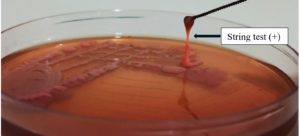
• The ability of the colony to stretch as a mucoviscous string.
• It is considered an HMV phenotype, when the string formed stretches in length >10mm.16
• String test positive for a hypervirulent strain of Klebsiella pneumoniae is shown in Figure.
Blood Hemolysis
• The plate hemolysis test was performed by inoculating the isolates on blood agar plates which contain 5% sheep blood.
• Hemolysis was identified after 24 hours of incubation at 37°C.17
Biofilm Forming Assay
• A microtiter plate method was used to assess biofilm production. The attachment of Klebsiella strains to an inert substratum was examined.
• The strains were maintained in BHIB at 37°C for 24 hours. 50μl from culture dilution was transferred into the individual wells of a 96-well, flat-bottomed polystyrene microtiter plate and incubated at 37 °C for 48 hours.
• Plank-tonic bacteria were removed after incubation, wells were gently rinsed thrice with sterile normal saline and methanol fixation was done for 20 min.
• Then each well was stained with crystal violet and washed. Biofilm-associated crystal violet was decolorized in 1 mL of ethanol. Finally, the optical density of the solution was read at 620nm.18
Blood parameters
CRP levels were measured using CRP-turbilatex method. It is a quantitative turbidimetric test for the measurement of CRP.19
Genomic DNA Extraction and amplification of 16S rRNA gene
The DNA Extraction kit (Qiagen blood mini Kit -HIMEDIA) was used to extract DNA from samples. The content and purity of DNA were determined by measuring absorbance at wavelengths of 280 nm. Gel electrophoresis was used to check the integrity of the genomic DNA.20
The 16SrRNA gene amplification was performed using Qiagen HotStarTaq VR Master Mix and universal primer pair forward primer F 5’-AGAGTTTGATCCTGGCTCAG-3’ and the reverse primer R 5’-ACGGTTACCTTGTTACGACTT-3’. The final volume of 25 ml PCR reaction mixture contains a master mix of 12.5 ml, forward and reverses primer 2 ml, DH2O 5.5 ml, and Template DNA 3 ml.
The PCR setting was 94°C for 15 min for initial denaturation, followed by 35 cycles of denaturation at 94°C for 1 min, 52°C for 1 min at annealing, extension at 72 °C for 1 min 30 s, and final polymerization extension of 72°C for 5 min (The Applied Biosystems Veriti 96- Well Thermal Cycler). The amplified PCR sample was taken to agarose gel electrophoresis (1%) by 1X TAE (Tris-Acetate-EDTA) buffer with ethidium bromide. The amplified bands were visualized on UV transilluminator and the image was taken under UV illumination using the gel documentation (Bio-Rad) method.21
The PCR amplified products have been purified with a QIAquick PCR purification kit. The Purified PCR gene amplicons by sequence, the reaction was performed in the PCR thermal cycler using the BigDye Terminator v3.1 Cycle Sequencing Kit (Applied Biosystems, USA). The purified air-dried samples were sequenced in ABI 3730Xl 96-capillary DNA Analyzers. The sequence quality was validated using Finch TV version 1. 5. 0.22
Sequencing
The characterization and recognition of Klebsiella strains were done by 16S rRNA sequences. The individual sequences were subjected to similarity search and comparison in the NCBI database using the similarity tool BLAST. The identification of strains was based on the maximum similarity hits obtained from BLAST. The 16S rRNA sequence strains were submitted to the GenBank nucleotide database of NCBI, the sequence was accepted and the accession number for each sequence was obtained separately.23
Multidrug Resistance Genes Detection
The following genes were identified using Polymerase chain reaction (PCR) and the specific primers are given in Table 1 and their amplification conditions are given in Table 2.
• ESBL-encoding genes (blaCTX–M)
• Carbapenemases genes (blaVIM, blaKPC, blaNDM, IMP)
• Biofilm gene (WCaG)
• Hyper virulent Kp genes -magA(K1),wzy(K2)
Table (1):
Primers used for detecting target genes.
| PCR | Target gene | Sequences of Primer (5’-3’) | Product Size (bp) |
|---|---|---|---|
| Resistant genes | |||
| 1. | CTX-M | F- CGCTTTGCGATGTGCAG R- ACCGCGATATCGTTGGT |
550 |
| 2. | NDM | F-GGGCAGTCGCTTCCAACGGT R-GTAGTGCTCAGTGTCGGCAT |
476 |
| 2. | IMP | F-TTGACACTCCATTTACDG R-GATYGAGAATTAAGCCACYCT |
139 |
| 2. | VIM | F-GATGGTGTTTGGTCGCATA R-CGAATGCGCAGCACCAG |
390 |
| 2. | KPC | F-CATTCAAGGGCTTTCTTGCTGC R-ACGACGGCATAGTCATTTGC |
538 |
| Biofilm gene | |||
| 3. | wcaG | F-GGTTGGKTCAGCAATCGTA R-ACTATTCCGCCAACTTTTGC |
169 |
| Hyper virulent gene | |||
| 4. | magA (K1) | F-GGTGCTCTTTACATCATTGC R-GCA ATG GCC ATT TGC GTT AG | 1282 |
| 5. | Wzy(K2) | F- GACCCGATA TTC ATA CTT GAC AGA G R -CCT GAA GTA AAA TCG TAA ATA GAT GGC |
641 |
Table (2):
PCR – Amplification conditions.
PCR |
Initial Denaturation |
Denaturation |
Annealing |
Elongation |
Final Elongation |
Cycle |
|---|---|---|---|---|---|---|
1 |
95°C 7 min |
95°C 1 min |
58°C 1 min 30 sec |
72°C 2 min |
72°C 5 min |
40 cycles |
2 |
95°C 5min |
95°C 2 min |
55°C 2 min |
72°C 1 min |
72°C 7 min |
35 cycles |
3 |
95°C 10 min |
95°C 5 min |
56°C 1 min 50 sec |
72°C 1 min |
72°C 10 min |
45 cycles |
4 & 5 |
95°C 7 min |
95°C 2 min |
59°C 1 min 35 sec |
72°C 2 min |
72°C 7 min |
40 cycles |
Amplification reactions were carried out and Electrophoresis and Visualization of DNA fragments using a Gel imaging system were performed.24
Statistical analysis
The statistical analysis was done with the SPSS software version 24.0. A p-value < 0.05 was considered to be statistically significant.
This study included 63 newborns with culture-proven sepsis caused by 11 different types of pathogens. The rate of neonatal sepsis cases in the NICU was 39.2 %. The higher proportion of the new-born with septicemia was male. 44 (70.3%) of newborns were belonging to Early onset sepsis (EOS) cases. Newborns with EOS had lower gestational age, decreased birth weight, and infection age at a younger age than infants with LOS (P < 0.05). EOS infants stayed in the hospital for substantially longer than those in the LOS group.
The majority of the isolates, 35(74.05%) were Gram-negative bacteria. K. pneumoniae n=17(26.9%) was the most common pathogen both in the EOS and LOS groups. Demographic and clinical presentations of neonatal sepsis are given in Table 3. The Antibiogram pattern of Klebsiella pneumonia is shown in Table 4.
Table (3):
Demographic and Clinical presentation of Neonatal sepsis.
| Variable | Other bacteria | K. Pneumoniae |
|---|---|---|
| Sex (male : Female) | 61:39 (%) | 59.6:40.4 (%) |
| Hospitalization duration (day)(mean ± SD) | 8 ± 2 | 12 ± 4 |
| Weight(g)(mean ± SD) | 2,850 ± 500 | 2,700 ± 800 |
| No. Of preterm neonates | 3 | 1 |
| No. of cases with PROM | 8 | 5 |
| White blood cell count | 10,110 ± 253 | 9,250 ± 560 |
| ESR(mean ± SE) | 17 ± 2.1 | 15 ± 5.1 |
| The onset of Septicemia(day)(mean ± SD) | 2 ± 2 | 1 ± 2 |
| Fever (mean ± SD) | 37.1 ± 0.7 | 37.3 ± 0.8 |
| No. of cases with meconium-stained liquor | 9 | 7 |
| CRP(positives: negatives) | 40:6 (86.9:13.1%) | 15:2(88.2:11.8%) |
| Present condition at sepsis onset | ||
| Hypothermia | 19 (41.3%) | 4 (23.5%) |
| Respiratory difficulty | 21 (45.6%) | 7 (41.1%) |
| Apnea | 8 (17.3%) | 1 (5.8%) |
| Jaundice | 4 (8.6%) | 1 (5.8%) |
| Prematurity | 12 (26%) | 4 (23.5%) |
| Convulsion and neurological alterations | 1 (2.1%) | – |
| Poor feeding | 14 (30.4%) | 2 (11.7%) |
(ESR- Erythrocyte sedimentation rate; SD-standard deviation; CRP-C-reactive protein).
Table (4):
Antibiotic-resistant pattern of Klebsiella pneumoniae isolates.
| Antibiotics | K. Pneumoniae Total no.: 17 |
Other bacteria Total no.: 46 |
p-value | ||
|---|---|---|---|---|---|
| No. | % | No. | % | ||
| Amikacin | 2 | 11.8 | 4 | 8.7 | 0.657 |
| Ciprofloxacin | 12 | 70.6 | 7 | 15.2 | <0.001*** |
| Cotrimoxazole | 15 | 88.2 | 9 | 19.6 | <0.001*** |
| Cefepime | 15 | 88.2 | 5 | 10.9 | <0.001*** |
| Ampicillin | 17 | 100 | 9 | 19.6 | <0.0001*** |
| Ceftriaxone | 15 | 88.2 | 5 | 10.9 | <0.001*** |
| Cefazolin | 10 | 58.8 | 3 | 6.5 | <0.001*** |
| Cefuroxime | 14 | 82.4 | 5 | 10.9 | <0.001*** |
| Cefotaxime | 13 | 76.5 | 3 | 6.5 | <0.001*** |
| Piperacllin-Tazobactum | 10 | 58.8 | 3 | 6.5 | <0.001*** |
| Tobramycin | 8 | 47.1 | 6 | 13.04 | 0.007** |
| Gentamicin | 11 | 64.7 | 8 | 17.4 | 0.001*** |
| Imipenem | 4 | 23.5 | 2 | 4.3 | 0.041* |
| Meropenem | 4 | 23.5 | 2 | 4.3 | 0.041* |
Note: Chi-square test was used and Fisher’s exact test was used when at least one of the expected cell counts was less than 5
If the p-value is less than 0.05 it is assigned with one star (*)
If the p-value is less than 0.01 it is assigned with two stars (**)
If the p-value is less than 0.001 it is assigned with three stars (***)
Infections with MDR-KP were found in both the Early and late-onset sepsis groups. The MDR-KP infection group had a lower birth weight, lower gestational age, and longer time of antibiotic exposure (P <0.05). There were no changes in maternal variables, neonate gender, or delivery pattern (P > 0.05). In addition, in the MDR group, the period of hospitalization was greater.
A high prevalence of ESBL was detected in the K. pneumonia isolates reaching 58.8%. Among the Klebsiella pneumoniae isolates, the prevalence of CRKP and MβL producers were 29.4% and 23.5% respectively. Sepsis risk factors including PROM and neutropenia were significantly associated with CRKP.
Two strains were Strong biofilm producers, three were moderate, five were weak and seven were non-biofilm producing Klebsiella pneumonia and four strains were positive for biofilm-producing wcaG gene. Nine isolates showed Beta hemolysis.
In Klebsiella pneumoniae isolates, a string test was done for phenotypic confirmation of hvKp and 7(41.1%) strains were positive. In these 7 hvKp strains, 3 positive and 4 were negative for magA( K1). 2 were positive and 5 were negative for gene wzy (K2).
In terms of ESBL genes, three isolates in our investigation carried blaCTX–M and regarding the carbapenemases, four isolates harbored blaVIM, two for IMP, one for NDM and KPC gene.
16s rRNA sequence analysis
In the current study, Klebsiella pneumoniae strains are exposed to 16S rRNA sequences. The different lengths of the nucleotide bases created by this study have been exposed to BLAST analysis. BLAST results in the 16S rRNA gene sequence showed almost all isolates had greater than 99.9% similarity and database reference sequences are given in Table 5.
Table (5):
BLAST analysis, similarity, and NCBI Gene bank accession numbers of Klebsiella pneumonia.
S.No |
Sample |
Bacterial strain |
Similarity |
Blast Result |
Accession No |
|---|---|---|---|---|---|
1 |
Blood |
K.pneumoniaehvkp1 |
100 % |
K.pneumoniae |
OM442431 |
2 |
Blood |
K.pneumoniaehvkp2 |
100 % |
K.pneumoniae |
OK635329 |
3 |
Blood |
K.pneumoniaehvkp3 |
100 % |
K.pneumoniae |
OM442431 |
4 |
Blood |
K.pneumoniaehvkp4 |
100 % |
K.pneumoniae |
OM066754 |
5 |
Blood |
K.pneumoniaehvkp5 |
100 % |
K.pneumoniae |
OM066754 |
Treatment
According to the sensitive data, all of the infants were given antibiotics. When necessary, other treatment procedures were used, such as entire parental nourishment and intensive care.
Neonatal sepsis has different etiological agents in developed and developing nations. Gram-negative organisms were the most common pathogens in this study, as well as earlier studies from India and Nigeria.25 Coagulase-negative staphylococcus (CONS) is frequently isolated from developed nations.26
Klebsiella pneumoniae is typically encountered in the neonatal critical care unit. It can be found in the hands of healthcare professionals as colonizers. In addition, outbreaks of neonatal septicemia caused by Klebsiella pneumoniae have been reported often in nurseries and NICUs.27
Klebsiella pneumoniae was found to be the common cause of early-onset sepsis in this investigation, although no Group B Streptococcus was found in any of the newborns. Although Group B Streptococcus was once thought to be a major cause of early-onset sepsis, new research reveals that the incidence of this pathogen is decreasing.28
Escherichia coli, Staphylococcus aureus, Coagulase-negative staphylococcus (CONS), and Klebsiella species are the common pathogens in LOS, which are mainly acquired in hospital settings. Klebsiella pneumoniae was the prevalent etiological agent of late-onset sepsis in this investigation.29
In our study, the majority of the newborns were male. It has been also observed in previous studies. This could be attributable to a gender bias in health practices. Furthermore, males have been found to have a higher prevalence of ESBL-producing K. pneumonia.30
Infants’ immune systems are immature, making them vulnerable to nosocomial infections. An upsurge in ESBL-producing K. pneumoniae sepsis has been observed over the world, with the NICU accounting for the majority of cases.31 Antimicrobial resistance in Klebsiella is mostly linked with the formation of ESBL. In 2017, the WHO included ESBL-producing Klebsiella as most dangerous superbugs along with Pseudomonas aeruginosa and Acinetobacter baumanni.32
Multidrug resistance was seen in our K. pneumoniae isolates, with carbapenem resistance increasing. There are a few antibiotics that can be used to treat CRKP: imipenem or meropenem. A combination of antibiotics can also be used along with the removal of invasive devices which can be beneficial. Polymyxin B can be used to treat multidrug-resistant CRKP.33
The majority of Klebsiella pneumoniae isolates evaluated in this study were resistant to many antibiotics except amikacin, meropenem, and imipenem. In our, study Klebsiella were resistant to second and third-generation cephalosporins. This shows the importance of testing gram-negative bacterial isolates for cephalosporin resistance and ESBL development regularly. Due to the increased prevalence of ESBL, increased resistance to aminoglycosides, which are widely used for empirical therapy, was also seen in our study.34
ESBL-producing K. pneumoniae isolates have been found in high numbers in several studies. Most of the isolates found in this study originated in neonatal and critical care units. Infections caused by ESBL-producing organisms in infants are usually acquired in hospitals and are associated with invasive procedures. These units frequently utilize ampicillin + gentamicin as a first-line antibiotic and cefotaxime as a second-line antibiotic.35
Carbapenemases are thought to be the primary mechanism that causes CRKP to exist, based on the genes. According to PCR analysis, four isolates harbored blaVIM. The majority of them possessed the blaVIM gene. Our genotypic profile contrasts with that of another study by Ghaith et al. who found a higher prevalence of blaOXA–48 and no blaVIM in any of their isolates.36
In terms of ESBL genes, three isolates in our investigation carried blaCTX–M. It is consistent with research by Amer et al. who found similar rates in isolates.37
Additional carbapenemases, such as IMP and VIM, were discovered in the isolates investigated. In the meantime, several studies have found VIM and KPC generating K. pneumoniae. In our study, four isolates were positive for blaVIM, two for IMP, and one for NDM and KPC gene.32,25
In conclusion, K. pneumoniae isolates in the NICU increased in frequency and antibiotic resistance. K. pneumoniae is a serious hazard to the healthcare system, and it necessitates strict infection control methods in healthcare settings, as well as antibiotic stewardship to prevent the overuse of antibiotics in neonatal sepsis. As the signs and symptoms are so varied, diagnosing and treating them can be difficult. Furthermore, the extended turnaround time of laboratory diagnosis adds to the difficulties, necessitating the continuation of empirical antibiotic therapy until the suspected sepsis is eradicated. To present, the growing appearance of multidrug-resistant organisms has resulted in only a few therapeutic options being available, delaying effective therapy. In hospital settings, developing preventative and management strategies should be implemented. Infection control activities such as antibiotic stewardship programs with continuous surveillance are required to track new CRKP infections as soon as possible, especially in high-risk units like NICUs.
ACKNOWLEDGMENTS
The authors would like to thank the Chettinad Academy of Research and Education, Kelambakkam, India for funding and support.
CONFLICT OF INTEREST
The authors declare that there is no conflict of interest.
AUTHORS’ CONTRIBUTION
All authors listed have made a substantial, direct, and intellectual contribution to the work, and approved it for publication.
FUNDING
This research was supported by the Chettinad Academy of Research and Education, Kelambakkam, India.
DATA AVAILABILITY
All datasets generated or analyzed during this study are included in the manuscript.
ETHICS STATEMENT
The study was conducted according to the guidelines of the Institutional Human Ethical Committee (No.010 /IHEC/Jan 2021) – Chettinad Academy of Research and Education, Kelambakkam, India.
- Nordberg V, Iversen A, Tidell A, Ininbergs K, Giske CG, Naver L. A decade of neonatal sepsis caused by gram-negative bacilli-a retrospective matched cohort study. Eur J Clin Microbiol Infect Dis. 2021;40(9):1803-1813.
Crossref - Liu L, Oza S, Hogan D, et al. Global, regional, and national causes of child mortality in 2000-13, with projections to inform post-2015 priorities: an updated systematic analysis. The Lancet. 2015;385(9966):430-440.
Crossref - Shane AL, Sanchez PJ, Stoll BJ. Neonatal sepsis. The Lancet. 2017;390(10104):1770-1780.
Crossref - Fuchs A, Bielicki J, Mathur S, Sharland M, Van Den Anker J. Antibiotic use for sepsis in neonates and children: 2016 evidence update. WHO Reviews. 2016.
- Araujo BC, Guimaraes H. Risk factors for neonatal sepsis: an overview. Journal of Pediatric and Neonatal Individualized Medicine (JPNIM). 2020;9(2):e090206.
- Arrington AS. Ventilator-Associated Pneumonia. In Healthcare-Associated Infections in Children. Springer, Cham. 2018:107-123.
Crossref - Church NA, McKillip JL. Antibiotic resistance crisis: Challenges and imperatives. Biologia. 2021;76(5):1535-1550.
Crossref - Riley MM. The rising problem of multidrug-resistant organisms in intensive care units. Critical Care Nurse. 2019;39(4):48-55.
Crossref - Mulani MS, Kamble EE, Kumkar SN, Tawre MS, Pardesi KR. Emerging strategies to combat ESKAPE pathogens in the era of antimicrobial resistance: a review. Front Microbiol. 2019;10:539.
Crossref - Satlin MJ, Chen L, Patel G, et al. Multicenter clinical and molecular epidemiological analysis of bacteremia due to carbapenem-resistant Enterobacteriaceae (CRE) in the CRE epicenter of the United States. Antimicrob Agents Chemother. 2017;61(4):e02349-16.
Crossref - Zamarano H, Musinguzi B, Kabajulizi I, et al. Bacteriological profile, antibiotic susceptibility and factors associated with neonatal Septicaemia at Kilembe mines hospital, Kasese District Western Uganda. BMC Microbiol. 2021;21(1):303.
Crossref - Chakraborty S, Mohsina K, Sarker PK, Alam MZ, Karim MI, Sayem SA. Prevalence, antibiotic susceptibility profiles and ESBL production in Klebsiella pneumoniae and Klebsiella oxytoca among hospitalized patients. Periodicum Biologorum. 2016;118(1).
Crossref - Younas S, Ejaz H, Zafar A, Ejaz A, Saleem R, Javed H. AmpC beta-lactamases in Klebsiella pneumoniae: An emerging threat to the pediatric patients. Pak Med Assoc. 2018;68(6):893-897. PMID: 30325907
- Aminul P, Anwar S, Molla MM, Miah MR. Evaluation of antibiotic resistance patterns in clinical isolates of Klebsiella pneumoniae in Bangladesh. Biosafety and Health. 2021;3(6):301-306.
Crossref - Dhungana K, Awal BK, Dhungel B, Sharma S, Banjara MR, Rijal KR. Detection of Klebsiella pneumoniae carbapenemase (KPC) and metallobetalactamae (MBL) producing Gram negative bacteria isolated from different clinical samples in a Transplant Center, Kathmandu, Nepal. Acta Scientific Microbiology. 2019;2(12):60-69.
Crossref - Shankar C, Nabarro LE, Anandan S, et al. Extremely high mortality rates in patients with carbapenem-resistant, hypermucoviscous Klebsiella pneumoniae blood stream infections. J Assoc Physicians India. 2018;66(12):13-16.
- Das A, Behera BK, Acharya S, et al. Genetic diversity and multiple antibiotic resistance index study of bacterial pathogen, Klebsiella pneumoniae strains isolated from diseased Indian major carps. Folia Microbiologica. 2019;64(6):875-887.
Crossref - Nirwati H, Sinanjung K, Fahrunissa F, et al. Biofilm formation and antibiotic resistance of Klebsiella pneumoniae isolated from clinical samples in a tertiary care hospital, Klaten, Indonesia. BMC Proceedings. 2019;13(11):1-8.
Crossref - Qazi M, Saqib N, Raina R. Risk factors and outcome of Klebsiella pneumonia sepsis among newborns in Northern India. IJRMS. 2019;7(5);
Crossref - Ghaffarian F, Hedayati M, Ebrahim-Saraie HS, Roushan ZA, Mojtahedi A. Molecular epidemiology of ESBL-producing Klebsiella pneumoniae isolates in intensive care units of a tertiary care hospital, North of Iran. Cell Mol Biol. 2018;64(7):75-79.
Crossref - He Y, Guo X, Xiang S, et al. Comparative analyses of phenotypic methods and 16S rRNA, khe, rpoB genes sequencing for identification of clinical isolates of Klebsiella pneumoniae. Antonie Van Leeuwenhoek. 2016;109(7):1029-1040.
Crossref - Singh SK, Mishra M, Sahoo M, et al. Antibiotic resistance determinants and clonal relationships among multidrug-resistant isolates of Klebsiella pneumoniae. Microbial Pathogenesis. 2017;110:31-36.
Crossref - Makharita RR, El-Kholy I, Hetta HF, et al. Antibiogram and genetic characterization of carbapenem-resistant gram-negative pathogens incriminated in healthcare-associated infections. Infect Drug Resist. 2020;13:3991.
Crossref - Danisvijay D, ShifaMeharaj SH, Sujhithra A, et al. Hypervirulent and Biofilm Specific Resistance Among Carbapenemase Producing Klebsiella pneumoniae Causing Respiratory Tract Infection. Klebsiella pneumoniae. 2020.
Crossref - Sands K, Carvalho MJ, Portal E, et al. Characterization of antimicrobial-resistant Gram-negative bacteria that cause neonatal sepsis in seven low-and middle-income countries. Nat Microbiol. 2021;6(4):512-523.
Crossref - Asante J, Amoako DG, Abia AL, et al. Review of clinically and epidemiologically relevant coagulase-negative staphylococci in Africa. Microb Drug Resist. 2020;26(8):951-970.
Crossref - Fernandez-Prada M, Martinez-Ortega C, Santos-Simarro G, Moran-Alvarez P, Fernandez-Verdugo A, Costa-Romero M. Outbreak of extended-spectrum beta-lactamase-producing Klebsiella pneumoniae in a neonatal intensive care unit: Risk factors and key preventive measures for eradication in record time. Anales de Pediatria. 2019;91(1):13-20.
Crossref - Johansson Gudjonsdottir M, Elfvin A, Hentz E, Adlerberth I, Tessin I, Trollfors B. Changes in incidence and etiology of early-onset neonatal infections 1997-2017-a retrospective cohort study in western Sweden. BMC Pediatr. 2019;19(1):490.
Crossref - Zou H, Jia X, He X, et al. Emerging Threat of Multidrug Resistant Pathogens From Neonatal Sepsis. Front Cell Infect Microbiol. 2021;11:621.
Crossref - Lee YQ, Kamar AA, Velayuthan RD, Chong CW, Teh CS. Clonal relatedness in the acquisition of intestinal carriage and transmission of multidrug resistant (MDR) Klebsiella pneumoniae and Escherichia coli and its risk factors among preterm infants admitted to the neonatal intensive care unit (NICU). Pediatr Neonatol. 2021;62(2):129-137.
Crossref - Pillay D, Naidoo L, Swe-Han KS, Mahabeer Y. Neonatal sepsis in a tertiary unit in South Africa. BMC Infect Dis. 2021;21(1):225.
Crossref - Wang N, Zhan M, Liu J, et al. Prevalence of Carbapenem-Resistant Klebsiella pneumoniae Infection in a Northern Province in China: Clinical Characteristics, Drug Resistance, and Geographic Distribution. Infect Drug Resist. 2022;15:569.
Crossref - Ibrahim ME, Abbas M, Al-Shahrai AM, Elamin BK. Phenotypic characterization and antibiotic resistance patterns of extended-spectrum β-lactamase-and AmpC β-lactamase-producing gram-negative bacteria in a referral hospital, Saudi Arabia. Can J Infect Dis Med Microbiol. 2019;2019.
Crossref - Bedzichowska A, Przekora J, Stapinska-Syniec A, et al. Frequency of infections caused by ESBL-producing bacteria in a pediatric ward-single-center five-year observation. Arch Med Sci. 2019;15(3):688-693.
Crossref - Ghaith DM, Zafer MM, Said HM, et al. Genetic diversity of carbapenem-resistant Klebsiella Pneumoniae causing neonatal sepsis in intensive care unit, Cairo, Egypt. Eur J Clin Microbiol Infect Dis. 2020;39(3):583-591.
Crossref - Amer R, El-Baghdady K, Kamel I, El-Shishtawy H. Prevalence of extended spectrum Beta-Lactamase Genes among Escherichia coli and Klebsiella pneumoniae clinical isolates. Egyptian Journal of Microbiology. 2019 Jan 1;54(1):91-101.
Crossref - Hassuna NA, AbdelAziz RA, Zakaria A, Abdelhakeem M. Extensively-drug resistant Klebsiella pneumoniae recovered from neonatal sepsis cases from a major NICU in Egypt. Front Microbiol. 2020;11:1375.
Crossref
© The Author(s) 2022. Open Access. This article is distributed under the terms of the Creative Commons Attribution 4.0 International License which permits unrestricted use, sharing, distribution, and reproduction in any medium, provided you give appropriate credit to the original author(s) and the source, provide a link to the Creative Commons license, and indicate if changes were made.


