


ISSN: 0973-7510
E-ISSN: 2581-690X
Balqees Yahya Najm1 ![]() , Sarab H. Khaleel2
, Sarab H. Khaleel2![]() and Hala Mahmmad Majeed3
and Hala Mahmmad Majeed3![]()
Raw milk is a nutrient-rich food that is considered a high-quality nutritional medium for many microorganism, including Escherichia coli. The aim of the present work was the diagnosis, by molecular methods, of Shiga toxins produced by E. coli strains isolated from cow milk samples collected from different farms in Al-Mahmmodia, Al yoosifya, Al lattiffya, Al howasha, and Arab Jboor in the government of Baghdad during the summer season. Milk samples were incubated in media for bacterial isolation. Isolates were identified using Gram staining and biochemical tests. Out of 50 samples, 15 (30%) showed the presence of E. coli. To confirm the identity of the isolates, their 16S rRNA genes were amplified using specific primers. The results showed that all isolates were E. coli. Shiga toxin-producing E. coli (STEC) were detected among the samples. The prevalence of stx1 genes was higher than that of stx2 among them. No STECs were found among six of the sample isolates, and none of these isolates was positive for stx1 and stx2. SDS-PAGE was used to determine the molecular weight of the toxin, and four selected E. coli bacteria producing Shiga-like toxins showed a clear band of approximately 70 kDa.
E. coli, Shiga Toxin, PCR, Milk Samples
Escherichia coli (E. coli) is a gram-negative, rod-shaped, flagellated, non-sporulating, and facultative anaerobic bacterium belonging to the Enterobacteriaceae family. According to virulence factors, bacteria from this species can be classified into several groups, including enterotoxigenic E. coli (ETEC), attaching and effacing E. coli (AEEC), enteropathogenic E. coli (EPEC), enterohemorrhagic E. coli (EHEC), and Shiga toxin-producing E. coli (STEC), also known as verotoxin-producing E. coli (VTEC).1,2 STEC/VTEC are able to produce one or two potent toxins called Shiga toxin (encoded by stx1 and stx2) and verotoxin (encoded by VT1 and VT2). 3,4 STEC O157:H7 was identified as the causative factor of a series of diarrhea outbreaks in Canada, Japan, the US, and the UK.5-7 Both O157 and non-O157 STEC strains are thought to be found mostly in cattle (Bettlheim, 2000). Consumption of undercooked meat, unpasteurized dairy products, and vegetables or water contaminated by ruminant feces can spread STEC-related food-borne diseases. Some Evidence has suggested that contact with an infected animal or human may also spread STEC-related diseases.8,9 Many E. coli serogroups produce Shiga toxins, including O157, O26, O103, O111, O145, O45, O91, O113, O121, and O128.10,11
There is a variety of culture approaches for STEC enrichment and detection,12 but none of them guarantees speed, accuracy, sensitivity, specificity, or safety. In contrast, molecular approaches such as the polymerase chain reaction (PCR) are safe, accurate, sensitive, and specific for detecting STEC.13
Recent increases in raw milk consumption have been attributed to milk’s high nutritional value, task benefits, and health advantages. Although comprehensive research on raw milk contamination with STEC has been undertaken in industrialized nations, relevant data from Asia, particularly the Middle East, are still lacking. In Baghdad, Iraq,14 detected that 39.5% cases of bloody diarrhea, among 200 children, were due to E. coli infections, especially to E. coli O157. Several studies on milk and contaminating milk microbes have been conducted worldwide.15 Furthermore, few studies on the molecular identification of STEC and other harmful pathogens in fresh beef milk over a defined time period have been reported in Iraq. This work was conducted to detect and characterize bacteria isolated from raw milk samples using molecular techniques and to determine the frequency of E. coli in such samples through PCR amplification of the 16S rRNA gene. In addition, this work aimed to isolate the Shiga toxin protein and determinate its molecular weight.
Sample Selection
In total, 50 unpasteurized milk samples were collected from cows at grazer breeding farms in Al-Mahmmodia, Al yoosifya, Al lattiffya, Al howasha, and Arab Jboor in the Baghdad government (10 mL of milk was taken from each cow, 10 cows per region) under sterile conditions in aseptic tubes. The samples were brought directly to the microbiology laboratory at Baghdad University.
Isolation of E. coli from the Sample Collection
To isolate E. coli, 0.1 mL of each of the 50 samples was inoculated in a diluted nutrient broth medium (Difco 60656 USA) and then plated on MacConkey (MC) with the help of a glass spreader. The plates were incubated at 37°C for 24 h; incubated samples were then sub-cultured onto eosin methylene blue (EMB) agar for the purification of the isolates. Colonies were examined by Gram staining using a light compound microscope with an oil immersion lens (100×).
The bacterial isolates were then subjected to a series of biochemical tests, including catalase, indole, and urease production; nitrate reduction; citrate utilization in Simmon’s citrate agar; and TSI, Voges-Proskauer, and methyl red tests.16
Molecular Analysis
To provide support to the biochemical tests used for the identification of E. coli isolates, a molecular analysis of the isolates was done through PCR.
Extraction and Preparation of Genomic DNA
DNA of the tested bacterial colonies was isolated following the method described by Wilson.17 DNA concentrations were determined spectrophotometrically at 260 nm using a UV-visible spectrophotometer; DNA purity was determined using the A260/A280 ratio, and values ranging from 1.5 to 1.8 were considered acceptable. DNA integrity was analyzed using agarose gel (0.8%) electrophoresis.18
Preparation of PCR Reaction Mix
For E. coli identification through PCR, the following 16S rRNA gene-specific primers were used: F5′ -CGAGTGGCGGACGGGTGAGT – 3′ and R5′ -TCGACATCGTTTACGGCGTGGA – 3′ (Promega), and a 727 bp amplification product was expected.19
The amplification reactions were performed in Eppendorf tubes using the AccuPower PCR PreMix (Bioneer; Table 1), following the method described by Schippa et al.20
Table (1):
Composition of each PCR mix.
| Reagent | Concentration | Volume |
|---|---|---|
| Taq DNA Polymerase | 2.5 U | 13 μL |
| dNTPs | 250 mM | |
| Tris-HCl (pH 9.0) | 10 mM | |
| KCl | 30 mM | |
| MgCl 2 | 1.5 mM | |
| Primers | 10 pmoles | 1 μL each |
| DNA sample | 50 ng | 3 μL |
| Distilled water | ——– | 5 μL |
| Total volume | ——– | 20 μL |
Reactions were performed in a thermal cycler (TC-3000/TECHNE/USA) under the following conditions: an initial denaturation step at 95°C for 5 min, 30 cycles of denaturation at 95°C for 30 s, primer annealing at 60°C for 30 s, and extension at 72°C for 1 min, and a final extension step at 72°C for 5 min.19
Amplification products were loaded in 1.2% agarose gel wells and electrophoresed in TBE (1×) buffer for 80 min at 100 V; gels were then stained with ethidium bromide, visualized using a UV-trans illuminator at 320 nm, and photographed with a digital camera.
Detection of Shiga Toxins Produced by E. coli using PCR
Isolates identified as E. coli were evaluated for potential Shiga toxin production by PCR using specific primers (Table 2) for stx1 and stx2 genes as described by Talukdar et al.21 with some modifications.
Table (2):
Primers used for detection of the Shiga toxin genes stx1 and stx2.
| Primer name |
Sequence 5′ – 3′ | Product size (bp) |
Reference | Company |
|---|---|---|---|---|
| Stx1F | CACAATCAGGCGTCGCCAGCGCACTTGCT | 606 | Maleki et al., 2017 | Promega |
| Stx1R | TGTTGCAGGGATCAGTCGTACGGGGATGC | |||
| Stex2F | CCACATCGGTGTCTGTTATTAACCACACC | 372 | Maleki et al., 2017 | Promega |
| Stex2R | GCAGAACTGCTCTGGATGCATCTCTGGTC |
PCR was performed using a thermal cycler under the following conditions: denaturation at 94°C for 5 min, 30 cycles of denaturation at 94°C for 1 min, annealing at 60°C for stx1 or 62°C for stx2 for 1 min, extension at 72°C for 1 min, and a final 5 min extension at 72°C.21
Extraction of Shiga Toxins from E. coli
After testing 15 E. coli milk isolates for the potential production of Shiga toxins by PCR, it was determined that only 9 had the corresponding genes. These 9 isolates were grown in LB medium for 48 h at 37°C in a shaking incubator; the cells were then centrifuged at 4°C for 10 min at 10,000 rpm, and the sediment was resuspended in 5 mL phosphate buffer and disrupted by sonication (using an Athena sonicator) at 70 Hz in cycles of 0.7 for seven periods and for 30 sec per round.22 Cell extracts were concentrated by the ammonium sulphate method described by Al-Zahrani23; four fractions were obtained using ammonium sulphate 10%, 20%, 30%, and 60%, and each fraction pellet was dissolved in 10 mL of TE buffer.
Molecular Weight Determination of the Shiga Toxin
For determining the molecular weight of the Shiga toxin, 20 μL of each fraction was mixed with an equal volume of treatment buffer (protein gel loading dye solution 4×). The mixture solutions were heated in a water bath at 100°C for 5 min; the samples were then loaded into the wells of a 12.5% sodium dodecyl sulfate (SDS)-polyacrylamide slab gel and electrophoresed as described by Ahmed 24.
Fifteen bacterial isolates were obtained from 50 milk samples collected from different regions in Baghdad and subjected to a series of tests, including morphological, biochemical, and molecular methods that facilitate the identification of E. coli. Regarding morphological characteristics, in MC agar plates, the colonies formed by the isolates had a similar morphological shape, but either appeared pink or red colored, whereas in EMB agar plates, smooth, spherical, black colonies could be observed. Gram-negative, pink-colored, small rod-shaped organisms either unclustered or clustered in pairs or short chains (according to microscopic examination of Gram-stained smears from MC and EMB agar plates under a light microscope, 100×) were selected.
The cells were subjected to several biochemical tests.
Biochemical analyses showed that all isolates were positive for catalase and indole production, TSI and methyl red tests, and nitrate reduction, but negative for Simmon’s s citrate and Voges-Proskauer tests and urease production.
The results showed that raw milk samples from the Al yoosifya region had the highest percentage of appearance of E. coli strains, reaching 46% of the total isolates, followed by Al latteffya with a 33%. Meanwhile, Al howasha and Arab Jboor had the lowest percentages (Table 3).
Table (3):
Number of E. coli isolates obtained from the five sampled regions in Baghdad.
Region |
No. of isolates (% from total) |
|---|---|
Al-Mahmmodia |
0 (0%) |
Al yoosifya |
7 (46%) |
Al howasha |
2 (13%) |
Arab Jboor |
1 (6%) |
Al latteffya |
5 (33%) |
Total |
15 (100%) |
Milk is one of the foods with the highest nutritional values. Whole milk, straight from the cow, contains approximately 3.5% milk fat. According to several studies, milk and milk products continue to play a significant role in human health; it has been proven to promote immunity and moderate hypertension and other malignancies25 as well as aid in weight loss procedures by improving satiety in dieters.
In this study, E. coli was identified in 15 raw milk samples using routine biochemical techniques. Our findings are comparable to those of Islam et al.26, who identified E. coli in samples of raw milk collected from Upazila marketplaces in the Bangladeshi districts of Jamalpur, Tangail, Kishoreganj, and Netrokona, with up to 75% milk samples positive for E. coli.27 isolated 31 E. coli strains from 60 different milk products. In addition, the prevalence of E. coli was 12% in a study by Tahira et al.27
To confirm that these isolates were E. coli, specific primers were used for the amplification of the 16S rRNA gene, and the results showed that all 15 DNAs from the selected bacteria had the expected fragment of approximately 727 bp (Figure 1).

Figure 1. PCR products of the 16S rRNA gene amplification from different bacterial isolates. Products, which are approximately 727 bp long, were electrophoresed in a 0.5% agarose gel. Left lane: 1000 bp DNA marker ladder; lanes 1–15: E. coli samples 1–15; lane 16, PCR negative control; lane 17: PCR positive control
The genotypic method used here for the identification of bacterial isolates is expensive, but it usually has good reproducibility. Our results are consistent with those reported by Islam et al.26 who identified E. coli in raw milk by PCR amplification of ribosomal RNA using specific primers (F5′ – GACCTCGGTTTAGTTCACAGA – 3′ and R5′ – CACACGCTGACGCTGACCA – 3′) that resulted in a single band of approximately 585 bp. Our results are also concordant with those of Al-Saadi 28, who identified E. coli using the same primer set that was used here.
This study also examined the presence of stx1 (Figure 2) and stx2 (Figure 3) in the genome of the 15 selected bacterial isolates. We found that only 9 isolates seemed to be STEC strains. Among the 9 isolates, only 5 (55.5%) were positive for stx1, whereas 4 (44.4%) were positive for stx2. Therefore, among the 15 originally selected isolates, 6 seem to lack both stx1 and stx2 genes in their genomes.

Figure 2. PCR amplification products using stx1 gene-specific primers. Left lane: 1 kb DNA ladder; lanes 1–9: amplification products from E. coli samples 1–9. Amplification products are seen as unique bands of roughly 606 bp that were amplified using DNA from the corresponding E. coli samples as templates; lane 9: PCR negative control; lane 10: PCR positive control
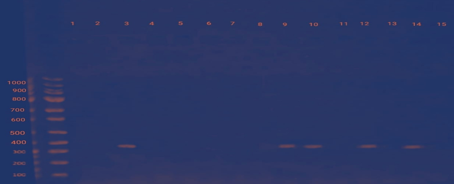
Figure 3. PCR-based stx2 gene discovery. Left lane: 1 kb DNA ladder; lanes 1–12: amplification products from E. coli samples 1–12. Amplification products are seen as unique bands of about 372 bp; lane 13: PCR negative control; lane 14: PCR positive control
To our knowledge, this is the first study on the occurrence of STEC in raw milk in Iraq.
Milk is an ideal environment for the growth of many microorganisms, including pathogens. Fresh milk, as we all know, is full of pathogenic and non-pathogenic germs that can be passed to humans through milking and consumption. Coliform bacteria are found in raw milk. Therefore, if milk is not properly boiled before consumption, it can be dangerous. Moreover, if correct hygiene procedures are not followed during milking, contaminated milk can cause diseases. In the majority of cases, milk containing E. coli may be collected from animals with subclinical mastitis.
STEC was detected among local E. coli isolates by PCR using stx1 and stx2 gene-specific primers. Our results showed in table 4 that local isolates produced Shiga toxins. The percentage of stx1 genes was higher than that of stx2. Interestingly, the genes were not found together in any of the isolates. However, in different locations, the presence of these genes was found to vary. 29,30 In a study conducted by Mamun et al.31 in Bangladesh, the stx1 gene was found in 5 (10.20%) of the 49 E. coli positive samples, while the stx2 gene was found in 26 (53.06%). In addition, 6 isolates (12.24%) tested positive for both the stx1 and stx2 genes, while the remaining 12 isolates (22.46%) tested negative for any of the genes.
Table (4):
Presence of Shiga toxin genes in selected E. coli isolates.
Isolate ID |
Shiga toxin stx1 gene |
Shiga toxin stx2 gene |
|---|---|---|
E. coli 1 |
+ |
– |
E. coli 2 |
– |
– |
E. coli 3 |
– |
+ |
E. coli 4 |
+ |
– |
E. coli 5 |
– |
– |
E. coli 6 |
+ |
– |
E. coli 7 |
+ |
– |
E. coli 8 |
+ |
– |
E. coli 9 |
– |
+ |
E. coli 10 |
– |
+ |
E. coli 11 |
– |
– |
E. coli 12 |
– |
+ |
E. coli 13 |
– |
– |
E. coli 14 |
– |
– |
E. coli 15 |
– |
– |
Our results are similar to those reported by Mohammadi et al.32 in Kermanshah, Iran. They isolated STEC from raw milk and found that 43.59% of the isolates carried stx1 genes, while 56.41% had stx2 genes.
According to our findings, the prevalence of STEC in raw milk samples was 17.47%. The samples were collected during summer, which has been linked to an increase in the number of cows with STEC (26). Therefore, STEC contamination in milk may be substantially less common during other periods of the year.
In the Apuila region, Parisi et al.33 found a lower prevalence of STEC (5.7%) in raw milk. Moreover, STEC prevalence in raw milk was reported to be 0.87% in Ontario and 3.9% in Germany, respectively.34,35 In a prior investigation from France, STEC was found in 21% of the 205 raw milk samples tested. All these data show prevalence levels different to ours.36 Geographical location, season, farm size, number of animals on the farm, hygiene, farm management techniques, variance in sampling, variation in types of samples assessed, and changes in detection methodologies utilized are all likely to contribute to the observed variation.
Based on virulence gene profiles and their association with clinical illness severity, at the Joint Food and Agriculture Organization of the United Nations, World Health Organization (FAO/WHO) Expert Meeting on Microbiological Risk Assessment, the potential risk to human health of STEC strains in food was categorized into five levels, being level 1 the highest and level 5 the lowest (FAO/WHO, 2018). O157 STEC, for example, is a well-known pathogen that can cause human illnesses ranging in severity from uncomplicated diarrhea to bloody diarrhea to severe outcomes of hemolytic uremic syndrome (HUS).37
Here, concentrated samples from different percentages of ammonium sulfate fractions were subjected to electrophoresis on 12.5% SDS-PAGE using 1× PAGE buffer under denaturing conditions. The gel was run at 500 mA/100 V until the dye reached the bottom. A commercial marker protein was used as a standard marker to estimate the approximate molecular weight of the protein of interest in the fractions. The gel was stained with Coomassie brilliant blue. It was observed that there distinct bands having a molecular weight equal to 70 kDa could be observed.
A protein of similar size to that of Shiga toxin was observed on SDS-PAGE (data not shown), but further analysis is required to confirm that it is an active Shiga toxin expressed by STEC isolates. Shiga toxin from local isolates in Baghdad city was purified partially by ammonium sulfate and concentrated by lyophilization; the killer protein was detected using SDS-PAGE, which exhibited a distinct band; our results are similar to those of Fathi et al.22 who purified Shiga toxin from E. coli, migrated it using SDS, and determined that it had a molecular weight of 70.5 kDa.
From the results obtained in this study, we can conclude that milk may be contaminated with E. coli. The findings of this study highlight the necessity for more stringent preventive measures, such as sterilization of dairy equipment, utensil washing, uniform milking hands, clean udders, and unhealthy animal eradication. Finally, milk must pasteurized or boiled before collection.
ACKNOWLEDGMENTS
None.
CONFLICT OF INTEREST
The authors declare that there is no conflict of interest.
AUTHORS’ CONTRIBUTION
All authors listed have made a substantial, direct and intellectual contribution to the work, and approved it for publication.
FUNDING
None.
DATA AVAILABILITY
All datasets generated or analyzed during this study are included in the manuscript.
ETHICS STATEMENT
This article does not contain any studies on human participants or animals performed by any of the authors.
- Holko I, Bisova T, Holkova Z, Kmet V. Virulence markers of Escherichia coli strains isolated from traditional cheeses made from unpasteurised sheep milk in Slovakia. Food Control. 2006;17(5):393-396.
Crossref - Wang,Q, Ruan X, Wei D, et al. Development of a serogroup specific multiplex PCR assay to detect a set of Escherichia coli serogroups based on the identification of their O-antigen gene clusters. MolCell Probes. 2010;24(5):286-290.
Crossref - Louie M, De Azavedo JC, Handelsman MY, et al. Expression and characterization of the eaeA gene product of Escherichia coli serotype O157:H7. Infect Immun. 1993;61(10):4085-4092.
Crossref - Paton JC, Paton AW. Pathogenesis and diagnosis of Shiga toxin-producing Escherichia coli infections. Clin Microbiol Rev. 1998;11(3):450-479.
Crossref - Karmali MA. Infection by verocytotoxin-producing Escherichia coli. Clin Microbiol Rev. 1989;2(1):15-38.
Crossref - Beutin L, Kaulfuss S, Cheasty T, et al. Characteristics and association with disease of two major subclonesof Shiga toxin (Verocytotoxin)-producing strains of Escherichia coli (STEC) O157, that are present among isolates from patients in Germany. Diagn Microbiol Infect Dis. 2002;44(4):337-346.
Crossref - Willshaw GA, Cheasty T, Smith HR, O’Brien SJ, Adak GK. Vero cytotoxin-producing Escherichia coli (VTEC) O157 and other VTEC from human infections in England and Wales. 1995-1998. J Med Microbiol. 2001;50(2):135- 142.
Crossref - Louie M, Read S, Louie L, et al. Molecular typing methods to investigate transmission of Escherichia coli O157:H7 from cattle to humans. Epidemiol Infect. 1999;123(1):17-24.
Crossref - Reilly A. Prevention and control of enterohaemorrhagic Escherichia coli (EHEC) infections: memorandum from a WHO meeting. WHO consultation on prevention and control of enterohaemorrhagic Escherichia coli (EHEC) infections. Bulletin of the World Health Organization. 1998;76:245-255.
- Lin A, Nguyen L, Lee T. et al., “Rapid O serogroup identification of the ten most clinically relevant STECs by Luminex microbead-based suspension array,” Journal of Microbiological Methods. 2011;87:105–110.
- Erickson MC, Doyle MP. Food as a vehicle for transmission of Shiga toxin-producing Escherichia coli. J Food Protect. 2007;70(10):2426-2449.
Crossref - Madic J, Vingadassalon N, de Garam CP, et al. Detection of Shiga toxin-producing Escherichia coli serotypes O26:H11, O103:H2, O111:H8, O145:H28, and O157:H7 in raw-milk cheeses by using multiplex real-time PCR. Appl Environ Microbiol. 2011;77(6):2035-2041.
Crossref - Solomakos N, Govaris A, Angelidis AS, et al. Occurrence, virulence genes and antibiotic resistance of Escherichia coli O157 isolated from raw bovine, caprine and ovine milk in Greece. Food Microbiol. 2009;26(8):865-871.
Crossref - Shebib,Z., Abdul Ghani, Z.G. and Mahdi, L.. First report of Escheria coli O157 among Iraqi children. Easterian medterian Health. Journal, (2003) vol.9Nos 1/2.
- Hassan J, Parvej MS, Rahman MB, et al. Prevalence and characterization of Escherichia coli from rectal swab of apparently healthy cattle in Mymensingh, Bangladesh. Microbes and Health. 2014;3(1):12-14.
Crossref - Soomro AH, Arain MA, Khasheli M, Bhutto, B. Isolation of Escherichia coli from raw milk and milk products in relation to public health sold under market conditions at Tandojam. Pak J Nutr. 2002;1(3):151-152.
Crossref - Wilson K. Preparation of Genomic DNA from Bacteria. In: Ausubel FM, Brent R, Kingston RE, Moore DD, Seidman JG, Smith JA and Struhl K. Eds., Current Protocols in Molecular Biology, Wiley & Sons, New York, 2.4.1-2.4.5.
- Maniatis T, Fritsch EF, Sambrook J. Molecular cloning. Cold Spring Harbor Laboratory, Cold Spring Harbor, New York, USA, 1982.
- Maleki D, Jahromy SH, Karizi SZ, Eslami P. The Prevalence of acrA and acrB genes among multipledrug resistant uropathogenic Escherichia coli isolated from patients with UTI in Milad Hospital, Tehran. Avicenna J Clin Microbiol Infect. 2017;4(1):39785.
Crossref - Schippa S, Lebba V, Barbaro M, et al. A distinctive microbial signature’ in celiac pediatric patients. BMC Microbiology. 2010;10: 175.
- Talukdar PK, Rahman M, Rahman M, et al. Antimicrobial resistance, virulence factors and genetic diversity ofEscherichia coli isolates from household water supplies in Dhaka, Bangladesh. PloS ONE. 2013;8(4):e61090.
Crossref - Fathi J, Ebrahimi F, Nazarian S, Tarverdizade Y. Purification of Shiga-like toxin from Escherichia coli O157:H7 by a simple method. Journal of Applied Biotechnology Reports. 2017;4(4):707-711.
- Burgess RR. Protein precipitation techniques. Methods Enzymol. 2009;463:331-42.
Crossref - Ahmed KD. The positive control of ILVC expression inE. coli K12. Ph.D. Thesis, Durhum Univ., England, 1989.
- Giovannucci E, Rimm EB, Wolk A, et al. Calcium and fructose intake in relation to risk of prostate cancer.Cancer Research. 1998;58:442-447.
- Islam MA, Kabir SML, Seel SK. Molecular detection and characterization of Escherichia coli isolated from raw milk sold in different markets of Bangladesh. Bangladesh Journal of Veterinary Medicine. 2016;14(2):271-275.
Crossref - Tahira B, Kaleem U , Abdul Samad, et al. I solation and molecular characterization of Shiga toxin producing E. coli O157:h7 in raw milk using Mpcr. IJPSR. 2017;. 8(7): 3107-3112.
- Al-Saadi ZHA, Abdullah RM. Phenotypic and molecular detection of Escherichia coli efflux pumps from UTI patients. Biochem. Cell. Arch. 2019; 19(1): 2371-2376.
Crossref - Adwan GM, Adwan KM. Isolation of Shiga toxigenic Escherichia coli isolated from raw beef in Palestine. Int J Food Microbiol. 2004;97(1):81-84.
Crossref - Parisi A, Miccolupo A, Santagada G, Pedarra C, Dambrosio A, Normanno G. Detection of verocytotoxin-producing Escherichia coli (VTEC) in minced beef and raw milk by colony blot hybridization.Food Control. 2010;21(5):770-773.
Crossref - Mamum MM, Parvej MS, Ahamed A, et al. Prevelance and characterization of Shiga toxigic Echerchiae coli in Broiler bird in Mymensingh. Bangl. J. Vet. Med. (2016). 14(1): 5-8
- Mohammadi P, Abiri R, Pezaei M, Salmanzadeh-Ahrabi S. Isolation of Shiga toxin-producing Escherichia coli from raw milk in Kermanshah, Iran. Iran J Microbiol.2013;5(3):233-238. PMCID: PMC3895560.
- Parisi A, Miccolupo A, Santagada G, Pedarra C, Dambrosio A, Normanno G. Detection of Vero cytotoxin-producing Escherichia coli (VTEC) in minced beef and raw milk by colony blot hybridization. Food Control. 2010;21(5):770-773.
Crossref - Klie H, Timm M, Richter H, Gallien P, Perlberg KW, Steinruck H. Detection and occurrence of verotoxinformingand/or Shigatoxin-producing Escherichia coli (VTEC and/or STEC) in milk. Berliner und Münchener Tierarztliche Wochenschrift. 1997;337-341.
- Steele ML, McNab WB, Poppe C, et al. Survey of Ontariobulk tank milk for foodborne pathogens. J Food Prot. 1997;60(11):1341-1346.
Crossref - Perelle S, Dilasser F, Grout J, Fach P. Screening food raw materials for the presence of the world’s most frequent clinical cases of Shiga toxin-encoding Escherichia coli O26, O103, O111, O145 and O157. IntJ Food Microbiol. 2007;113(3):284-288.
Crossref - FAO/WHO. Shiga toxin-producing Escherichia coli (STEC) and food: attribution, characterization, and monitoring. In Microbiological risk assessment series, 2018;31. http://www.fao.org/3/ca0032en/CA0032EN.pdf (Accessed: June 8th, 2021).
© The Author(s) 2022. Open Access. This article is distributed under the terms of the Creative Commons Attribution 4.0 International License which permits unrestricted use, sharing, distribution, and reproduction in any medium, provided you give appropriate credit to the original author(s) and the source, provide a link to the Creative Commons license, and indicate if changes were made.