


ISSN: 0973-7510
E-ISSN: 2581-690X
Genus Streptomyces contributes to almost 80% of the world’s antibiotics under actinomycetes group. Pigments produced by this genus also been widely reported to have application in different field. This study was aimed in isolating a bio pigment having antimicrobial, anticancer and quorum quenching properties. To achieve the aim soil samples from the regions of Western Ghats around Coimbatore (TN, India) were collected which holds biological diversity and rich in mineral wealth. The aim was attained through screening of antagonisms for potential strain and the morphological, biochemical, physiological and molecular characterization was investigated sequentially. Further, the biological studies were evaluated. Totally 161 Actinomycetes strains were isolated from the collected samples out of which potent 27 were pigmented Streptomyces sp. The potential strain was screened through antagonism principle and the morphological, biochemical, physiological and molecular characterization revealed that the strain is Streptomyces hygroscopicus AVS7 (KP732441). The crude pigmented secondary metabolites were extracted using polar solvent methanol from the biomass of the strain AVS7. The chemically characterized metabolite was a carboxylic acid derivative compound. The crude methanolic metabolites extract which was reddish brown in color showed maximum zone of inhibition against 6 different pathogenic organisms from antimicrobial assessment. The crude extract showed quenching activity of the quorum sensing molecules produced by Staphylococcus aureus. From this it is evident that the crude secondary metabolites have the potentiality of being an antibiotic. The bioactive crude extract was found to have biological properties at significant range and further bioprospecting activity will be evaluated in future studies.
Antagonist, MIC, pigments, quorum quenching, Streptomyces sp.
Bio pigment production is now one of the emerging fields of research possessing potential applications in various industrial settings. Colour plays a special role in the food and textile industry1. The industry is now able to produce some microbial pigments for applications in food, cosmetics and textiles. In nature, a single microorganism produces pigments in different colors (fungi, yeasts, actinomycetes and bacteria) are quite common2. These pigments produced by microorganisms also have an antimicrobial activity and inhibit the disease causing pathogens by eliminating or preventing growth of the pathogen. Among the producers of commercially important metabolites or antibiotics, bacteria have proved to be profilic source with surprisingly small group taxa for the vast majority of compounds3. Among these microorganisms actinomycetes are found to be the most economically and biotechnologically priceless prokaryotes. They are widely distributed in terrestrial and aquatic ecosystems. In soil, these organisms play a crucial role in recycling of refractory biomaterials by decomposition of complex mixtures like polymers in dead plant and animal materials and also capable of producing several secondary metabolites4. Actinomycetes have provided important bioactive compounds of high commercial value and are being a source to be routinely screened for new bioactive substances. These searches have been remarkably successful and about 45% of naturally occurring antibiotics have been used in medicinal fields are isolated from actinomycetes. Considering these natural compounds in the market, today about 130 to 140 microbial products and a similar number of derivatives (including semi-synthetic antibiotics) are applied in human medicine, mostly in chemotherapy and veterinary medicine. About fifteen to twenty compounds are used in agricultural fields mainly as herbicides, pesticides, plant protecting agents and food additives. The majority of these compounds are produced by Streptomyces species. The high percentage of new compounds derived from new target oriented screening methods is also of actinomycetal origin. These antibiotics from Streptomyces sp are used as anti tumoral medicine, immunosuppressors, anti viral agents and also possess the antioxidant property 5. The Streptomyces sp produced pigments are very much useful in replacing the synthetic dyes. Metabolites of Streptomyces sp also found to possess quorum quenching property inhibiting the cell to cell communication of bacterial colonies. The subsequent discovery of compounds that inhibit the cell-to-cell communication, dubbed anti-quorum sensing (anti-QS) agents could provide a novel method of combating infection6, 7. In the present study we have isolated and characterized Streptomyces sp from Western Ghats terrestrial soil having antimicrobial activity against Gram-positive as well as Gram-negative bacteria and fungi, anticancer properties and quorum quenching was also evaluated.
Isolation and screening of isolates from soil sample
The samples were collected from eleven different terrestrial regions Valparai [Stanmore, Selali, Mamika estate, Waverly estate, Eettiyar estate, and Valparai town (1, 2)], Topslip and Marudhamalai hills [(BU1, BU2, BU3) Bharathiar University campus, Coimbatore], TamilNadu, India. For systematic screening of pigmented Actinomycetes to explore the Western Ghats region SCN medium8 was used for the isolation and enumeration of actinomycetes. Samples were serially diluted at tenfold and plated on starch casein nitrate agar plates supplemented with nystatin (20mg/L) in triplicates to minimize the fungal growth. The samples were incubated at 30ºC for 7 days for sporulation and pigmentation. Eventually, mixtures of colonies were formed and they were purified onto a new SCN agar plates until discrete colonies were formed and subsequently stored at -80ºC in 20% glycerol. The glycerol stocks of strains were taken out and streaked on SCN agar for identification and characterization studies. The pigment producing strains were screened for their antagonistic activity against pathogens following cross streak method. Pigmented Actinomycetes isolates were streaked along the diameter on SCN agar plates and incubated at 30° C for six days and after incubation, 12 hrs grown cultures of clinical pathogens (procured from PSG Hospitals, Coimbatore, TN) of Gram positive organisms such as Staphylococcus aureus, Staphylococcus epidermidis, Enterococcus faecalis, Shigella sp and Gram negative organisms such as Escherichia coli, Pseudomonas aeruginosa, Klebsiella pneumoniae, Salmonella typhi, Proteus vulgaris and Bacillus subtilis. These pathogens are facultative anaerobes and majorly cause Gastro Intestinal Infections, Urinary Tract Infections and nosocomial infections. The above stated pathogens were streaked perpendicular to the central strip of actinomycetes culture. Plates were again incubated at 37° C for 24 hrs and zone of inhibition was measured.
Determination of morphological and biochemical characteristics
The selected strain AVS7 was characterized morphologically following the directions given by the International Streptomyces Project (ISP) 9, 10, 11. The morphology of aerial hyphae, substrate mycelium and spore chain arrangement were determined by light microscopy and Scanning Electron Microscopy (SEM) by cover slip technique, and examined after seven days of growth at 30° C. The strain grown on different media (ISP 1 to ISP 7 and SCN) was investigated by following directions given in the Bergey’s Manual of Systematic Bacteriology. The carbon sources utilization and melanoid production were characterized for the strain AVS7 based on the method of Shirling and Gottlieb 12. In addition to this, biochemical and physiological characters were determined according to the method of Schofield and Schaal 13 and Tresner14.
Molecular characterization
Total genomic DNA was isolated based on the method of Edwards15 with slight modification to obtain consistent cell lysis and DNA recovery. For isolation of DNA, 72hours old broth culture was prepared in SCN medium. About 1.5 ml of broth culture was centrifuged at 10,000 rpm for 10min. Supernatant was discarded and 500 µl of solution I (10mM Tris HCl, 10mM MgCl2, 10mM KCl2, 2mM EDTA) was added, vortex and centrifuged at 10,000 rpm for 10 minutes. Supernatant was discarded and 500 µl of solution II (Sol I + 0.4mM NaCl) was added to the pellet, vortexed and centrifuged at 10,000 rpm for 10 min. To the pellet 500 µl of solution III (10mM KCl2, 0.4mM NaCl, 10mM of H2KPO4 & Na2HPO4) was added, vortexed and 30 µl of lysozyme was also added. Mixture was gently tapped and the tube was incubated at 37°C for 15min. 50µl of solution IV (10% SDS) was added and mixed well by inverting. The mixture was incubated at 55°C for 15min. To this 500µl of solution V (5M NaCl) was added and centrifuged at 10,000rpm for 5min. The supernatant was transferred to fresh microfuge tube and then 500µl of isopropyl alcohol was added and centrifuged at 10,000rpm for 10 min. The supernatant was discarded and 500 µl of 70% ethanol was added and centrifuged at 10,000 rpm for 10 minutes. Finally the supernatant was discarded and the pellet was dried. The pellet was suspended in 20 µl of TE buffer and stored at 20ºC. The Gel electrophoresis was performed and DNA was obtained. The amplification of 16 s ribosomal RNA was done using thermocycler (PTC 150, USA) with universal primers and Taq polymerase using standard protocols. The amplified fragments were sequenced and the sequence was compared with reference species of bacteria for similarity using NCBI (National Centre for Biotechnological Information) BLAST available at http:/ www.ncbi.nlm.nih.gov/. The sequences were taken for phylogenetic tree construction using PHYLIP package. The 16s rRNA sequence was submitted at GenBank, NCBI, USA.
Production and extraction of pigments
The glycerol stock of AVS7 spores were spread on SCN agar plates and incubated at 30°C for seven to ten days and allowed for good sporulation. The seed medium (SCN) was prepared by inoculating two loops of spores from SCN plates and cultured in Erlenmeyer flasks for the growth of vegetative inoculums. The flasks were incubated at 30°C for three days on a rotary shaker (180rpm). The culture of 5% (v/v) inoculums was transferred into 100mL aliquots of production medium (SCN broth) in 20x250mL Erlenmeyer flasks and incubated at 30°C for seven days on a reciprocating shaker (180rpm). The culture was centrifuged at 5000rpm for ten mins at 20°C; the cell free culture filtrate and biomass was separated. The pigment from mycelia biomass was extracted using polar and non polar solvents. The total pigment from biomass was extracted, filtered and concentrated. The cell free culture filtrate was separated using equal volume of polar and non polar solvents, filtered and concentrated. The crude extract was subjected to GC- MS to find out the metabolites profile and FT- IR analysis was performed to determine the functional groups in the crude extract. This was carried out to elucidate the aliphatic and aromatic compounds present in the crude metabolite profile.
Biological activity of crude extract
Determination of antimicrobial assay
The antimicrobial assay and Minimum Inhibitory Concentrations (MIC) assay were assessed by well diffusion method 16 using crude biomass extract and culture filtrate against facultative anaerobic pathogens such as S. aureus, S. epidermidis, E. faecalis, E. coli, P. aeruginosa, K. pneumoniae, S. typhi, P. vulgaris, Shigella sp, Vibrio cholerae and B. subtilis. Fifty micrograms of cell free culture filtrate extract and crude biomass extract was added in respective wells on Mueller Hinton Agar (MHA) plates which were previously seeded with the above stated pathogens. The diameter of zone of inhibition was measured after 24h of incubation at 37° C for bacteria. The crude extract which exhibited highest antimicrobial profile was prepared at different concentrations ranging from 0 to 1000µg/µL. The lowest concentration of the total pigment extract which showed activity was considered to be MIC of the extract.
Quorum quenching property
Inhibition of protease and lipase
Protease activity was determined using skim milk agar (SMA) plate17. S. aureus was inoculated in Tryptic Soy Broth (TSB) as control and TSB with S. aureus, crude methanolic extract as test. After incubation at 37ºC at 180 rpm the filtrate was centrifuged at 12000 rpm for 10 mins and supernatant was collected and filled in respective wells on SMA plates. The digested substrate forms clear areas or zones surrounding the wells if proteolytic enzymes were produced. If the extract inhibits the activity of proteolytic enzymes there will be absence of zone around the sample added wells. Tribuytrin agar medium (TBA) was used to detect the inhibition of lipase for crude extract. If the microorganisms tested have a lipolytic activity, an opaque halo could be easily observed around the colonies. 40µL of S. aureus in control and in presence of crude extract were added to the respective wells in TBA plates. The plates were incubated at 37ºC for 48 h and observed for the inhibition of lipolytic activity through the absence of zone of clearance.
Cytotoxicity studies
The human breast cancer cell line (MCF-7) was obtained from National Centre for Cell Science (NCCS), Pune and grown in Eagles Minimum Essential Medium (EMEM) containing 10% fetal bovine serum (FBS). All cells were maintained at 37° C, 5% CO2, 95% air and 100% relative humidity. Maintenance cultures were passaged weekly, and the culture medium was changed twice a week. The monolayer cells were detached with trypsin-ethylenediaminetetraacetic acid (EDTA) to make single cell suspensions and viable cells were counted using a hemocytometer and diluted with medium containing 5% FBS to give final density of 1×10⁵ cells/ml. one hundred microlitres per well of cell suspension were seeded into 96-well plates at plating density of 20,000 cells/well and incubated to allow for cell attachment at 37°C, 5% CO2, 95% air and 100% relative humidity. After 24 h the cells were treated with serial concentrations of the test samples. They were initially dissolved in neat dimethyl sulfoxide (DMSO) and diluted to twice the desired final maximum test concentration with serum free medium. Additional four, 2 fold serial dilutions were made to provide a total of five sample concentrations. Aliquots of 100 µl of these different sample dilutions were added to the appropriate wells already containing 100 µl of medium, resulted the required final sample concentrations. Following drug addition the plates were incubated for an additional 48 h at 37° C, 5% CO2, 95% air and 100% relative humidity. The medium containing without samples were served as control and triplicate was maintained for all concentrations.
MTT assay
3-[4,5-dimethylthiazol-2-yl]2,5-diphenyltetrazolium bromide (MTT) is a yellow water soluble tetrazolium salt. A mitochondrial enzyme in living cells, succinate-dehydrogenase, cleaves the tetrazolium ring, converting the MTT to an insoluble purple formazan. Therefore, the amount of formazan produced is directly proportional to the number of viable cells. After 48h of incubation, 15µl of MTT (5mg/ml) in phosphate buffered saline (PBS) was added to each well and incubated at 37°C for 4h. The medium with MTT was then flicked off and the formed formazan crystals were solubilized in 100µl of DMSO and then measured the absorbance at 570 nm using micro plate reader. The % Growth inhibition was determined using the following formula. % Growth inhibition = 100- Abs (sample)/Abs (control) x100. Nonlinear regression graph was plotted between % Cell inhibition and Log10 concentration and IC50 was determined using GraphPad Prism software.
Isolation and screening of Actinomycetes
Totally 161 different Actinomycetes strain were isolated from eleven different soil samples collected at Western Ghats, Coimbatore, TamilNadu, India. Out of that 27 were pigmented Streptomyces sp. The pathogens P .aeruginosa, K. pneumoniae and E. faecalis were highly susceptible to the strain AVS 7 with the zone of inhibition of 15mm, 14mm and 12mm respectively.
Determination of morphological and biochemical characteristics
Morphological observations of spore chains under light microscopy with a magnification of 400x and 1000x, the strain on SCN agar was soft and ovoid in shape with smooth surfaces. The elongated chain of spores was flexible; the same was also supported by the images magnified through Scanning Electron Microscopy (SEM) (Fig. 1). The color of the aerial mycelium was pinkish grey and the strain AVS7 produced diffusible reddish brown pigment.
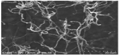 Fig. 1: SEM image of AVS7 spores
Observation of well developed irregular branched vegetative mycelium and no fragmentation of substrate were seen. The arthrospores were formed in chains with numerous spores and the aerial mycelium was branched monopodially. The physiological and biochemical characteristics were displayed in table 1 and this determines the genus level of the organism as Streptomyces. The strain hydrolysed starch, casein and was found to be producers of enzyme substances citrate, catalase, urease and nitrate reduction. The isolate AVS7 fermented reducing and non reducing sugars lactose, maltose, rhamnose and dextrose. Considerable production of pigment was observed only in starch casein nitrate medium, ISP 2 and ISP 3. Maximum diffusible pigments were seen in SCN medium while less diffusible pigments were observed with ISP 2. Other media used such as ISP 1, ISP 4, ISP 5, ISP 6 and ISP 7 did not induce the production of any pigment but growth of the organism was good in the media used. The organism Streptomyces sp was found to produce maximum spores in SCN medium with the range of pH from pH 7 to 9 and the strain was tolerable upto 5% NaCl concentrations.
Molecular characterization
A 1121-bp gene sequence was obtained with an amplification using 16SrRNA gene primers for the strain AVS 7. A BLAST search of the GenBank database with this sequence showed its 99% similarity to that of Streptomyces hygroscopicus. Phylogenetic tree was constructed with sequence of Genus Streptomyces members using PHYLIP package. The sequence was submitted in GenBank and obtained a temporary ID (KP732441).
Production and extraction of pigments
Fermentation process of the strain S. hygroscopicus AVS7 was carried on at 30ºC for 7 days at 180rpm. The pigment production was observed from 3rd day of incubation and the pigment started to explore throughout the incubation period and after 7th day, the pigment production was normal. Accordingly, the fermented broth was harvested on the 7th day for further extraction and purification. Cell free culture filtrate and biomass was separated using centrifugation process and the pigments were extracted using polar solvent ethyl acetate and methanol respectively. The methanolic biomass crude extract contained more pigments than cell free culture filtrate crude extract.
IR spectrum has proven to be the most effective way to give the information about the functional groups present in the compound. Figure 2 shows the FT-IR report for the purified fraction. In that, the stretching frequencies of the IR spectrum recorded 2923.87 & 2855.34 corresponds to Aliphatic –CH stretching and 750.0 corresponds – (CH2) n- in hydrocarbon. No peaks at 3500-3000 which clearly confirms that there is no –OH, NH and aromatic hydrogen in this compound. No peak at around 1600-1500 which further confirms the absence of aromatic carbons. The Gas chromatography – Mass spectrometry was analysed for the crude metabolites extract. The chromatogram was obtained with 4 major peaks which are given in figure 3. The highest peak was with the retention time 31.12 and Rf value was 6 with relative abundance 149. The molecular mass was 193.05. From this we can approximately conclude that the compound is a aliphatic keto compound and a carboxylic acid derivative.
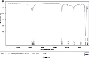 Fig. 2: FT- IR analysis of AVS7 crude extract
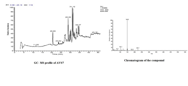 Fig. 3: GC- MS analysis of AVS7 crude extract
Biological activity of the crude extract
Determination of antimicrobial activity
The crude biomass extract and culture filtrate showed good antimicrobial potentials against Gram positive and Gram negative clinical pathogens. S. aureus was found to be highly susceptible to both methanolic biomass extract and culture filtrate extract with the zone of inhibition of 2.6 cm and 1.8cm respectively. Both the extracts showed inhibition against P. aeruginosa with the inhibition zone of 1.0cm and 1.2 cm respectively. The methanolic biomass extract showed an inhibition zone of 1.6 cm and 2.2 cm against K. pneumoniae and V. cholerae (Table 1). The methanolic biomass extract was having highest antimicrobial profile so it was taken to determine the quorum quenching property. To assess the quorum quenching property it is necessary to find out the MIC of the extract. MIC of the crude methanolic biomass crude extract was assayed. 20µg/µL of the pigment extract was found to be effective against all the clinical pathogens. S. aureus and Vibrio cholerae were sensitive to a minimum dose of 10 microlitres of the crude extract.
Table (1):
Antimicrobial profile.
| S. No. | Pathogens | Zone of inhibition(cm) | |
|---|---|---|---|
| Methanolic extract | Culture filtrate | ||
| 1 | Staphylococcus aureus | 2.6 | 1.8 |
| 2 | Klebsiella pneumoniae | 1.6 | 0.9 |
| 3 | Stapylococcus epidermidis | 1.0 | 0.7 |
| 4 | Pseudomonas aeruginosa | 1.2 | 1.0 |
| 5 | Enterococcus faecalis | 1.0 | – |
| 6 | Bacillus subtilis | 0.7 | – |
| 7 | E .coli | 0.9 | – |
| 8 | Proteus vulgaris | – | – |
| 9 | Shigella sp | – | – |
| 10 | Salmonella typhi | – | – |
| 11 | Vibrio cholerae | 2.2 | 1.2 |
‘-’ No zone of inhibition; cm centimeter; < 0.5cm no inhibition
Quorum quenching property
Inhibition of protease and lipase
Absence of zone of clearance was observed in the assay for proteolytic and lipolytic activity after the stipulated incubation period. The absence of zone of clearance with respect to the control indicated quenching or inhibition of quorum sensing molecules by the crude extract. This means that the crude extract have the ability to stop the QS system which is an essential role in synchronising gene expression and functional co-ordination among bacterial communities.
Cytotoxicity evaluation
The potential strain AVS7 exhibited anti cancerous activity at a significant range (Fig 4) and the 50% inhibitory activity was found at 3.66µg/ml. The regression was 0.9912 which was a significant one and 72% of cell death occurred at 10µg/ml (Table 2). This crude metabolite was found to be efficient anti cancer product.
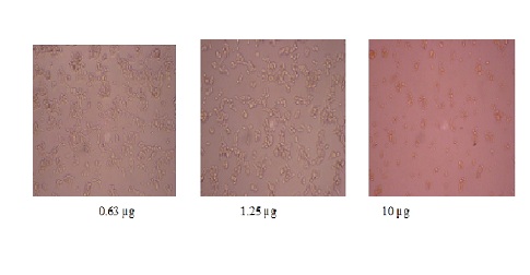
Fig. 4: Cytotoxicity analysis of crude extract of AVS7
Table (2):
Cytotoxicity of AVS7 metabolites.
Conc (µg) |
% Cell Inhibition |
|---|---|
0.63 |
10.01021 |
1.25 |
24.31052 |
2.5 |
40.34729 |
5 |
60.7763 |
10 |
72.01226 |
Terrestial actinomycetes are the well explored group in bacteria. Most of the antibiotics discovered from actinomycetes belong to genus Streptomyces. Apart from the available drugs we wanted to isolate a novel compound from the terrestrial Streptomyces sp obtained from the unexplored Western Ghats region. Hence the soil samples were collected from the Western Ghats (Nilgiri cluster) and about 121 actinomycetes strains were isolated. Among these 27 were pigmented strains. Screening of these pigmented strains revealed the potency of the isolate AVS7 and this primary screening unveiled that the SCN media was a good base for the production of antimicrobial compounds. From the results of morphology, biochemical, physiology and molecular characterization we found out the AVS7 is Streptomyces hygroscopicus. The spores were numerous on aerial mycelium and they form a chain arrangement. The strain showed a good growth on SCN agar amended with NaCl and the isolate can be placed in intermediate tolerant group and this investigation was similar to the report of Valan et al 18. Most of the antimicrobial compounds were extracted in methanol and ethyl acetate and this has been reported in many investigations. Similarly, in our study also crude extract from the biomass of S. hygroscopicus AVS7 was extracted in methanol. Many of the antibiotics were extracted from Streptomyces sp and antibiotics like daunamycin and irunamycin were extracted using methanol. This antibiotic has the antimcirobicidal effect on pathogenic bacteria19, as our crude was also extracted in methanol this may also contains a potential antibiotic. As the extract had the antimicrobial potentiality by inhibiting P. aureginosa, E. faecalis and K. Pnuemoniae and major activity was against Gram positive bacteria. Similar report was identified in aerobic filamentous bacterium from soil showed activity against Gram positive bacteria20. So by this means SCN broth induced the production of antimicrobial metabolites which are pigmented. The same was observed in the research of Valan et al18 that the novel Streptomyces sp from Western Ghats was found to have the highest antibacterial effect in MNG broth. This study revealed the excellence of antimicrobials produced by the Streptomyces sp depends mainly upon the media constituents and physiological effectiveness. The influence of antimicrobial compounds was showing the highest activity when SCN was used as sole media. Same piece of work was reported by Holmalahti et al21 who showed that the nature of medium composition strongly affected antimicrobial compounds production in different organisms. This antimicrobial compounds as crude metabolites were strongly produced and characterized for the presence of functional groups and GC- MS and FT-IR analysis revealed that the metabolite with the highest peak was a carboxylic acid derivative of aliphatic keto compound with the mass of 193.05. These metabolites have the highest antimicrobial profile and developed resistance against the pathogenic organisms. Most of the pathogenic organism resist available antibiotics and develop resistance against it through biofilm formation. A quest arised to identify the interaction of the pigmented secondary metabolites over quorum sensing molecules. Hence it was analysed through protease and lipase assay. However, the contribution of these proteases to the pathogenicity of Staphylococcus sp was highly speculative until now and deserves further investigation. The role for proteases may involve in protection against antimicrobial peptides such as the neutrophil defensins and the platelet microbicidal proteins. These peptides seem to play important roles in host defense. Because these antimicrobial peptides are subjected to proteolytic inactivation of the extracellular proteases production by the staphylococci that may represent a bacterial defense system22. Since the arlR-arlS locus modifies extracellular proteolytic activity, it is tempting to speculate that this locus might be involved in the virulence of S. aureus. It is not yet known whether ArlR acts directly on protease gene transcription or through interactions with other genes. Promisingly inhibition of these quorum sensing molecules of Gram positive organisms observed through the assays as observed by the earlier reports23. These sensing was quenched by the antimicrobial metabolites in crude form. Simialrily, lipase is also an important lipolytic enzyme of S. aureus that contributes significantly to the pathogenesis of staphylococcal infection24. The fact that organisms from deep infections are generally Lip+ suggests that lipase plays an important role in tissue invasion23, 25. Generally, the Gram positive organisms secrete proteases and lipases which act as cell to cell communicating agents. Absence of these molecules in the assay plates indicated the prevention of cell to cell communication of the pathogen by the crude biomass pigment extract. In this regard, the antimicrobial compounds disconnect the communication between the cells which may results in forming the pathogenecity. The mode of prevention may be either due to the inhibition in the production or by quenching the produced quorum sensing molecule is yet to be determined. Majorily, the quenching of these molecules will lead us to discovery of novel antibiotics against skin diseases as lipase was quenched by this crude compound. The understanding of the mechanism will lead to the judicial usage of the compound in inhibiting the pathogens. This research implied that the crude metabolite has the anti cancerous potentiality and the isolated culture have potency to be exploited as the sources of novel cytotoxic molecules showed highest activity at least concentration. Further research will be focused on knowing the chemical nature of the purified biomolecule for their antimicrobial and anticancer activities.
In conclusion, the Western Ghats terrestrial soil had majority of Streptomyces sp than any other actinomycetes species. The isolated S. hygroscpoicus AVS 7 produced red pigment which possessed the antimicrobial as well as possesses the quorum quenching property. So we suggest that this red pigment can be taken as antibiotic in medical fields. Further the crude extract has to be purified and studied for characterization of the pigmented compound also to be done to make it a potential drug.
- Clydesdale, F.M. Color as a factor in food choice, Crit. Rev. Food Sci., 1993; 33: 83–101.
- Bull, A.T. Microbial diversity and bioprospecting. ASM press., Washington, D.C., 2004; 191–203.
- Demain, A.L. Pharmaceutically active secondary metabolites of microorganisms. Appl. Microbiol. Biotechnol., 1999; 52: 455–63.
- Penka, M., Sava, T., Nadezhda, D., Valentina, C., Stefka, A.N., Nevena, B. characterization of coil actinomycetes from Antartica. J. of cult. Collections, 2000-2002; 3: 3-14.
- Hentzer, M., Wu, H., Andersen, J.B, Riedel, K., Rasmussen, T.B., Bagge, N., Kumar, N., Schembri, M.A., Song, Z., Kristoffersen, P. Attenuation of Pseudomonas aeruginosa virulence by quorum sensing inhibitors. The EMBO Journal., 2003; 22: 3803-3815.
- Smith, R.S., Iglewski, B.H. Pseudomonas aeruginosa quorum sensing as a potential antimicrobial target. J. of Clini. Invest., 2003; 112: 1460-1465.
- El Nakeeb, M.A., Lechevalier, A. Selective isolation of aerobic actinomycetes. Appl. Microbiol., 1963; 75-77.
- Shirling, E.B., Gottlieb, D. Methods for characterization of Streptomyces species. Int. J. Syst. Bacteriol., 1966; 16: 313-34.
- Shirling, E.B., Gottlieb, D. Cooperative description of type cultures of Streptomyces. V. Additional description. Int. J. Syst. Bacteriol., 1972; 22: 265-394.
- Williams, S.T., Good fellow, M., Wellington, E.M.H., Vickers, J.C., Alderson, G., Sneath, P.H.A., Sackin, M.J., Mortimer, A.M. A probability matrix for identification of Streptomyces. J. Gen. Microbiol., 1983; 129: 1815-1830.
- Shirling, E.B, Gottlieb, D. Methods manual: International Cooperative Project for Description and Deposition of type cultures of streptomycete. Int. J. Syst. Bacteriol., 1964; 1-27.
- Schofield, G.M., Schaal, K.P. Carbohydrate fermentation patterns of facultatively anaerobic actinomycetes using micromethods. Fems Microbiology Letters, 1980; 8: 67-69.
- Tresner, H.D., Hayes, J.A., Backns, E.J. Differential tolerance of Streptomyces to sodium chloride as a taxonomic aid. Appl. Microbiol., 1968; 16: 1134-1136.
- Edwards, U., Rogall, T., Blocker, H., Emde, M., Bottger, E. C. ().Isolation and direct complete nucleotide determination of entire genes. Characterization of a gene coding for 16S ribosomal RNA. Nucleic Acids Res., 1989; 17: 7843-7853.
- Cappuccino, J.G., Sherman, N. Microbiology, A laboratory manual Pearson Education, Inc., New Delhi. 2004; 282-283.
- Martley, F.G., Jarvis, W., Bacon, D.F., Lawrence, R.C. Typing of coagulase-positive Staphylococcus sp by proteolytic activity on buffered caseinate agar with special reference to bacteriophage nontypable strains. Infection and Immunity., 1970; 4: 439-442.
- Hedstrom, S.A., Nilson-Ehle, P. Trioleoyl glycerol lipolysis by Staphylococcus aureus strain from recurrent furunculosis, pyomyositis, impetigo and osteomyelitis. Acta Pathol. Microbiol. Immunol. Scand., 1983; 91(13): 169-173.
- Projan, S.J., Novick, R.P. The molecular basis of pathogenicity. In K.B. Crossley and G.L. Archer (Eds.), 1997. The Staphylococci in human disease. New York: Churchill Livingstone.
- Dufosse, L., Microbial Pigments. Encyclopedia Microbiol, 2009; 4: 457-471.
© The Author(s) 2017. Open Access. This article is distributed under the terms of the Creative Commons Attribution 4.0 International License which permits unrestricted use, sharing, distribution, and reproduction in any medium, provided you give appropriate credit to the original author(s) and the source, provide a link to the Creative Commons license, and indicate if changes were made.