


ISSN: 0973-7510
E-ISSN: 2581-690X
 , Hermansah Hermansah3 , Agustian Agustian3, Nurmiati Nurmiati4 and Tri Widya Edelwis5
Cellulolytic and lignocellulolytic microbes play an important role in the decomposition of plant biomass, but their isolation in tropical rainforest soil is often hindered by microbial competition, fungal growth dominance, and variations in pH and nutrient content that affect the viability of the isolates. This study aimed to isolate and characterize lignocellulose-degrading microorganisms from the HPPB soil of Andalas University, while also evaluating their relationship with environmental parameters. Isolation was carried out using CMC Agar and Tanat Agar on three soil composites, accompanied by soil chemical analysis. The results show a clear fertility gradient, with nitrogen decreasing from 34-15 mg/kg, phosphorus from 38-19 mg/kg, and potassium from 88-42 mg/kg. Microbial populations exhibit functional specialization: Composite B has the highest population of cellulolytic bacteria (26.9 × 10⁴ CFU/g), while Composite C is dominated by lignocellulolytic bacteria (50.0 × 10⁴ CFU/g). Based on colony diameter, bacteria SP2 (84.1 mm) and SP3 (69.7 mm) are the most active cellulolytic isolates, while fungi SP6 (90.0 mm) and SP8 (80.2 mm) are the dominant lignin decomposers. Enzyme dynamics over 28 days showed a sequential pattern: bacteria reached peak cellulase activity earlier, with SP1 increasing from 1.13-2.64 U/mL on day 7, while fungi worked more intensively in the later phase, marked by LiP activity of isolate SP6 reaching 0.1172 U/mL on day 28. Molecular analysis of the five selected isolates confirmed that bacteria SP1, SP2, and SP3 belong to the genus Bacillus, while fungal isolates JSP8 and JSP6 are closely related to Trichoderma and Penicillium, which are three key genera in the lignocellulosic decomposers of tropical ecosystems. Overall, these results affirm the close relationship between soil conditions, substrate composition, and the functional specialization of microbes, while also demonstrating the applicative potential of the isolates as bioactivators for biomass decomposition and organic waste processing.
Bioactivators, Cellulolytics, Lignocellulolytics, Soil Nutrients, Tropical Rainforests
Cellulolytic and lignocellulolytic microbes play a key role in the decomposition of plant organic matter, especially cellulose and lignin, which are the main structural components of plant biomass.1 This degradation process by microbes releases bound carbon back into the atmosphere in the form of CO2, while providing nutrients for plant growth through mineralization.2 Without microbial activity, the accumulation of undecomposed plant biomass would disrupt the balance of the carbon cycle.3 Recent studies have shown that lignocellulose-degrading microbes contribute up to 60% of total soil CO2 emissions in forest ecosystems.4
Tropical rainforests are known as hotspots of soil microbial diversity due to high humidity, stable temperatures, and the availability of abundant organic substrates.5 However, the isolation of cellulolytic and lignocellulolytic microbes from these ecosystems faces complex challenges, one of which is high competition between microbes, reducing the abundance of target species.6 Fungal growth is more dominant, making it difficult to isolate bacteria on one selective medium so that several purifications are needed to separate bacterial isolates and fungal isolates. There is still limited selective media to distinguish microbial activity that produces specific cellulase and ligninase enzymes.7 Soil conditions with varying acidity levels that inhibit the growth of some potential isolates,8 especially cellulolytics and lignocellulolytics.
Although the potential of cellulolytic and lignocellulolytic microbes from tropical rainforests has been isolated, their potential as bioactivators is still very limited and preliminary studies on soil nutrients in tropical rainforest areas and environmental factors that control their distribution must also need more in-depth studies.9 There are no references to the effect of soil pH on the selectivity of microbial activity,10 how the role of nutrients (N, P and K) in modulating degrading microbial communities.11 There are still few references to microbial data from tropical rainforests, both primary and secondary forests,12 especially in the West Sumatra region of Indonesia in the area of the Biology Education and Research Forest, Andalas University, Padang, Indonesia.
The purpose of this study is to isolate cellulolytic and lignocellulolytic microbial bioactivators from tropical rainforest soils using selective media, namely CMC agar and Tanat Agar and then analyze the initial relationship between environmental parameters, soil nutrient content with the abundance of potential cellulolytic and lignocellulolytic isolates. This research will provide a database of potential isolates for biodecomposer, bioremediation and bioenergy applications.13
Study area and sampling
Sampling was conducted at six points (T1-T6) in the tropical rainforest in the Biology Education and Research Forest (HPPB) with an altitude of 310-320 meters above sea level. The location has a humid climate (rainfall >3000 mm/year) and dominant woody vegetation. Soil samples were taken from areas with varying fertility status (Low to Very Low), then analyzed for isolation of cellulolytic and lignocellulolytic microbes. Detailed coordinates and characteristics of the sampling points were carried out at six points (T1-T6) with the following location characteristics: Point T1: Located at coordinates S 00°54’44.59″, E 100°28’10.12″ with an elevation of 310 meters above sea level (masl), oriented towards the West, Point T2: Located at S 00°54’44.37″, E 100°28’11.41″ (320 masl), facing Southwest, Point T3: Located at S 00°54’44.10″, E 100°28’11.64″ (318 masl), facing West, Point T4: Coordinates S 00°54’43.69″, E 100°28’11.42″ (314 masl), heading Northwest, Point T5: Located at S 00°54’43.23″, E 100°28’11.59″ (311 masl), oriented North, Point T6: Located at S 00°54’44.17″, E 100°28’12.02″ (319 masl), oriented East (Figure 1).
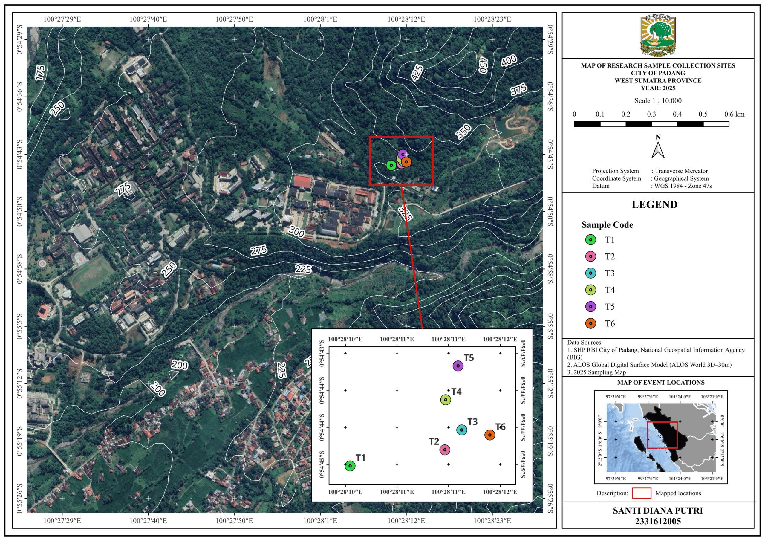
Figure 1. Spatial distribution of tropical rainforest sampling points in the Biology Education and Research Forest (HPPB), Andalas University, Padang, West Sumatra, Indonesia (Source: OpenStreetMap)
Soil Sampling Method Tropical Rainforest HPPB Area, Andalas University
Soil sampling was conducted at six points (T1-T6) at HPPB Andalas University using a composite purposive sampling method. Samples are grouped into three pairs of composites based on proximity of location and environmental characteristics: (1) Composite A (T1 + T2) with an elevation of 310-320 masl and a West/Southwest cardinal direction (elevation of 10 m), (2) Composite B (T3 + T4) at an elevation of 314-318 masl with a West/Northwest direction (ִelevation of 4 m), and (3) Composite C (T5 + T6) at an elevation of 311-319 masl with a North/East direction (elevation of 8 m). Each sampling point was taken in the depth layer (0-10 cm) using a sterile soil core with a diameter of 5 cm with three replications. Samples from two points in one composite group were then homogenized and stored in sterile double zipplock bags (500 g) at 4 °C for further analysis.
Isolation of Cellulolytic and Lignocellulolytic Microbes
Isolation of cellulolytic and lignocellulolytic microbes was carried out using two selective media, namely CMC agar and Tanat agar. A soil sample weighing 1 g was serially diluted (10-1-10-5) using 9 mL of sterile aquadest, then 1 mL of suspension from each dilution was inoculated into the media using the pour plate technique. After 72 hours of incubation, the growing colonies were purified three times using the streak plate method. Cellulolytic microbes have been selected on CMC agar8 (1% CMC, 0.1% peptone, 0.05% yeast extract, 0.5% NaCl, 2% agar; pH 6.8-7.0), then after incubation at 35 °C for 72 hours, the plate is stained with 0.1% Congo Red and washed with 1 M NaCl to reveal a clear zone as an indicator of cellulase activity, as used in the cellulolytic isolation method.4,13 Lignocellulolytic microorganisms were detected using Tanat agar (PDA + 0.1% tannic acid), where the brown color change or clarity around the colonies serves as an indicator of ligninase activity such as tannase, laccase, or peroxidase, according to the tannin-based selection approach and explanation of the lignocellulose degradation mechanism.3 Isolates that consistently show zone changes are selected for further characterization.
Chemical Analysis of Tropical Rainforest Soils in the HPPB Area of Andalas University
Soil chemical analysis was conducted to characterize the environmental conditions of microbial growth. Soil pH measurements were made in situ using a portable pH meter with a soil:water ratio of 1:2. Temperature parameters were measured at a depth of 0-10 cm using a digital soil thermometer. Soil nutrient content of N, P and K using Digital Display Nitrogen-Phosporus-Potassium-Nutrient Rapid Tester with Precision Test Method with sample preparation as follows: Measurement of nitrogen (N), phosphorus (P), and potassium (K) levels in the soil is carried out in the following stages: First, the tool is turned on using the power button. An air-dried soil sample was used to ensure initial consistency. The soil was then mixed with distilled water at a ratio of 2:1 (water:soil) in a sterile container to form a homogeneous paste. The tool was inserted vertically into the mixture at a depth of 6-8 cm. After a stabilization time of 10 seconds, the device displays the N, P, and K concentration values simultaneously. This method minimizes the interference of moisture factors through standardization of sample preparation, so that the results obtained represent the actual content of nutrients in the soil (true N, P, and K values).
Enzymatic characterization in the decomposition of Litter, Weeds, and Biochar: Cellulase and Lignin Peroxidase (LiP) activities
Litter, weeds, and biochar (1:1:1) that have been sterilized are mixed with soil microbial isolates as inoculum cultured in 100 ml of liquid CMC media, namely SP1, SP2, SP3, SP6, SP8 for 28 days. The dynamics of changes in cellulase and LiP enzyme activity were observed every seven days during the 28 day observation period as follows: Cellulase activity was assessed by quantifying reducing sugars released from CMC hydrolysis using the DNS method. The reaction mixture contains 1 mL of 0.05 M citrate buffer (pH 4.8), 1 mL of 1% CMC, and 1 mL of enzyme, while the control uses water as a substitute for the enzyme. All experiments were conducted in five replications (n = 5). The mixture was incubated at 50 °C for 30 minutes, followed by the addition of 1.5 mL of DNS and heating at 100 °C for 10 minutes. Absorbance was measured at 575 nm and quantified using a glucose standard curve (0-1 mg/mL). Enzyme activity is calculated based on the concentration of glucose produced, corrected for dilution and reaction time14 LiP activity was determined based on the degradation of aniline blue and expressed as U/mL, where one unit corresponds to the conversion of 1 µmol substrate per minute. The 3.2 mL reaction mixture consisted of 50 mM sodium tartrate buffer (pH 2.5), 0.01% aniline blue, enzyme extract, and H2O2, followed by incubation at 25 °C for 30 min. The reaction was terminated with DNS, and absorbance was recorded at 620 nm using an enzyme-free blank. Each treatment was performed in five replicates (n = 5). Activity was calculated from the change in absorbance using the molar extinction coefficient of aniline blue (ε = 17,000 M-1 cm-1), with adjustments for reaction time, enzyme volume, and dilution factor.15
Morphological characterization of isolates
Macroscopic observation of the isolate includes colony morphology (circular, irregular, filamentous, or rhizoid).16 The color and pigmentation of the colonies are recorded to detect cellular pigments or pigments that diffuse into the agar. In addition, the description of colony size in millimeters, according to the descriptive guidelines used in the evaluation of microbial cultures on solid media.
Molecular identification of microbial isolates
Molecular identification was carried out on isolates showing the highest enzyme activity by sequencing the 16S rRNA gene for bacteria and the ITS region for fungi.2 Bacterial DNA was amplified using the 8F and 1510R primers, while fungal amplification used ITS universal primers.7 PCR products of the expected fragment sizes were purified and sequenced using the Sanger method.9 The generated DNA sequences were analyzed using BLAST to determine the closest taxonomic match, and identities above 98%-99% were used to confirm the species level.17 All validated sequences have been submitted to the GenBank database, and their accession numbers are included in the manuscript to facilitate transparent and reproducible taxonomic identification.18
Data analysis
Data analysis was conducted using STAR (Statistical Tools for Agricultural Research) with five replications per treatment (n = 5). If there are significant differences between treatments, further testing is conducted using Duncan’s Multiple Range Test (DMRT) at a 5% significance level. Additionally, the relationship between soil environmental parameters (pH, C-organic, N-total, and the availability of P and K) and microbial abundance is analyzed using a Pearson correlation heatmap to identify the environmental factors that have the greatest contribution to microbial response and microbial enzyme activity.
Sampling site characteristics
Based on Table 1, there has been no statistically validated correlation between environmental variables. Soil pH generally decreases with increasing elevation, although Component B has the lowest pH (6.0). This phenomenon may be due to the fact that nutrients are more easily filtered and the microclimate is cooler. This observation is purely descriptive and lacks validation through inferential statistical analysis. The observed pH range (6.0-6.5) in the tropical highland region, where intense weathering and leaching due to high rainfall increase acidity, is ecologically significant because soil pH greatly controls nutrient availability, microbial activity, and organic matter decomposition. Moreover, the narrow variation in environmental temperature (25-26 °C) with a relatively uniform microclimate, although statement 14 explains that temperature can affect carbon and nitrogen dynamics in the soil, the narrow temperature range at this location limits its explanatory power regarding microbial distribution. Differences in wind direction have an impact due to exposure to sunlight and moisture retention, both of which are known as important factors in the heterogeneity of soil properties.
Table 1. Chemical Characteristics of Composite Soil at the Research Site: Coordinates, Elevation, Cardinal Direction, Elevation Difference (Elevation), Environmental Temperature, and Environmental pH
Composite (Sampling Point) |
Coordinates |
Elevation (above sea level) |
Cardinal Direction |
DElevation |
e-T (°C) |
e-pH |
|---|---|---|---|---|---|---|
Composite A (T1 + T2) |
S 00°54’44.59″ E 100°28’10.12″ |
310 + 320 |
West + Southwest |
10 m |
26 |
6.5 |
Composite B (T3 + T4) |
S 00°54’44.10″ E 100°28’11.64″ |
318 + 314 |
West + Northwest |
4 m |
25 |
6 |
Composite C (T5 + T6) |
S 00°54’43.23″ E 100°28’11.59″ |
311 + 319 |
North + East |
8 m |
25 |
6 |
Description: e-T refers to ambient environmental temperature, and e-pH refers to environmental pH conditions
Analysis of soil nutrient content of biological education and research forest (HPPB), University of Andalas
The results of the macronutrient (N, P, K) content analysis at the three sample composite points revealed significant spatial variations in soil fertility status. The data showed a consistent pattern of decreasing nutrient concentrations from Composite A (T1 + T2) towards Composite C (T5 + T6). The entire study site showed serious nitrogen (N) deficiency, with levels ranging from 15-34 mg/kg (“Very Low” to “Low” category). This phenomenon is consistent with the typical characteristics of tropical rainforest soils, which have fast N mineralization rates but are susceptible to nutrient losses through intensive leaching processes.19 This condition is exacerbated by the high rainfall in tropical ecosystems that accelerates the leaching process, especially in soils with sandy textures.20
Phosphorus (P) analysis revealed a more complex variability in fertility status, ranging from the “High” category in Composite A (38 ± 0.9 mg/kg) to “Moderate” in Composites B and C. The high P levels in Composite A are strongly suspected to be related to the optimal phosphate-solubilizing microbial activity at pH 6.5,21 as well as the accumulation of organic matter at the site. However, the 50% decrease in P levels in Composite C (19 ± 0.5 mg/kg) reflects the spatially limited availability of available P, a characteristic commonly found in old tropical soils.22 This uneven pattern of P distribution could potentially affect vegetation composition, where low-P adaptive species will tend to dominate in zones with lower P content.23
Potassium (K) showed a similar downward trend to N, with Composite C having the lowest levels (42 ± 1.7 mg/kg) which is classified as “Very Low”. This low K availability can be explained by two main mechanisms: first, the high intensity of nutrient leaching in tropical rainforest ecosystems,20 and second, the immobilization of K by the dominant 1:1 type clay minerals (kaolinite) in acidic soils.24 These findings have important implications for ecosystem restoration management, where interventions through the addition of organic fertilizers or the implementation of agroforestry systems with N-fixing legumes are potential solutions.25
Spatial variation of npk and ecological implications in tropical rainforests
Analysis of macronutrient content in the three composite soil samples (Table 2) revealed a clear trend of declining soil fertility from Composite A (T1 + T2) to Composite C (T5 + T6). Nitrogen (N) decreased from 34 mg/kg in Composite A, 24 mg/kg in Composite B, to 15 mg/kg in Composite C. Phosphorus (P) also exhibited a similar decreasing pattern, from 38 mg/kg in Composite A, 29 mg/kg in Composite B, to 19 mg/kg in Composite C. Potassium (K) declined from 88 mg/kg in Composite A, 65 mg/kg in Composite B, to 42 mg/kg in Composite C. This spatial variation appears to be associated with site characteristics, such as elevation and cardinal direction, which influence soil nutrient dynamics. The relatively low N concentrations, particularly in Composite C, are consistent with previous reports of low N retention in tropical soils due to a combination of high denitrification rates (>25 °C, +2.4 mg N/kg/day) at these temperatures (Table 1: 25-26 °C)26 and low cation exchange capacity (CEC < 4 cmol(+)/kg) in coarse-textured soils.27 The decrease in P content with elevation is also influenced by soil pH (6.0-6.5),26 where microbial phosphatase activity is optimal at pH 6.3-6.8,28 as well as by soil organic matter content. The decline in K reflects the dominance of kaolinite minerals, which generally have a low CEC (3-15 cmol(+)/kg).29 Ecologically, the variation in N, P, and K is significant because it controls nutrient availability, microbial activity, and organic matter decomposition in tropical rainforest soils. The observed gradient of macronutrients provides insight into soil heterogeneity at the study sites and its implications for ecosystem productivity.
Table 2. Nutrient Composition (N, P, K) and Fertility Classification of Composite Soil Samples at HPPB, University of Andalas
Composite (Sampling Point) |
N (mg/kg) |
P (mg/kg) |
K (mg/kg) |
Soil Fertility Status |
|---|---|---|---|---|
Composite A (T1 + T2) |
34 ± 0.9a |
38 ± 0.9a |
88 ± 2.0a |
Low (N & K), High (P) |
Composite B (T3 + T4) |
24 ± 1.0b |
29 ± 0.7b |
65 ± 2.7b |
Very Low (N), Medium (P), Low (K) |
Composite C (T5 + T6) |
15 ± 0.9c |
19 ± 0.5c |
42 ± 1.7c |
Very Low (N & K), Medium (P) |
Microbial Population Dynamics on Selective Media (CMC Agar and Tanat Agar)
Soil microbial populations in the HPPB Tropical Rainforest were analyzed based on their ability to degrade cellulitic (CMC Agar) and lignocellulolytic (Tanat Agar) can be seen in Table 3. The following data shows the average number of microbial colonies (×104 CFU/g) along with the standard deviation at each sampling point.
Table 3. Microbial Population on CMC (Cellulytic) and Tanat Agar (Lignocellulolytic) selective media (×104 CFU/g)
Sample Point |
CMC Agar Mean ± SD (×104 CFU/g) |
Tanat Agar Mean ± SD (×104 CFU/g) |
|---|---|---|
Composite A (T1 + T2) |
17.7 ± 77.6b |
1.0 ± 0.0c |
Composite B (T3 + T4) |
26.9 ± 157.5a |
8.4 ± 31.8b |
Composite C (T5 + T6) |
8.4 ± 82.8c |
50.0 ± 0.0a |
Description: Values are presented as mean ± SD (×10⁴CFU/g). Different superscript letters (a-c) within the same column indicate significant differences at P < 0.05
Based on Table 3, information was obtained about significant differences in microbial growth characteristics between CMC and Tanat Agar media. In CMC media, the variability of cellulolytic microbial population showed a very high standard deviation value, especially in sample T3 + T4 (26.9 ± 157.5 ×10⁴ CFU/g), where the SD value exceeded the mean value. This phenomenon is consistent with the findings of Casey et al.30 who reported that the natural heterogeneity of lignocellulosic substrates can cause significant variation in colony numbers. The high coefficient of variation (CV > 100%) in all CMC samples indicates the need for optimization of the isolation method, including the possibility of adding a longer acclimatization period or modifying the media composition.
In contrast, Tanat Agar medium showed better population stability, with SD of 0.0 at T1 + T2 and T5 + T6, indicating consistent growth of lignocellulolytic microbes. This finding is consistent with previous studies that reported the effectiveness of tannins as selective agents capable of suppressing the growth of non-target microorganisms.31 A marked difference was observed in the T5 + T6 sample, where the microbial population in Tanat Agar (50.0 × 10⁴ CFU/g) was significantly higher than in CMC Agar (8.4 × 10⁴ CFU/g). This may be attributed to site-specific adaptation of the microbial community to lignin degradation components, as previously reported regarding functional specialization of lignocellulolytic microbes.29 The distribution of cellulolytic and lignocellulolytic microbes is shown in Figure 2.
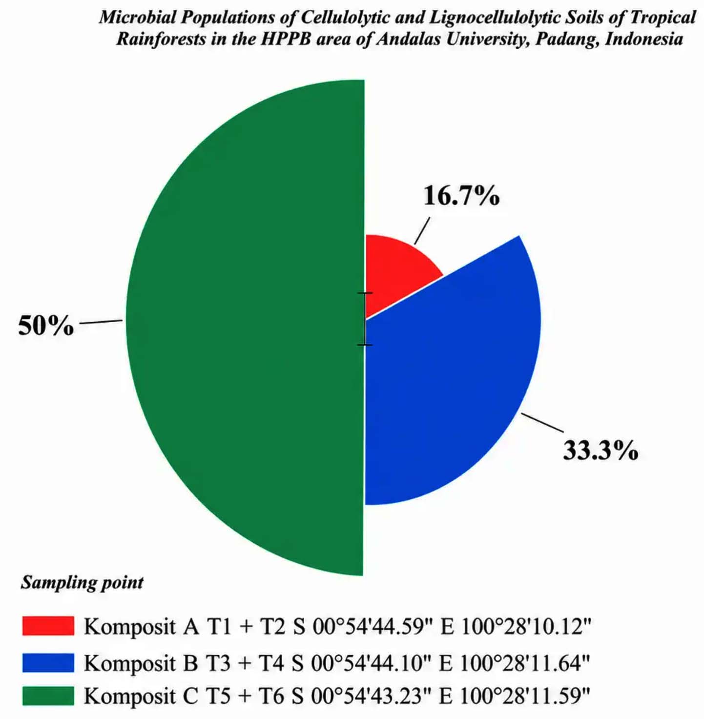
Figure 2. Distribution (%) of Cellulolytic and Lignocellulolytic Microbial Populations in Tropical Rainforest Soils of the HPPB Area of Andalas University Based on Sampling Points
The methodological implications of these findings inform the importance of a multisampling approach to overcome the heterogeneity of natural substrates, especially when using CMC media. Optimization of culture conditions, including the addition of pH buffers and modification of incubation time, may be necessary to improve the reproducibility of results. In addition, these findings underscore the need for complementary analyses such as enzyme activity assays and molecular identification to gain a more comprehensive understanding of microbial population dynamics.
The main limitations of this study include the relatively short incubation duration and the lack of molecular characterization of the isolates. For further research, it is recommended to apply a multi-omics approach that integrates metagenomic data, proteomics, and enzyme activity analysis to uncover the underlying mechanisms that control the population dynamics of lignocellulolytic microbes in different types of selective media.
The growth characteristics of microbial populations on selective media, as presented in Figure 3, show that Panel A illustrates cellulolytic microbial colonies on CMC Agar medium at a 10-4 dilution, while Panel B shows lignocellulolytic microbial colonies on Tanat Agar medium at the same dilution; visual observation indicates differences in microbial composition and colony density between the two media, with CMC Agar (Panel A) displaying more prominent bacterial colonies, appearing as numerous small cream-colored colonies, although fungal growth, represented by larger filamentous colonies, is also present, whereas on Tanat Agar (Panel B), fungal colonies are clearly dominant, forming large white circular colonies, and bacterial colonies are relatively sparse, suggesting that Tanat Agar medium is more conducive to fungal growth, whereas CMC Agar medium favors bacterial proliferation, providing descriptive insights into the selective growth preferences of cellulolytic and lignocellulolytic microorganisms under the given experimental conditions.
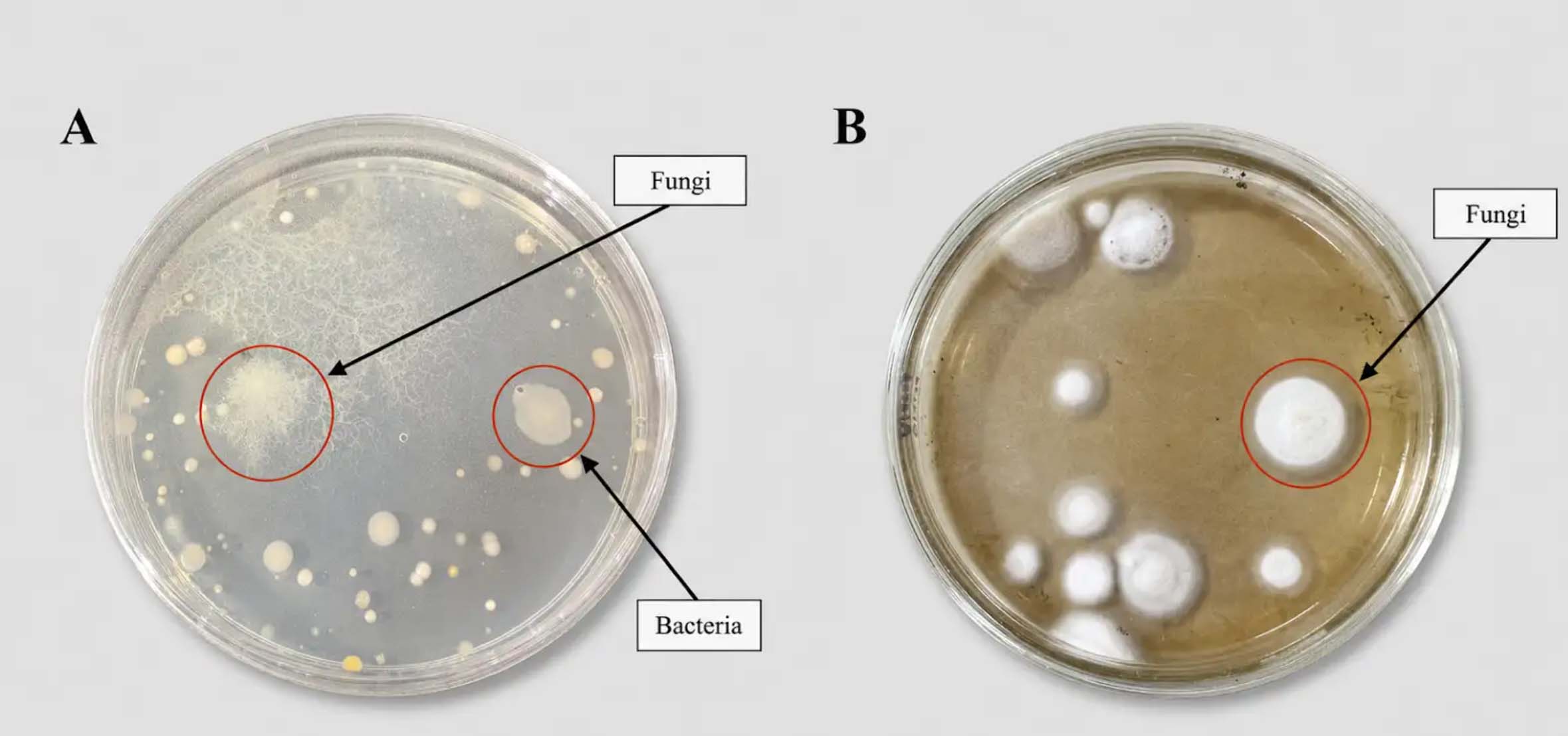
Figure 3. (a) Cellulolytic microbial population on CMC Agar medium (10-4); (b) Lignocellulolytic microbial population on Tanat Agar medium (10-4)
Pearson correlation matrix with significance levels between soil physicochemical parameters and microbial functional populations
The relationship between environmental factors and the total populations of cellulolytic and lignocellulolytic microbes is illustrated in the Pearson correlation matrix shown in Figure 4, where correlation coefficients (r) are represented by color gradients.
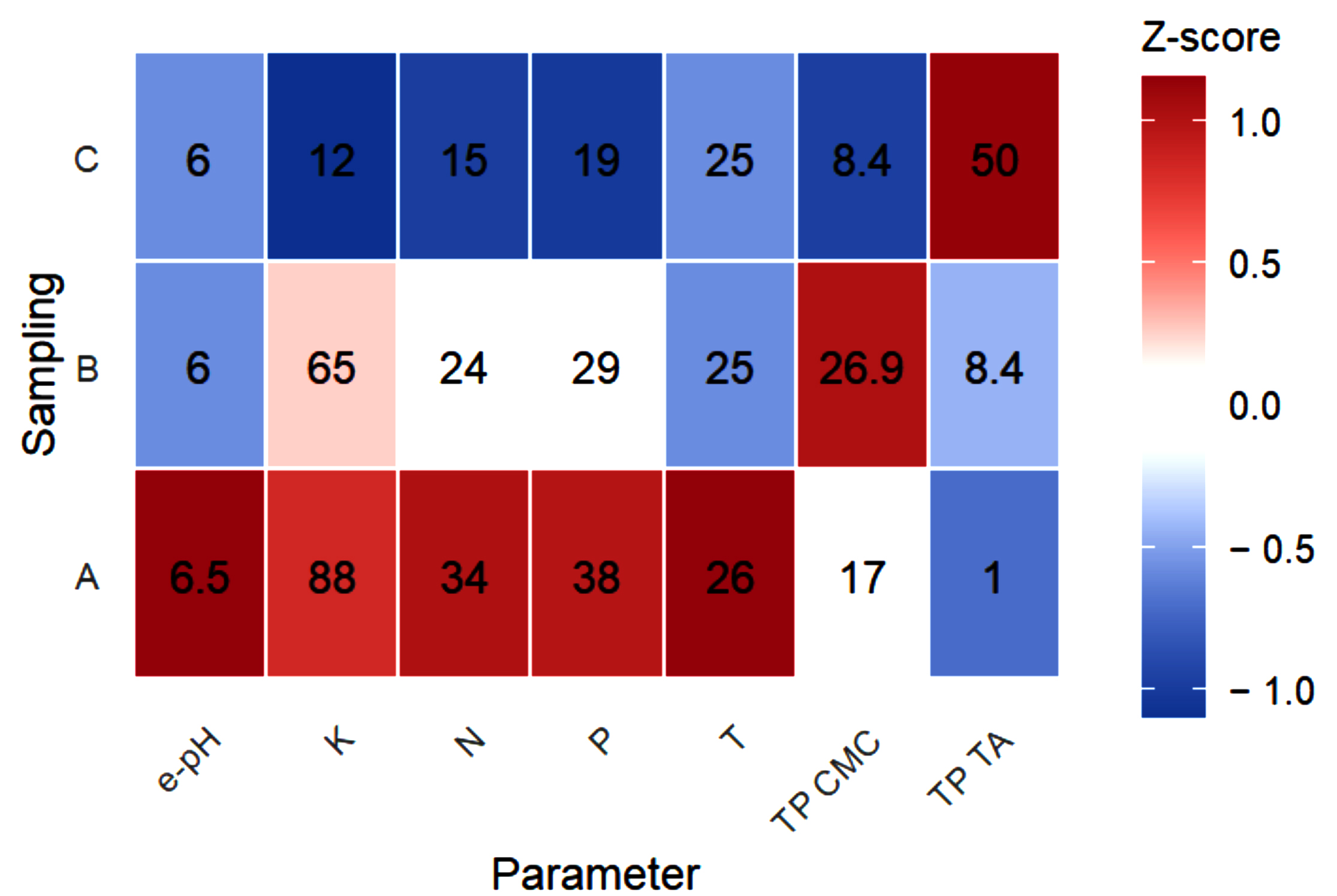
Figure 4. Pearson Correlation Matrix of Soil Physicochemical Parameters (T, pH, N, P, K) and Functional Microbial Populations (TP CMC, TP TA) (104 CFU/g). Variables measured are as follows: temperature (T, °C), soil pH (e pH), nitrogen (N, mg/kg), phosphorus (P, mg/kg), potassium (K, mg/kg), total cellulolytic microbial population
(TP CMC, ×104 CFU/g), and total lignocellulolytic microbial population (TP TA, ×104 CFU/g)
The Pearson correlation results between environmental parameters and functional microbial populations show an informative ecological pattern, although not statistically significant due to sample limitations (n = 3). The total population of lignocellulolytic microbes (TP TA) exhibited a very strong negative correlation with N, P, and K, indicating that the lignin-decomposing group tends to decrease in soils with high nutrient availability. This pattern is consistent with findings in tropical forests that the addition of N or P can suppress ligninase enzyme activity and shift microbial composition, especially in phosphorus-poor forests.32 Nutrient manipulation studies in China have also reported that N input decreases the biomass of certain microbes and alters community balance, while P has a greater impact on P-limited ecosystems.32 On the other hand, cellulolytic microbes (TP CMC) show a moderate positive correlation with K, in line with the role of potassium-solubilizing microbes (KSM) that can enhance K availability and the activity of cellulose-degrading enzymes, including CMCase, as reported in various KSM inoculation studies.33 The relationship between pH and temperature appears weaker, but remains physiologically relevant. Hydrolytic enzymes in tropical rainforest soils are known to have an optimum pH under acidic conditions, and edaphic changes can shift enzyme activity, as demonstrated in a soil enzyme study in Panama.32,34 Long term warming has also been reported to alter the composition of microbial communities and carbon degradation.35 Thus, although this correlation is not statistically significant, its ecological pattern remains consistent as an initial reference for the study.
Microbial isolation and enzyme activity based on colony diameter on specific media (CMC Agar and Tanat Agar)
Colony growth diameter observations indicated that isolate SP5 showed no growth on either selective medium (CMC Agar and Tanat Agar). This finding highlights the need for further investigation into several possible explanations: (1) cell dormancy, (2) specific nutrient requirements not provided by standard media, or (3) unique ecophysiological adaptations to the isolate’s native environment. This also underscores the complexity of microbial interactions with both abiotic and biotic factors in tropical rainforest ecosystems, which are currently mapped only spatially. Additional details are provided in Table 4.
Table 4. Colony Growth Characteristics and Lignocellulolytic and Cellulolytic Enzyme Activities of HPPB Soil Microbial Isolates on Selective Media
N |
Isolate |
Sampling Location |
Type of Bioactivator |
Colony Diameter on CMC Agar (mm) |
Colony Diameter on Tanat Agar (mm) Lignocellulase |
Enzyme activity based on (Diameter) Colony |
Enzyme Activity based on (Diameter) Colony Cellulase |
|---|---|---|---|---|---|---|---|
1 |
SP1 |
T3-T4 HPPB |
Bacteria |
18.9 ± 0.7 |
0 ± 0 |
– |
+ |
2 |
SP2 |
T5-T6 HPPB |
Bacteria |
84.1 ± 1.5 |
0 ± 0 |
– |
+++ |
3 |
SP3 |
T3-T4 HPPB |
Bacteria |
69.7 ± 1.2 |
9.7 ± 0.2 |
+ |
++ |
4 |
SP4 |
T1-T2 HPPB |
Bacteria |
30.8 ± 1.3 |
0 ± 0 |
– |
+ |
5 |
SP5 |
T1-T2 HPPB |
Bacteria |
0 ± 0 |
0 ± 0 |
– |
– |
6 |
SP6 |
T1-T2 HPPB |
Fungi |
56.6 ± 1.2 |
90.0 ± 0.8 |
+++ |
++ |
7 |
SP7 |
T3-T4 HPPB |
Fungi |
39.2 ± 0.9 |
80.2 ± 0.9 |
+++ |
+ |
8 |
SP8 |
T5-T6 HPPB |
Fungi |
50.9 ± 1.4 |
12.4 ± 0.5 |
+ |
++ |
Description: Enzyme score: (-) absent, (+) low, (++) medium, (+++) high. SD: Standard deviation of three replicates. Incubation conditions: 36 °C, 72 hours, aerobic
The results provided information on the diversity of lignocellulose degradation ability in HPPB soil microbial isolates that showed different substrate specialization and biotechnological potential. Bacterial isolates SP2 and SP3 showed optimal growth on CMC Agar media with colony diameters of up to 84.1 ± 1.5 mm and 69.7 ± 1.2 mm respectively with high cellulase activity ( + + + and + + ). This finding is in line with the research of Ren et al.18 who reported that cellulolytic bacteria in tropical ecosystems tend to develop large colony growth strategies as a form of competitive adaptation in obtaining nutrients. However, SP2 which is only active on CMC Agar and does not show growth on Tanat Agar, this supports the concept of “metabolic trade-off” where microbes optimize one metabolic pathway at the expense of other capabilities.17
Furthermore, on Tanat Agar medium, fungal isolates SP6 and SP7 demonstrated lignocellulose-degrading capability, with colony diameters exceeding 80 mm and exhibiting the highest lignocellulase activity (+++). This pattern is consistent with the characteristics of white-rot fungi, particularly in relation to their complex ligninolytic enzyme system.35 The dual growth capability of SP6 and SP7 on both media supports the hypothesis that tropical soil fungi have generally evolved lignocellulolytic capabilities as a survival strategy in highly competitive environments.17
From a biotechnological perspective, fungal isolates SP6 and SP7 exhibited strong lignocellulolytic activity on Tannic Acid (TA) agar, indicating their potential to degrade complex lignocellulosic substrates. Ligninolytic fungi are widely recognized for producing oxidative enzymes involved in lignin depolymerization and biomass conversion.36 In contrast, isolate SP2 showed prominent cellulolytic activity on CMC agar, suggesting its ability to utilize cellulose as a carbon source through enzymatic degradation. Microbial cellulases play an important role in biomass transformation and industrial bioconversion processes.37
The differences in functional activities among isolates reflect microbial adaptation to environmental conditions in tropical rainforest ecosystems. Such functional traits contribute to organic matter decomposition and carbon cycling processes.38 The occurrence of lignocellulolytic fungi (SP6 and SP7) suggests a role in the degradation of plant-derived materials, whereas SP2 may contribute to cellulose decomposition and nutrient turnover in soil environments.39
Nevertheless, the potential application of these isolates in enzyme production or lignocellulosic waste processing requires further validation under controlled and industry-relevant conditions.40 In addition, isolate SP5 did not exhibit growth on either selective medium, which may indicate dormancy, specific nutritional requirements, or unsuitable cultivation conditions. Therefore, further physiological and molecular studies are necessary to clarify its ecological role and biotechnological potential. Overall, these findings highlight the diversity of microbial functional strategies in tropical rainforest ecosystems, as shown in Figure 5.
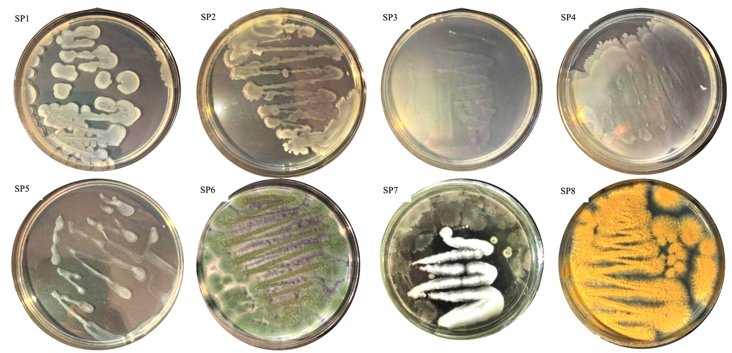
Figure 5. Cellulolytic and lignocellulolytic isolates from soil of HPPB area of Andalas University, Padang, West Sumatra, Indonesia
Visually, the bacterial colonies SP1, SP2, SP3, and SP4 exhibited similar coloration, ranging from white to cream, whereas the fungal isolates displayed more diverse pigmentation, with SP6 appearing green, SP7 white, and SP8 ranging from yellow to orange (Figure 5).
Molecular identification of bacterial and fungal isolates
The results of the molecular identification of bacterial and fungal isolates based on BLASTn analysis of the consensus sequences of the 16S rRNA gene (bacteria) and ITS (fungi) are presented in Table 5. Out of the eight isolates analyzed, only five produced consensus sequences of adequate quality for molecular identification. The displayed information includes sequence length, closest species match, percentage identity, query coverage, and final taxonomic decision. This data serves as a strong basis for validation before linking microbial identity with its functional potential, particularly related to lignocellulose degradation capability, soil microbial community dynamics, and their roles in decomposition processes and nutrient cycling in tropical environments.
Table 5. Molecular Identification Summary of Bacterial and Fungal Isolates
Isolate |
Consensus Length |
Top Hit (BLASTn) |
Identity (%) |
Query Coverage |
Taxonomic Assignment |
|---|---|---|---|---|---|
SP1 |
~1,420 bp |
Bacillus sp. |
98%-100% |
High |
Species level |
SP2 |
~1,450 bp |
Bacillus sp. |
>98% |
High |
Genus level (species-level overlap) |
SP3 |
~200-300 bp |
Bacillus group |
>97% |
Low (short sequence) |
Genus level only |
SP8 |
~750 bp |
Trichoderma sp. |
≥98% |
High |
Species level (strong) |
SP6 |
~800 bp |
Penicillium/ Aspergillus group |
≥98% |
High |
Genus level |
Note: Molecular identification of selected isolates based on BLASTn analysis, including sequence length, closest match, identity percentage, query coverage, and taxonomic assignment. Bacterial isolates were identified as Bacillus spp., while fungal isolates were classified as Trichoderma sp. and Penicillium/Aspergillus group.
Molecular identification shows that all bacterial isolates (SP1, SP2, and SP3) belong to the genus Bacillus, which is a group of bacteria that dominate tropical soils and play an active role in the decomposition of organic matter, nutrient cycling, and enzymatic degradation of lignocellulosic substrates.18,37 High sequence identity values (≥98%) and strong query coverage in isolates SP1, SP2, and SP3 support the classification at the species level. In line with several references that Bacillus spp. often dominate nutrient-rich microhabitats, have high metabolic flexibility, and are capable of adapting to environmental fluctuations typical of tropical forest ecosystems.10,38 Moreover, the presence of Bacillus among isolates with high enzymatic activity is consistent with many studies that show the genus has a strong ability to produce cellulase, xylanase, and lignin-degrading enzymes, which are very important in the biomass decomposition process.30,37
For the fungal isolates JSP8 and JSP6, BLASTn analysis revealed close affiliations with the genera Aspergillus, Trichoderma, and Penicillium, which are recognized as important fungal groups involved in lignocellulose decomposition in tropical ecosystems. JSP8 was strongly identified as Trichoderma spp., consistent with the role of this genus as a major producer of cellulase, hemicellulase, and other lignocellulose-degrading enzymes, as well as its ability to compete in soil environments rich in plant polymers.7 Meanwhile, JSP6 was closely associated with Penicillium, which contributes to litter decomposition, nutrient mineralization, and the enzymatic breakdown of complex lignocellulosic polymers in tropical forest soils.19 Overall, these molecular findings confirm that the obtained isolates correspond to key microbial taxa involved in lignocellulose degradation and biochemical processes in tropical rainforest soils. These findings are further supported by their roles in fungal-mediated lignin biodegradation39 and oxidative enzyme systems, including lignin peroxidase, manganese peroxidase, and laccase.41
Dynamics of cellulase and ligninolytic enzyme activities during Litter-Weed-Biochar decomposition and their biotechnological implications
The activity of cellulase and lignocellulase (LiP) enzymes from the eight purified isolates showed that only five isolates had optimal capabilities and were able to grow during purification. After the decomposition process of the litter-weeds-biochar for 28 days, the ability of the microbial isolates to degrade cellulose and lignocellulose components and their applicative potential in biomass-based biotechnology can be observed. Enzyme activity in each isolate can be seen in Tables 6 and 7.
Table 6. Comparative Temporal Profiling of Cellulase Activity Microbial Isolates from Tropical Rainforest Soils
| Isolate | Cellulase Enzyme Activity (U/ml) on the Day | ||||
|---|---|---|---|---|---|
| H0 (Day 0) | H7 (Day 7) | H14 (Day 14) | H21 (Day 21) | H28 (Day 28) | |
| SP1 (Bacteria) | 1.13a | 2.64a | 1.65b | 0.6028e | 0.7300b |
| SP2 (Bacteria) | 1.15a | 1.70cd | 1.70b | 0.8956a | 0.7600ab |
| SP3 (Bacteria) | 1.55a | 2.26ab | 1.70b | 0.7747abc | 0.0120c |
| SP6 (Fungi) | 1.41a | 1.71cd | 1.81b | 0.7733abcd | 0.7878ab |
| SP8 (Fungi) | 1.36a | 0.97e | 1.63b | 0.7517bcd | 0.8256a |
Note: Cellulase enzyme activity of microbial isolates was measured over 28 days of litter–weed–biochar decomposition. Bacterial isolates SP1–SP3 were identified as Bacillus sp. (SP1 and SP2) and Bacillus group (SP3), while fungal isolates SP6 and SP8 belonged to the Penicillium/Aspergillus group (SP6) and Trichoderma sp. (SP8).
Different superscript letters within the same row indicate significant differences (p < 0.05), as determined by ANOVA followed by Duncan’s Multiple Range Test (DMRT)
Table 7. Comparative Temporal Profiling of Lignocellulolytic Activity Microbial Isolates from Tropical Rainforest Soils
| Isolate | Lignocellulase Enzyme Activity (U/ml) on the Day | ||||
|---|---|---|---|---|---|
| H0 (Day 0) | H7 (Day 7) | H14 (Day 14) | H21 (Day 21) | H28 (Day 28) | |
| SP1 (Bacteria) | 0.0802abc | 0.1096bc | 0.0890cd | 0.0788b | 0.0692d |
| SP2 (Bacteria) | 0.0728bc | 0.1198a | 0.1018a | 0.0828b | 0.0692d |
| SP3 (Bacteria) | 0.0862abc | 0.0998d | 0.1004a | 0.0820b | 0.0760cd |
| SP6 (Fungi) | 0.0914ab | 0.1148ab | 0.0960ab | 0.0672c | 0.1172a |
| SP8 (Fungi) | 0.0800abc | 0.1100bc | 0.0902bcd | 0.0930a | 0.0852b |
Note: Lignocellulolytic enzyme activity of microbial isolates was measured over 28 days of litter–weed–biochar decomposition. Bacterial isolates SP1–SP3 were identified as Bacillus sp. (SP1 and SP2) and Bacillus group (SP3), while fungal isolates SP6 and SP8 belonged to the Penicillium/Aspergillus group (SP6) and Trichoderma sp. (SP8).
Different superscript letters within the same row indicate significant differences (p < 0.05), as determined by ANOVA followed by Duncan’s Multiple Range Test (DMRT)
Statistical analysis explains that the significance of the data shows that cellulase activity in bacterial isolates (SP1, SP2, SP3) is faster on the 7th day, while fungal isolates (SP6, SP8) are optimal on the 14th day. This pattern is consistent with the literature, which states that cellulase production by soil microorganisms generally reaches its peak within 5-7 days of incubation, coinciding with the availability of easily accessible cellulose substrate in the early fermentation phase. Relevant to controlled fermentation studies that report that the peak of enzymatic activity can occur in the first week of incubation, especially in soil cellulolytic microfungi and bacteria.40 Bacteria are more effective in the initial decomposition of simpler fractions such as cellulose and hemicellulose, while fungi, especially the white rot fungi group, are more dominant in lignin oxidation with enzymes such as LiP, MnP, and laccase. Bacteria are more effective in the initial decomposition of simpler fractions such as cellulose and hemicellulose, while fungi, especially the white rot fungi group, are more dominant in lignin oxidation with enzymes such as LiP, MnP, and laccase. Research in the tropical forest soil of Puerto Rico also shows that bacteria can grow on substrates containing lignin and enhance the activity of enzymes such as phenol oxidase and peroxidase, although at the same time, cellulase activity decreases. Additionally, research in the Amazon and Atlantic forest regions found that tropical environments are an important source of lignocellulolytic bacteria and actinomycetes with great potential for biotechnological applications.41 The focus of biotechnology sees the combination of bacterial and fungal isolates synergizing to become more efficient in biomass bioconversion. Studies on lignocellulose degradation emphasize the importance of microbial consortia, where bacteria provide the initial stage of carbohydrate depolymerization, followed by fungi with the more complex oxidation of lignin. In line with reports on the gradual increase in degradation efficiency, and relevant for industrial applications such as bioethanol production, lignocellulosic biorefineries, and biomass waste processing.42
Tropical rainforest microbes from the HPPB area of Andalas University show very high potential as cellulolytic and lignocellulolytic bioactivators, demonstrated by the dominance of cellulolytic populations in Composite B (26.9 × 10⁴ CFU/g) and lignocellulolytic populations in Composite C (50.0 × 10⁴ CFU/g), as well as superior enzymatic performance in bacteria SP1 and SP2, which each reached cellulase activity of up to 2.64 U/mL on the 7th day. In the fungal group, isolate SP6 became the most effective lignin decomposer with the highest LiP activity of 0.1172 U/mL on the 28th day, confirming the sequential working pattern of bacteria fungi that accelerates cellulose hydrolysis while enhancing lignin degradation. Molecular identification confirms that bacteria SP1, SP2, and SP3 belong to the genus Bacillus, which is known to be dominant in tropical soils, adaptive, and highly competent in cellulose and lignin degradation; while fungal isolates JSP8 and JSP6 are closely related to Trichoderma and Penicillium, two main genera of lignocellulose decomposers. The integration between enzymatic performance and taxonomic identity confirms that these isolates are key microorganisms with strong potential as bioactivators for accelerating biomass degradation and processing cellulose and lignocellulose based organic waste.
ACKNOWLEDGMENTS
The authors gratefully acknowledge the financial support from LPPM Universitas Andalas, through Research Contract No. 92/UN16.19/PT.01.03/PDD/2025. The authors also thank all colleagues and collaborators who contributed to this study.
CONFLICT OF INTEREST
The authors declare that there is no Conflict of interest.
AUTHORS’ CONTRIBUTION
All authors listed have made substantial, direct and intellectual contributions to the work, and approved it for publication
FUNDING
None.
DATA AVAILABILITY
All datasets generated or analyzed during this study are included in the manuscript.
ETHICS STATEMENT
Not applicable.
- Sanchez PA, ed. Properties and Management of Soils in the Tropics. 2nd ed. Cambridge University Press; 2019.
Crossref - Baldrian P, López‑Mondéjar R, Kohout P. Forest microbiome and global change. Nat Rev Microbiol. 2023;21(8):487‑501.
Crossref - Kumar R, Singh S, Singh OV. Bioconversion of lignocellulosic biomass: biochemical and molecular perspectives. J Ind Microbiol Biotechnol. 2021;35(5):377-391.
Crossref - Xue X, Ren C, Zhao C, et al. Litter removal and nitrogen deposition alter soil microbial community composition and diversity in a typical rubber (Hevea brasiliensis) plantation of Hainan, China. Appl Soil Ecol. 2025;208.
Crossref - Tripathi BM, Stegen JC, Kim M, Dong K, Adams JM, Lee YK. Soil pH mediates the balance between stochastic and deterministic assembly of bacteria. ISME J. 2018;12(4):1072-1083.
Crossref - Wang Y, Chen Y, Gao X, et al. Unveiling the driving role of pH on community stability and function during lignocellulose degradation in paddy soil. Front Microbiol. 2024;15:1338842.
Crossref - Filiatrault-Chastel C, Navarro D, Haon M, et al. AA16, a new lytic polysaccharide monooxygenase family identified in fungal secretomes. Biotechnol Biofuels. 2019;12(1):1-15.
Crossref - Lund PA, De Biase D, Liran O, et al. Understanding how microorganisms respond to acid pH is central to their control and successful exploitation. Front Microbiol. 2020;11:556140.
Crossref - Purahong W, Wubet T, Lentendu G, et al. Life in leaf litter: novel insights into community dynamics of bacteria and fungi during litter decomposition. Mol Ecol. 2016;25(16):4059–4074.
Crossref - Cao H, Chen R, Wang L, et al. Soil pH, total phosphorus, climate and distance are the major factors influencing microbial activity at a regional spatial scale. Sci Rep. 2016;6(1):25815.
Crossref - Waring BG, Averill C, Hawkes CV. Differences in fungal and bacterial physiology alter soil carbon and nitrogen cycling: insights from meta‑analysis and theoretical models. Ecol Lett. 2013;16(7):887–894.
Crossref - Qu X, Li X, Bardgett RD, Kuzyakov Y, et al. Deforestation impacts soil biodiversity and ecosystem services worldwide. Proc Natl Acad Sci USA. 2024;121(13):e2318475121.
Crossref - Dar MA, Shaikh AF, Pawar KD, et al. Evaluation of cellulose degrading bacteria isolated from the gut-system of cotton bollworm, Helicoverpa armigera and their potential values in biomass conversion. PeerJ. 2021;9:e11254.
Crossref - Nero G, Kivirand K, Ben Othman S, Rinken T. Amperometric method for the determination of cellulase activity and its optimization using response surface method. J Anal Sci Technol. 2022;13(21).
Crossref - Gałązka A, Jankiewicz U, Orzechowski S. The role of ligninolytic enzymes in sustainable agriculture: applications and challenges. Agronomy. 2025;15(2):451.
Crossref - Miller E. Colony morphology. In: Microbiology Laboratory Manual. Open Oregon Educational Resources; 2024. Accessed February 28, 2026. https://openoregon.pressbooks.pub/microbiologylaboratorymanual/chapter/background-theory-5/
- McDonald MD, Owusu-Ansah C, Ellenbogen JB, et al. What is microbial dormancy? Trends Microbiol. 2023;32(2):142-150.
Crossref - Ren W, Xu X, Long H, et al. Tropical cellulolytic bacteria: potential utilization of sugarcane bagasse as low-cost carbon source in aquaculture. Front Microbiol. 2021;12:745853.
Crossref - Hättenschwiler S, Coq S, Barantal S, Handa IT. Leaf traits and decomposition in tropical rainforests: revisiting some commonly held views and towards a new hypothesis. New Phytologist. 2011;189(4):950–965.
Crossref - Quesada CA, Lloyd J, Schwarz M, et al. Variations in chemical and physical properties of Amazon forest soils in relation to their genesis. Biogeosciences. 2010;7(5):1515-1541.
Crossref - Turner BL, Condron LM, Richardson SJ, Peltzer DA, Allison VJ. Soil organic phosphorus transformations during pedogenesis. Ecosystems. 2007;10(7):1048–1063.
Crossref - Vitousek PM, Menge DNL, Reed SC, Cleveland CC. Biological nitrogen fixation: rates, patterns and ecological controls in terrestrial ecosystems. Philos Trans R Soc Lond B Biol Sci. 2013;368(1621):20130119.
Crossref - John R, Dalling JW, Harms KE, et al. Soil nutrients influence spatial distributions of tropical tree species. Proc. Natl. Acad. Sci. U.S.A. 2007;104(3):864-869.
Crossref - Liang G, Stark J, Waring BG. Mineral reactivity determines root effects on soil organic carbon. Nat Commun. 2023;14:4962.
Crossref - 25. Batterman SA, Hedin LO, Van Breugel M, et al. Key role of symbiotic dinitrogen fixation in tropical forest secondary succession. Nature. 2013;502:224–227.
Crossref - Elrys AS, Zhu Q, Jiang C, et al. Global soil nitrogen cycle pattern and nitrogen enrichment effects: Tropical versus subtropical forests. Glob Chang Biol. 2023;29(7):1905‑1921.
Crossref - Weston NB, Troy C, Kearns PJ, Bowen JL, Porubsky W, Hyacinthe C, Meile C, Van Cappellen P, Joye SB. Physicochemical perturbation increases nitrous oxide production from denitrification in soils and sediments. Biogeosciences. 2024;21(21):4837–4851.
Crossref - Margalef O, Sardans J, Fernández‑Martínez M, et al. Global patterns of phosphatase activity in natural soils. Sci Rep. 2017;7(1):1337.
Crossref - Chen R, Zhu J, Jiang L, et al. Soil phosphorus availability controls deterministic and stochastic processes of soil microbial community along an elevational gradient in subtropical forests. Forests. 2023;14(7):1475.
Crossref - Casey JR, Follows MJ. A steady‑state model of microbial acclimation to substrate limitation. PLoS Comput Biol. 2020;16(8):e1008140.
Crossref - Benatti ALT, Polizeli MdLTM. Lignocellulolytic biocatalysts: the main players involved in multiple biotechnological processes for biomass valorization. Microorganisms. 2023;11(1):162.
Crossref - Liu L, Zhang T, Gilliam FS, et al. Interactive effects of nitrogen and phosphorus on soil microbial communities in a tropical forest. PLoS ONE. 2013;8(4):e61188.
Crossref - Dong K, Chen Z, Ma S, et al. Effects of nitrogen and phosphorus additions on soil particulate organic carbon through altering microbial decomposition in two tropical montane rainforests. Plant Soil. 2025;514(2):2551-2565.
Crossref - Xing F, Zhu Y, Jia Y, et al. Multi-environment meta-analysis reveals the mechanism of potassium-solubilizing microorganisms on crop yields. Front Plant Sci. 2025;16:1659478.
Crossref - Turner BL. Variation in pH optima of hydrolytic enzyme activities in tropical rain forest soils. Appl Environ Microbiol. 2010;76(19):6485-6493.
Crossref - Bugg TDH, Rahmanpour R. Enzymatic conversion of lignin into renewable chemicals. Curr Opin Chem Biol. 2015;29:10-17.
Crossref - Lynd LR, Weimer PJ, van Zyl WH, Pretorius IS. Microbial cellulose utilization: fundamentals and biotechnology. Microbiol Mol Biol Rev. 2002;66(3):506-577.
Crossref - Malik AA, Martiny JBH, Brodie EL, Martiny AC, Treseder KK, Allison SD. Defining trait-based microbial strategies with consequences for soil carbon cycling under climate change. ISME J. 2019;14(1):1-9.
Crossref - Leonowicz A, Matuszewska A, Luterek J, Ziegenhagen D, Wojtaś‑Wasilewska M, Cho NS, Hofrichter M, Rogalski J. Biodegradation of lignin by white rot fungi. Fungal Genet Biol. 1999;27(2-3):175‑185.
Crossref - Bibi S, Hussain S, Ahmad W, Ali U, Bahroz K. Determination of optimal conditions for the cellulase production ability of selected soil-borne fungal isolates under submerged fermentation. Biotechnol Lett. 2025;48(1):7.
Crossref - Higuchi T. Microbial degradation of lignin: role of lignin peroxidase, manganese peroxidase, and laccase. Proc Jpn Acad Ser B. 2004;80(5):204 214.
Crossref Biotechnol Lett. 2026;48(7):7.
Crossref - Hsin KT, Lee HT, Huang YC, et al. Lignocellulose degradation in bacteria and fungi: cellulosomes and industrial relevance. Front Microbiol. 2025;16:1583746.
Crossref
© The Author(s) 2026. Open Access. This article is distributed under the terms of the Creative Commons Attribution 4.0 International License which permits unrestricted use, sharing, distribution, and reproduction in any medium, provided you give appropriate credit to the original author(s) and the source, provide a link to the Creative Commons license, and indicate if changes were made.