


ISSN: 0973-7510
E-ISSN: 2581-690X
 , Bharati Pandey4, Diwas Pradhan3, Suman Kapila5 and Chand Ram Grover1
 The safety assessment of probiotic bacterial strains has become increasingly essential due to potential health concerns linked to their use under various medical conditions. In this study, we applied in vitro methods, in accordance with FAO/WHO and ICMR/DBT guidelines, to assess the safety profile of two native probiotic strains, Lactiplantibacillus plantarum HD48 and HD51 in accordance with the guidelines set by FAO/WHO and ICMR/DBT. Different assays were conducted to evaluate mucin degradation and blood hemolysis activity, antimicrobial susceptibility, the presence of virulence factors and production of biogenic amines. Lactiplantibacillus plantarum HD48 and HD51 were found to be non-hemolytic and non-mucinolytic. High-performance liquid chromatography (HPLC) analysis revealed the strains did not produce biogenic amines, such as cadaverine, histamine, tyramine and putrescine. Lpb. plantarum HD48 and HD51 were found to be susceptible to most of the antibiotics tested, with the exception of resistance to vancomycin and nalidixic acid; HD48 also showed intermediate susceptibility to norfloxacin and chloramphenicol, and HD51 showed intermediate susceptibility to vancomycin and nalidixic acid. Targeted PCR assay confirmed the absence of the screened genes associated with antibiotic resistance and virulence factors. Further, the strains HD48 and HD51 did not cause any significant (P > 0.05) changes in cell viability of Caco-2 cells as determined by the MTT assay (95%-99%). In conclusion, the results indicate a favourable in vitro safety profile of Lpb. plantarum HD48 and HD51. Therefore may be potentially suitable for a range of food applications and collectively these findings suggest that Lactiplantibacillus plantarum HD51 and HD48 are safe for use and warrant further validation through in vivo studies in both healthy and diseased models.
Probiotics, Lactiplantibacillus plantarum, Safety Studies, In Vitro Toxicity, Cytotoxicity, Biogenic Amines
Since the emergence of humans, microorganisms have profoundly shaped human physiology and health-some foster our well-being, while others, as pathogens, can jeopardize it.1 Recognized for their advantageous functions, lactic acid bacteria (LAB) colonize people from birth and undergo dynamic adaptation as they mature.2,3 These microbes are dietary staples, commonly found in fermented foods and have garnered increased attention with the rise of the “probiotic” concept. According to definition, probiotics are live microorganisms which, when administered in adequate amounts, confer a health benefit on the host.4 Contemporary research ranging from comprehensive reviews to meta-analyses demonstrates that specific LAB strains (e.g., Lactiplantibacillus plantarum) exhibit wide-reaching health effects, including antimicrobial activity, immunomodulation, metabolic regulation, antioxidant defence, detoxification, and gastrointestinal protection.1
Additionally, a systemic meta-analysis has highlighted Lpb. plantarum’s beneficial effects on gastrointestinal, cardiovascular, oral, and infectious diseases.5 LAB also been implicated in the modulating molecular pathways tied to aging such as mTOR, AMPK, and inflammatory cytokines pointing to potential roles in healthy longevity, though more human trials are needed.6 Lastly, advanced reviews underscore the technological challenges of ensuring probiotic viability and gut colonization, while affirming the well-supported health benefits of consuming 108-1011 CFU/day.1 Recent advances in microbiome research have intensified interest in probiotic-based interventions for prevention, management, and the treatment of various health disorders. The application of the probiotics as live biotherapeutic agents has been explored across diverse demographic groups, including both adults and infants, targeting a wide spectrum of physiological and clinical conditions.7 Despite their growing use, concerns regarding the safety profile of probiotics remain unresolved. Contradictory outcomes from clinical trials, observational studies, and experimental investigations have cast uncertainty on their consistent efficacy and safety across different health contexts.4,8 Furthermore, emerging evidence indicates potential risks associated with probiotic use, including opportunistic infections, production of deleterious metabolites or enzymes, acquisition or transfer of antimicrobial resistance genes, activation of virulence traits, gastrointestinal disturbances, and immune overstimulation, particularly in immunocompromised or critically ill individuals.9,10
Despite the long history of safe usage of lactic acid bacteria (LAB) in food and health applications, the rapid growth of the probiotic industry and heightened consumer awareness have driven the need for stringent safety evaluations and scientifically validated evidence supporting health claims associated with probiotic products. This has underscored the importance of conducting comprehensive safety assessments prior to introducing novel probiotic strains into food systems or clinical applications. A critical component of this process is evaluating benefit to risk ratio before human administration of other candidate strain.11
To support this, integrated in vitro methodologies have become foundational tools in preliminary safety assessment of the probiotic candidates for subsequent in vivo, preclinical and clinical validation.12
Lactiplantibacillus plantarum is a versatile and widely studied species within the Lactobacillaceae family, famous for its health benefits and probiotic qualities. Among the numerous strains of Lpb. plantarum, HD48 and HD51 have garnered attention for potential therapeutic applications. However, the safe utilization of these strains necessitates a rigorous preclinical evaluation to ensure they do not harbor harmful traits that could compromise their safety in human consumption or therapeutic use.
Evaluated the natural lactobacilli ability to bioadsorb lead, which might offer a way to bioremediate lead poisoning in advance. Following standardization of the lead bioadsorption test, 10 probiotic lactobacilli with >80% lead retention in the pellet were selected. According to statistical correlation study on the lead bioadsorption assay, factors affecting lead, and lead resistance profile, Lpb. plantarum HD51 is the most efficient lead adsorber. Scanning electron microscopy of Lpb. plantarum HD51 demonstrated that the binding occurred on the bacterial cell wall. Thus, Lpb. plantarum HD51 may be used as a biotherapeutic agent to reduce the body’s lead load by sequestering lead from the human body.13
Following the confirmation of the functional and therapeutic efficacy of Lactiplantibacillus plantarum HD48 and HD51 under both in vitro and in vivo conditions, it becomes essential to ensure that the strain complies with internationally recognized safety standards before its formulation as a food supplement or nutraceutical. To this end, we employed a comprehensive array of in vitro probiotic-specific assays to evaluate the presence or absence of safety related genes linked to antibiotic resistance, virulence factors, and biogenic amine production, providing critical insights into the strains overall safety and their potential for safe human application, in line with current standards for probiotic strain qualification.4,14
Bacterial strains and culture conditions
In the present study, Lactiplantibacillus plantarum HD48 and HD51 strains were obtained from the stock cultures maintained at the Synbiotic Functional Foods and Bioremediation Research Laboratory, Dairy Microbiology Department, National Dairy Research Institute (NDRI), Haryana. The cultures were activated in the sterile de Man, Rogosa and Sharpe broth (MRS; HiMedia, India; Cat. No. GM369-500G) and incubating at 37 °C for 16-18 hours. Pathogenic bacterial strains used as positive controls in the assays included Proteus vulgaris NCDC 73, Serratia marcescens NCDC 108, Staphylococcus aureus ATCC 29213, Escherichia coli NCDC 135, Pseudomonas aeruginosa NCDC 105, Enterococcus faecalis NCDC 114, and Bacillus cereus NCDC 66, all of which were procured from the National Collection of Dairy Cultures (NCDC) and propagated in the Brain Heart Infusion broth (BHI; HiMedia, India; Cat. No. GM369- 500G) at 37 °C for 18-24 hrs. The Staphylococcus aureus ATCC 25923 strain was supplied by the Bioremediation and Synbiotic Functional Foods Research Laboratory, Dairy Microbiology Division, ICAR-NDRI, Karnal. It was grown in BHI broth at 37 °C for 18-24 hrs. Salmonella arizonae ATCC 13314 was supplied by the Rumen Biotechnology Laboratory, Animal Nutrition Division, ICAR-NDRI, Karnal. It was grown in BHI broth at 37 °C for 18-24 hrs.
In vitro safety assessment
Hemolytic activity
An active culture of Lactiplantibacillus plantarum HD48 and HD51 strains were streaked onto blood agar base (HiMedia, India; Cat. No. GM369-500G) supplemented with 5% defibrinated sheep blood (Hemostat Laboratories; Cat. No. GM369-500G) and incubated at 37 °C for 24 hrs. The plates were subsequently examined for hemolytic activity, with the formation of clear zones around colonies indicating β-hemolysis. Observations were classified as α-hemolysis (green zones around colonies), β-hemolysis (clear zones around colonies), or γ-hemolysis (no hemolytic zones). Staphylococcus aureus ATCC 29213 was used as the positive control.
Biogenic amines production test
Colorimetric assay
Qualitative methodologies were used to evaluate the probiotic Lpb. plantarum HD48 and HD51 strains synthesis of biogenic amines. These probiotic strains were first subcultured four times in MRS broth enriched with 0.005% pyridoxal-5-phosphate and 0.1% of certain amino acids, such as arginine, tyrosine, lysine, and histidine (Hi-Media, Mumbai; Cat. No. GM369-500G). Biogenic amine production was qualitatively evaluated using a colorimetric method. The test strains, along with positive controls Enterococcus faecalis ATCC 19434, Salmonella ATCC 35640, and Pseudomonas aeruginosa ATCC 27853, were streaked into decarboxylase agar plates containing different precursor amino acids and incubated at 37 °C for 48 hours. This allows visualization of biogenic amine production through color changes on the plates. These reference strains were used as positive controls because each is established in the literature as a producer of the biogenic amine(s) relevant to the corresponding precursor amino acid tested, consistent with previously published decarboxylase-screening protocols. Additionally, Lpb. plantarum HD48 and HD51 strains were inoculated (2%) into Moeller decarboxylase broth (HiMedia, Mumbai; Cat. No. GM369-500G) and then incubated at 37 °C for 48 hours. The formation of biogenic amines was further confirmed by monitoring pH changes reflected in a color shift from pale yellow to purple, indicating the production of alkaline biogenic amines. These qualitative assays together provided a comprehensive evaluation of the biogenic amine-producing capability of probiotic strains.
Quantification of biogenic amines with HPLC
The preparation and quantification of biogenic amines by HPLC were conducted following the protocols of Singracha et al. and Kim et al.15,16 Probiotic strains Lpb. plantarum HD48 and HD51 were sub-cultured four consecutive times overnight in MRS broth supplemented with 0.005% pyridoxal-5-phosphate and 0.1% individual precursor amino acids such as histidine, lysine, tyrosine and arginine (HiMedia, Mumbai). After incubation, 5 mL of cell-free supernatant was obtained by centrifugation at 6,000 rpm for 10 minutes. This supernatant was vortexed with 25 mL of 0.1N hydrochloric acid for 5 minutes, followed by centrifugation at 10,000 rpm for 15 minutes at 4 °C. The clear aqueous phase was collected, and the remaining residue was re-extracted again, then filtered through Whatman No. 4 filter paper. For derivatization, 1 mL of the filtrate was combined with 500 µL of saturated sodium carbonate and 1 mL of dansyl chloride (10 mg/mL in acetone; Supelco, Bellefonte, PA, USA) in a screw-capped glass tube, followed by thorough vortexing. The mixture was incubated for 30 minutes at 70 °C in a hot water bath to expedite the process. To get rid of extra dansyl chloride, 100 µL of 30% ammonium hydroxide was added. The samples were filtered through a 0.45 µm membrane filter and brought up to 5 mL with acetonitrile prior to HPLC injection. The produced samples were stored at -20 °C before analysis. Biogenic amines were identified and quantified using retention duration and peak comparisons with positive controls and standard solutions (1 mg/mL) of tyramine, histamine, putrescine, and cadaverine (Sigma Aldrich, USA). Particular HPLC conditions are given in Table 1.
Table 1. HPLC conditions used for quantification of biogenic amines production
| Parameters HPLC Column | Conditions | ||
|---|---|---|---|
| RP-HPLC-UV/VIS (Shimadzu Corporation, Japan) | |||
| Ascentis® C18 column (5 µm particle size, 250 × 4.6 mm, 100 Å pore size; Supelco, Bellefonte, USA) | |||
| Time (min) | HPLC water (%) | Acetonitrile (%) | |
| Mobile Solvent | 0 | 40 | 60 |
| 1 | 40 | 60 | |
| 20 | 0 | 100 | |
| 25 | 0 | 100 | |
| 26 | 40 | 60 | |
| 30 | 40 | 60 | |
| Flow rate | 0.8 mL/min | ||
| Column temperature | 30 °C | ||
| Injection volume | 20 µL | ||
| Detector | UV 250 nm | ||
Mucin degradation test and growth in liquid medium
Mucin (0.5% & 1.0%) and 1% glucose were added to the basal medium (Table 2) together with 2% (w/v) agar-agar to create solidified agar plates. After applying an additional 10 µL of the test cultures to the agar surface, the plates were incubated for 24 hours at 37 °C. Following incubation, 0.1% amido black (HiMedia, Mumbai; Cat. No. GM369-500G) was dissolved in 3.5 M acetic acid for 30 minutes to stain the plates. The plates were cleaned with 1.2 M acetic acid to get rid of any extra discoloration. A favorable result for mucolytic activity was shown by the darkening surrounding the colonies. Fecal flora served as a positive control for this test.
Table 2. Composition of basal medium
Composition |
Gram (w/v) |
|---|---|
Tryptone |
7.5 |
Yeast extract |
7.5 |
Meat extract |
3.0 |
NaCl |
3.0 |
Dipotassium phosphate (K2HPO4·2H2O |
5.0 |
Monopotassium phosphate (KH2PO4) |
0.5 |
Magnesium sulphate (MgSO4·7H2O) |
0.5 |
Mucin |
0.01 |
Agar |
1.5 |
pH |
7.2 ± 0.2 |
Growth in liquid medium
MRS broth was prepared with mucin (0.3% & 1%) and glucose (0.5% & 1%) as the carbon source for the assay. After adding 100 µL of the probiotic strains Lpb. plantarum HD48 and HD51 to 10 mL of the produced MRS broth, the mixture was incubated for 24 hours at 37 °C. By measuring the absorbance (600 nm) at different time intervals (0, 12, 24, 36, and 48 hours), bacterial growth was tracked. The blank was the absorbance of the baseline MRS broth devoid of inoculum. The positive control in this experiment was fecal flora.
Antibiotic resistance profiling
Antibiotic susceptibility test (AST)
The phenotypic antibiotic resistance of the probiotic strains was evaluated using the antibiotic susceptibility test (AST) as recommended by the Clinical and Laboratory Standards Institute (CLSI, 2012), following the disc diffusion assay17 with minor modifications. A total of 25 antibiotics impregnated on ICOSA-hedral antibiotic discs (HiMedia, Mumbai; Cat. No. GM369-500G) were used. Overnight cultures of Lpb. plantarum strains HD48 and HD51 were spread uniformly on Mueller-Hinton agar (MHA) plates (200 mm diameter), and antibiotic discs (Annexure 2) including ICOSA G-1 Plus, ICOSA Universal 1, ICOSA Universal 2, Octa Disc G Plus-17, and Octa Disc G-VII Minus (HiMedia, Mumbai) were placed on agar surface. The plates were incubated at 37 °C for 24 hrs, and the diameter of the inhibition zones was measured using an antibiotic zone scale. In the absence of standard interpretive criteria for Lpb. plantarum, results were categorized based on previously published reports. Zone diameters ³20 mm were considered susceptible, 15-19 mm as intermediate and £14 mm as resistant (Table 3).
Table 3. Biogenic amine production by Lactiplantibacillus plantarum HD48 and HD51 strains
| Cultures used | Lysine | Histidine | Ornithine | Tyrosine |
|---|---|---|---|---|
| pH values of decarboxylase media | ||||
| Lpb. plantarum HD48 | 5.08 ± 0.2 | 4.93 ± 0.1 | 5.19 ± 0.1 | 5.38 ± 0.12 |
| Lpb. plantarum HD51 | 5.13 ± 0.1 | 5.23 ± 0.0 | 5.12 ± 0.4 | 5.93 ± 0.4 |
| Salmonella ATCC 35640 | 6.47 ± 0.2 | – | – | 6.84 ± 0.1 |
| E. faecalis ATCC 19434 | – | 6.13 ± 0.4 | 5.53 ± 0.0 | – |
| HPLC method | ||||
| Cadaverine | Histamine | Putrescine | Tyramine | |
| Lpb. plantarum HD48 | N/D | N/D | N/D | N/D |
| Lpb. plantarum HD51 | N/D | N/D | N/D | N/D |
Detection of antibiotic resistance and virulence genes using PCR-based method
The presence of the genes associated with the antibiotic resistance and virulence factors in Lactiplantibacillus plantarum strains HD48 and HD51 were investigated based on a comprehensive literature review. The DNA from Lpb. plantarum HD48 and HD51 strains were extracted and purified by using standard protocol. A targeted conventional PCR approach was employed for gene detection. A standard PCR reaction mixture was prepared by combining the master mix, specific primers, and nuclease-free water, followed by brief centrifugation to ensure homogeneity. Subsequently, 7.5 µL of the master mix was carefully dispensed into 200 µL PCR tubes containing 1 µL of template DNA and subjected to amplification in a thermal cycler (BIO-RAD S1000™ Thermal Cycler). The target genes, primer sequences, aneling temperature (Tm) and amplicon sizes are presented in Table 4.
Table 4. Primer sequences and their annealing conditions used in the PCR-based identification of virulence or Antibiotic resistance genes
| Target gene | Primer sequence (5′-3′ ) | Annealing condition | Amplicon size (bp) | |
|---|---|---|---|---|
| TetO | F | AACTTAGGCATTCTGGCTCAC | 62 °C | 515 bp |
| R | TCCCACTGTTCCATATCGTCA | |||
| TetM | F | CTAAGATATGGGCTCTAACAA | 54 °C | 576 bp |
| R | GTTAAATAGTGTTCTTGGAG | |||
| ant (6′)-Ia | F | ACTGGCTTAATCAATTTGGG | 56 °C | 577 bp |
| R | GCGTTTCCGCCACCTCACCG | |||
| catA8 | F | GGATATGAACTGTATCCTGCT | 58 °C | 461 bp |
| R | AATGAAACATGGTAACCATCAC | |||
| ermB | F | GAAAAGRTACTCAACCAAATA | 55 °C | 639 bp |
| R | AGTAACGGTACTTAAATTGTTTAC | |||
| msrA | F | GCAAATGGTGTAGGTAAGACAACT | 52 °C | 399 bp |
| R | ATCATGTGATGTAAACAAAAT | |||
| ermA | F | TCTAAAAAGCATGTAAAAGAA | 53 °C | 645 bp |
| R | CTTCGATAGTTTATTAATATTAGT | |||
| ermC | F | GCTAATATTGTTTAAATCGTCAAT | 47 °C | 642 bp |
| R | GCTAATATTGTTTAAATCGTCAAT | |||
| Coa | F | GCGCTAGGCGCATTAGCAGTTGC | 61 °C | 173 bp |
| R | CGCTGGTTCTCTAGATTTTCAATTATTCCCC | |||
| GelE | F | TATGACAATGCTTTTTGGGAT | 55 °C | 213 bp |
| R | AGATGCACCCGAAATAATAATATA | |||
| NucA | F | GATGGCTATCAGTAATGTTTCGAAAGGGC | 60 °C | 561 bp |
| R | ACATAAGCAACTTTAGCCAAGCCTTGACG |
Evaluation of in vitro adhesion and cytotoxicity of HD48 and HD51 in eukaryotic cell line
Each 96-well plate was seeded with 1 × 10⁵ Caco-2 cells. To encourage cell attachment, the plate was kept in a CO2 incubator at 37 °C for a whole day. The wells were cultivated for 24 hours after different concentrations of Lpb. plantarum HD48 and HD51 (108-1011 CFU/mL) dissolved in DMEM were added in triplicate (0.1 mL/well). The cells that received simply DMEM treatment were used as a control. Following the removal of the wasted medium, PBS (pH 7.4) was used to gently wash the cells. The MTT reduction assay was then used to gauge the Caco-2 cells’ viability.17,18
Statistical analysis
The findings are represented as mean ± SEM (Standard Error of the Mean). Data from the hemolytic activity, mucin degradation, and antibiotic susceptibility assays were subjected to Student’s t-test, whereas data from the biogenic amine production and cytotoxicity (MTT) assays were analysed by one-way analysis of variance (ANOVA) followed by Duncan’s multiple range test for pairwise comparisons of means. IBM SPSS Statistics software (Version 26, New York) and GraphPad Prism (Version 5.01, USA) were used for statistical analysis, and P £ 0.05 was considered significant.
Hemolytic activity
An important criterion in safety assessment for potential use of the probiotic strains is by assessing the hemolytic assay. In this study, the strains Lpb. plantarum HD48 and HD51 did not exhibit any nor hemolytic activity, and no colour change were observed around the colonies (Figure 1). In contrast, Staphylococcus aureus NCDC 1110 (positive control) demonstrated-hemolysis by demonstrating a clear zone around the streaked area.
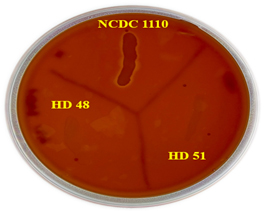
Figure 1. Hemolytic activity of probiotic Lactiplantibacillus plantarum HD48 and HD51: NCDC 1110: Staphylococcus aureus NCDC 1110; HD48: Lactiplantibacillus plantarum HD48; HD51: Lactiplantibacillus plantarum HD51
Mucin degradation assay
The mucin degradation activity of Lpb. plantarum HD48 and HD51 were done by petri plate method and liquid medium. Using a different substrate (1% mucin and glucose) as a carbon source, the mucolytic activity of test strains was measured on petri dishes dyed with and without amido black. Lpb. plantarum HD48 and HD51 showed no mucolytic zone, whereas faecal flora (positive control) created a distinct lysis zone surrounding the colony in every medium (Figure 2). Furthermore, on medium containing mucin and glucose, the autoclaved fecal (negative control) microbiota sample was unable to create a lysis zone surrounding the inoculation area.
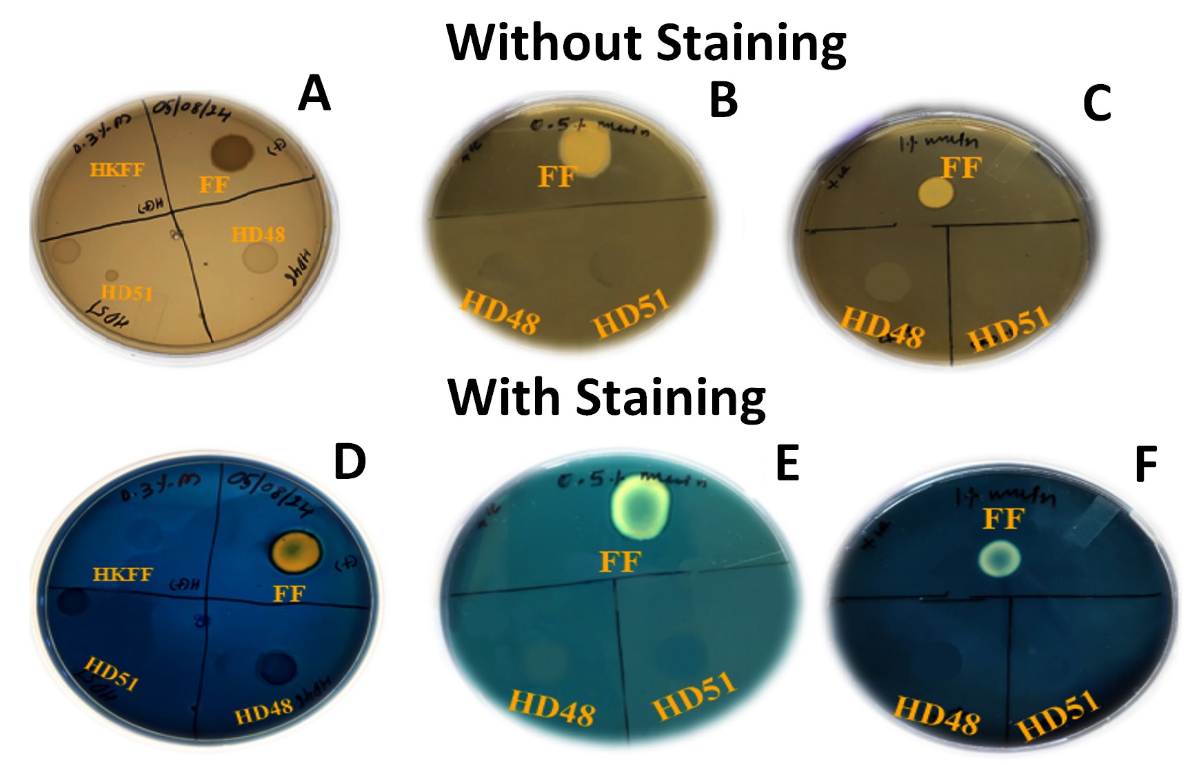
Figure 2. Mucin degradation activity of probiotic Lactiplantibacillus plantarum HD48 and HD51: Positive control (Fecal flora); HD48: Lactiplantibacillus plantarum HD48; HD51: Lactiplantibacillus plantarum HD51
Furthermore, the growth of Lpb. plantarum HD48 and HD51 were evaluated in liquid medium (without a carbon source), as well as in liquid medium supplemented with mucin and glucose (0.5% and 1%), and absorbance were recorded at 600 nm. The results showed that Lpb. plantarum HD48 and HD51 growth were significantly enhanced (P < 0.05) in medium containing 0.5% and 1% glucose medium (Figure 3). On the other hand, very little growth was seen in media with 0.5% and 1% mucin. The feces microbiota has a greater potential to break down mucin than the probiotic strain, as seen by the faecal flora (positive control) flourishing in the liquid medium in both carbon sources (mucin and glucose). Overall, our findings suggest that Lpb. plantarum HD48 and HD51 may not use mucin as the only carbon source because they are not mucolytic and do not have the usual enzyme needed for mucin destruction.
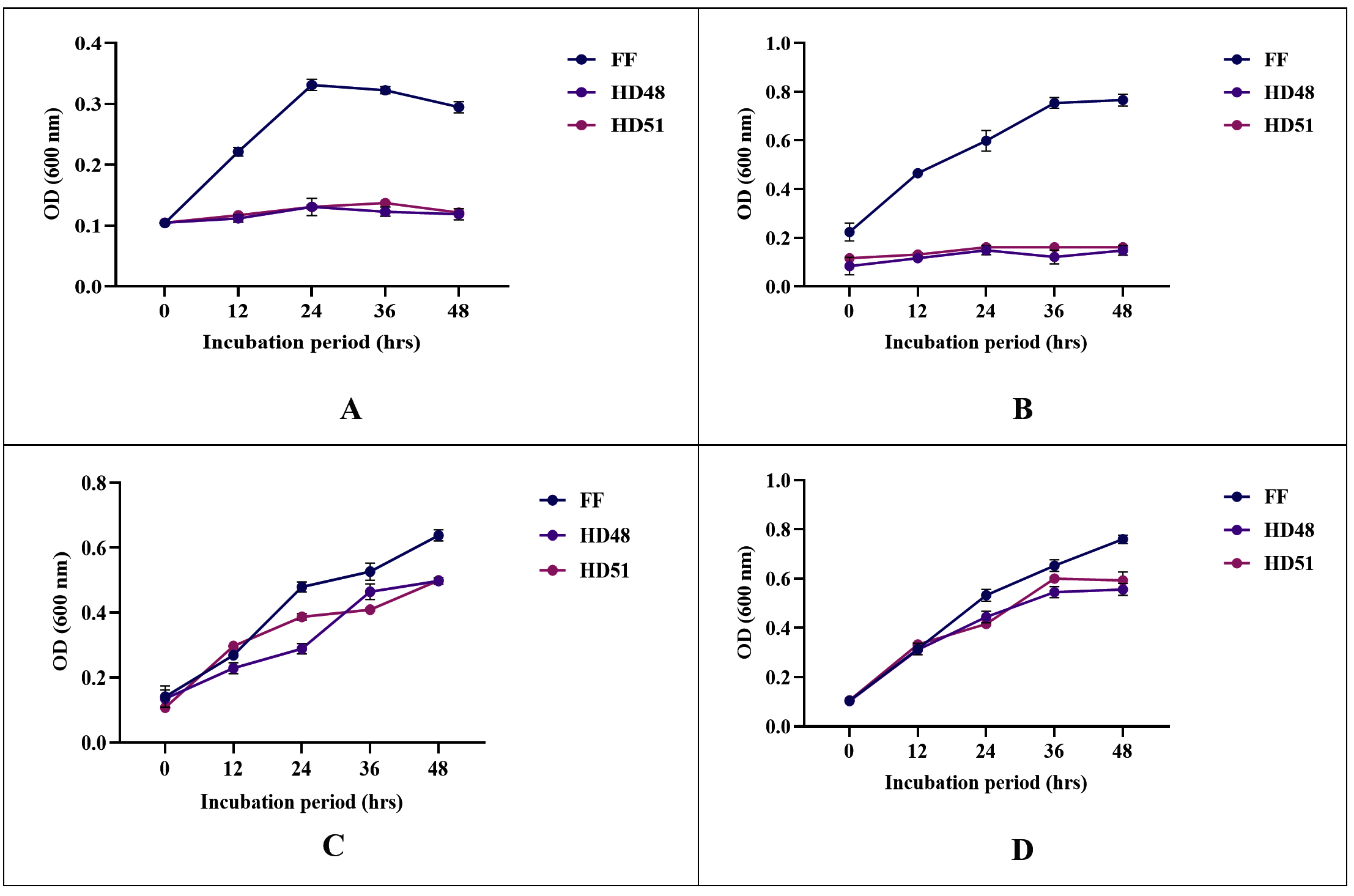
Figure 3. Growth patterns of probiotic Lactiplantibacillus plantarum HD48 and HD51 in culture medium containing glucose and mucin as a carbon source. (A) 0.5% mucin; (B) 1% mucin; (C) 0.5% mucin + 0.5% glucose; (D) 1% mucin + 1% glucose. Positive control FF: Fecal flora; HD48: Lactiplantibacillus plantarum HD48; HD51: Lactiplantibacillus plantarum HD51
Biogenic amines production test
Lpb. plantarum HD48 and HD51 were tested at various phases for their capacity to decarboxylate amino acids, such as tyrosine, lysine, histidine, and arginine, to form biogenic amines, such as tyramine, cadaverine, histamine, and putrescine. Following a 72 hrs incubation period in the decarboxylase liquid media, the growth of the corresponding test stains, Lpb. plantarum HD48 and HD51, was comparable to that of the standard control medium. No colour change was observed in the medium, indicating that growth of Lpb. plantarum HD48 and HD51 did not cause an increase in pH (Figure 4), while the medium containing Salmonella ATCC 35640 (positive control) colour change from yellow to purple with a significant difference P < 0.05 when comparing with Lpb. plantarum HD48 and HD51 strains. Therefore, it was determined that both probiotic strains tested negative for every precursor of biogenic amino acids.
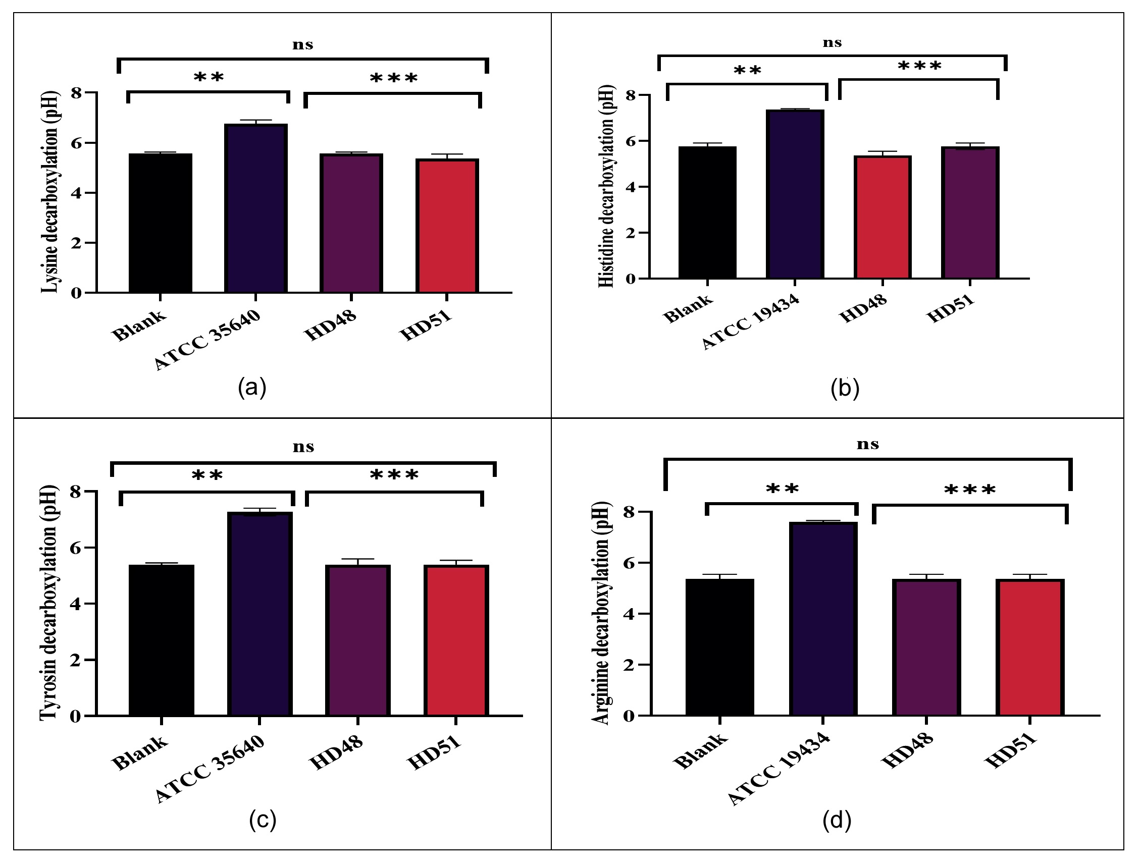
Figure 4. Biogenic amines production by qualitative method w.r.t the change in colour and pH of the decarboxylase broth media. (a). (A) Blank (MRS); (B) Salmonella ATCC 35640; (C) Lpb. plantarum HD48; (D) Lpb. plantarum HD51: (b). (A) Blank (MRS); (B) E. faecalis ATCC 19434; (C) Lpb. plantarum HD48; (D) Lpb. plantarum HD51. (c). (A) Blank (MRS); (B) Salmonella ATCC 35640; (C) Lpb. plantarum HD48; (D) Lpb. plantarum HD51. (d). (A) Blank (MRS); (B) E. faecalis ATCC 19434; (C) Lpb. plantarum HD48; (D) Lpb. plantarum HD51. Significant difference was considered at P < 0.05
Evaluation of in vitro cytotoxicity assay of Lpb. plantarum HD48 and HD51 on eukaryotic cell line
When exposed to MTT (3-(4,5-dimethylthiazol-2-yl)-2,5-diphenyltetrazolium bromide) dye from the lowest (107 CFU/mL) to highest (1011 CFU/mL) does after 24 hr, no significant difference were observed in cell viability of Caco-2 cells 9 (Figure 5) in comparison with control (untreated cells). Caco-2 cells maintained nearly about 95%-99% viability after exposure to Lpb. plantarum HD48 and HD51 even at the higher dose (1011 CFU/mL). Our results, showed that HD48 and HD51 strains caused no significant changes (P > 0.05) in the cell viability of Caco-2 cells, which are assessed by the MTT assay.
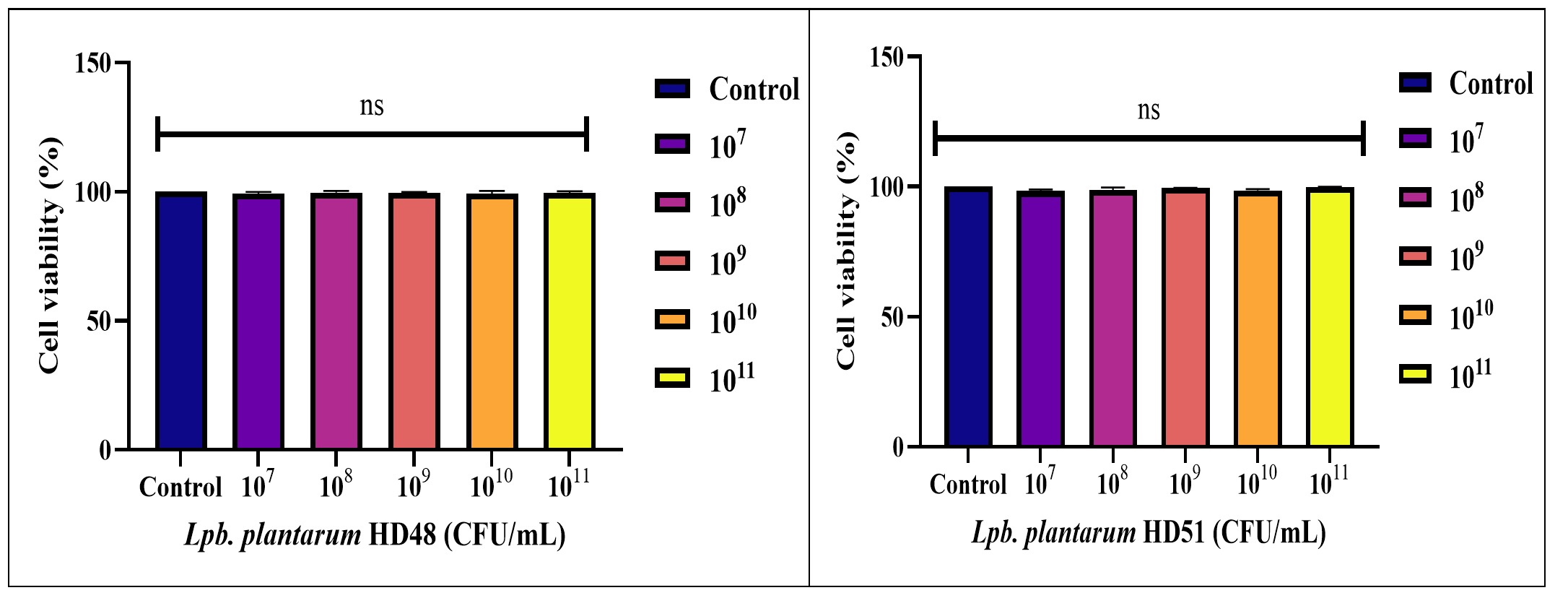
Figure 5. Determination of cell cytotoxicity of probiotic Lactiplantibacillus plantarum HD48 and HD51 Values are expressed as mean ± SD (n = 3) from three independent experiments. The values with superscripts (ns) represents no significant (P > 0.05) differences (Analysed by one way ANOVA using Dunntett test)
Antibiotic resistance profiling of Lactiplantibacillus plantarum HD48 and HD51 antibiotic susceptibility test
In this study, two probiotic Lactobacillus strains Lpb. plantarum HD48 and HD51 were assessed for their antibiotic susceptibility using the disk diffusion method with ICOSA discs (HiMedia, India), consisting of ICOSA I and ICOSA II, each containing set of 20 antibiotic discs arranged in a circular ring with a diameter of the 6 mm. Lpb. plantarum HD48 and HD51 were found susceptible toward all the tested antibiotics. However, strain showed resistance to vancomycin and nalidixic acid (Figure 6). Additionally, Lpb. plantarum HD48 showed intermediate zone of inhibition to Norfloxacin (17.4 ± 0.1 mm) and Chloramphenicol (18.3 ± 0.05 mm). Lpb. plantarum HD51 showed intermediate zone of inhibition to vancomycin (18.0 ± 0.05 mm) and Nalidixic acid (16.5 ± 0.5 mm).
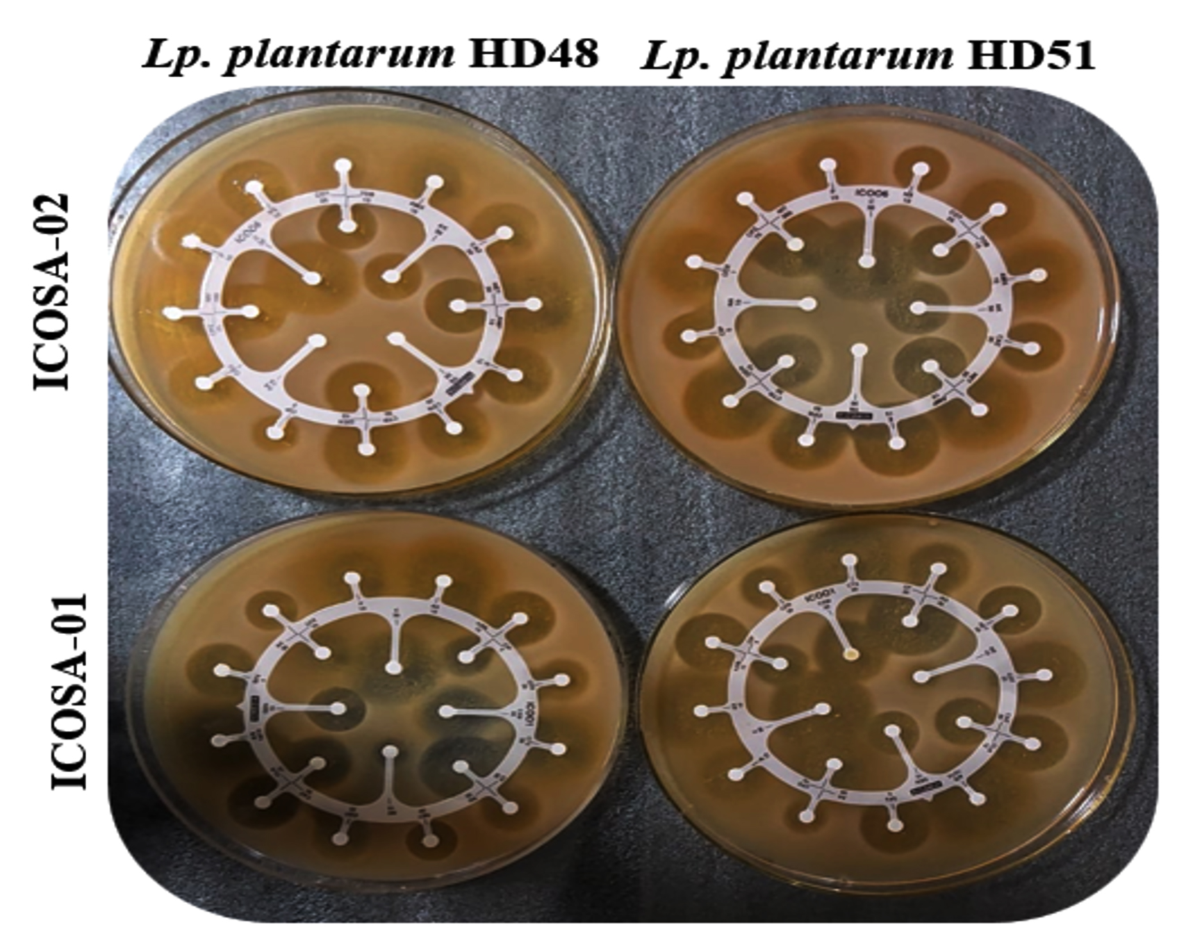
Figure 6. Antibiotic susceptibility profile of probiotic Lactiplantibacillus plantarum HD48 and HD51 using ICOSA discs (01 and 02)
PCR-based identification of antibiotic resistance and virulence genes
A total of 12 antibiotic resistance genes (ARGs) were assessed, confirming resistance to various antibiotics. None of these genes were detected in Lpb. plantarum HD48 and HD51 (Figure 7). Similarly, detection of virulence gene that encode coagulase (Coa), nuclease (nucA), gelatinase (gelE) and cytolysin (cylA) was checked in Lpb. plantarum HD48 and HD51, and no virulence genes were detected (Figure 8). However, different Enterococci strains DNA used as a positive control showed strong bands for the targeted potential antimicrobial and virulence factor genes. This indicates that the culture can be used safely without significant concerns about antimicrobial resistance (AMR). It should be noted that this PCR-based screening was limited to a defined panel of target genes; the absence of amplification for these specific primers does not rule out resistance mediated by undetected or novel genetic determinants, and whole-genome sequencing would provide a more comprehensive assessment of antimicrobial resistance and virulence potential.
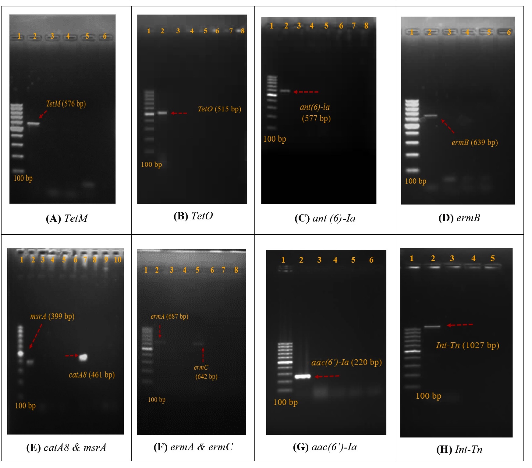
Figure 7. Gel images of antibiotic resistance gene. The description is as Lane 1. Ladder (100bp) in all images; Lane 2. Positive isolates of gel (A) 2. TetM (B1(F) Enterococci); (B) 2. TetO (B1(F) Enterococci); (C) 2. ant (6’)-Ia (B1(F) Enterococci); (D) ermB (Km (H) Enterococci); (E) 2. msrA & 7. CatA8 (B1(F) Enterococci); (F) 2. ermA (KnX(D) Enterococci); 5. ermC (Km(H) Enterococci) (G) aac(6’)-le-aph(2″)-Ia) (B1(F) Enterococci) and (H) Int-Tn (B1(F) Enterococci) Lane 3. Negative control; Lane 4. Lpb. plantarum HD48 and Lane 5. Lpb. plantarum HD51
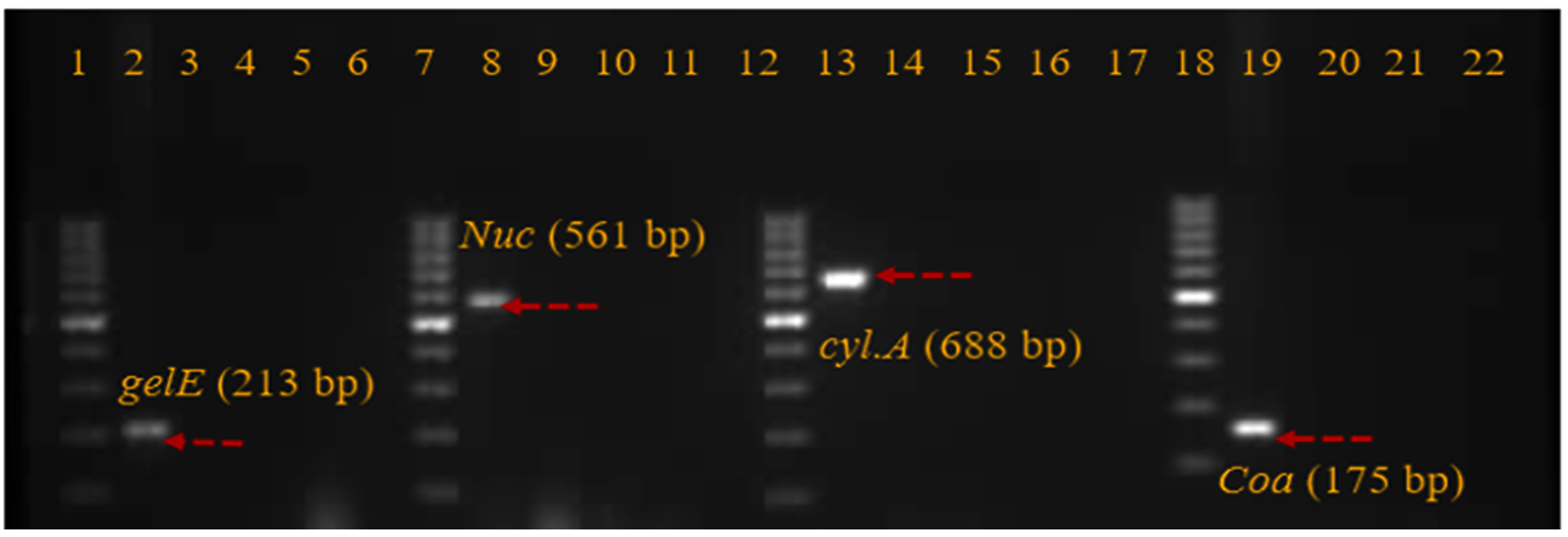
Figure 8. Gel image of virulence genes showing absence in Lpb. plantarum HD48 and HD51. The description of lane is here: 1. Ladder (100 bp); 2. Gelatinase (G1 (C) Enterococci); 3. negative control; 4. HD48; 5. HD51; 6. blank; 7. Ladder (100 bp); 8. Nuclease (S. aureus); 9. negative control; 10. HD48; 11. HD51; 12. Ladder (100 bp); 14. Cytolysin (Ar (D) Enterococci); 15. negative control; 16. HD48; 17. HD51; 18. Ladder (100 bp); 19. Coagulase (S. aureus); 20. negative control; 21. HD48 and 22. HD51
Lactiplantibacillus plantarum strains HD48 and HD51 met all essential in vitro safety standards for probiotic candidates, demonstrating no hemolysis, biogenic amine production, mucin degradation, cytotoxicity, transferable antibiotic resistance genes, or classical virulence factors. The present study was conducted to evaluate the safety of Lpb. plantarum HD48 and HD51 strains using an in vitro approach. This comprehensive safety profile aligns with prevailing regulatory and scientific guidelines for human probiotics.
Hemolysin, an immunogenic toxin, inflicts damage upon entering the bloodstream by lysing red blood cells, potentially causing anemia and edema in host.16 Lactobacillus species naturally colonize the guts of humans, animals and are extensively incorporated into functional foods for their probiotic benefits. Yet, under specific circumstances, they can act as opportunistic pathogens, posing risks to consumer health and food safety. Consequently, evaluating hemolytic properties remains a vital safety parameter for prospective probiotics. In this investigation, Lpb. plantarum strains HD48 and HD51 displayed no visible hemolysis on the blood agar plates, confirming their lack of hemolytic activity. By comparison, Staphylococcus aureus NCDC 110 produced distinct hemolytic zones, evidencing active hemolytic enzymes. These results underscore the safety profile of the Lpb. plantarum strains relative to known pathogens, aligning with regulatory expectations for non-hemolytic probiotics.
Mucin degradation within the gut can facilitate the translocation of both commensal and pathogenic microorganisms across the mucosal barrier and into the bloodstream. Because probiotic preparations are usually delivered to mucin-secreting sites in the gastrointestinal tract, therefore critical that candidate probiotic strains do not erode or disrupt this protective mucin layer, which serves as a key defence against opportunistic gut pathogens.19 In the current study, we evaluated the mucin-degrading potential of our isolates using hog gastric mucin as the substrate. Hog gastric mucin was selected because its chemical composition and structural organization closely resemble those of human gastric mucin, making it a widely accepted surrogate for in vitro assessments. The probiotic strains were incubated with this mucin preparation under controlled conditions, and their mucinolytic activity was systematically monitored. Our findings clearly showed that the tested strains lacked detectable mucinolytic activity. In contrast, control experiments using normal human faecal microbiota confirmed the presence of mucin-degrading activity, consistent with the known capabilities of the resident gut community. The absence of mucin degradation by our strains indicates that they are unlikely to compromise the integrity of the mucosal barrier following administration. These results are in line with observations of Budzinski et al.,3 who similarly reported that probiotic Lactobacillus strains are non mucolytic. Collectively, these data support the mucosal safety of the evaluated probiotic candidates and reinforce their suitability for use in mucin-rich regions of the gastrointestinal tract. Mucin degradation in the gut allows both commensal and pathogenic microbes to breach the mucosal barrier and access the bloodstream. Given that probiotics are often delivered to mucin-secreting tissues, it is critical that these strains lack mucinolytic activity. The mucin layer serves as a vital defence against opportunistic gut pathogens.19 In this study, we employed hog gastric mucin as the substrate due to its close chemical and structural resemblance to human gastric mucin. Our assays revealed no mucinolytic activity in the tested strains, in stark contrast to normal human fecal microbiota, which exhibited clear mucin degradation. These results align with findings that probiotic Lactobacillus strains are inherently non-mucolytic.20 This safety profile underscores the strains’ suitability for human applications, as they preserve mucosal integrity without compromising the gut’s protective barrier.
In vitro cytotoxicity assays, such as the MTT assay, are routinely employed to evaluate the potential toxicity of drugs, chemicals, and microbial strains in human or animal cell culture models.21 Using this approach, we investigated the effect of Lpb. plantarum HD48 and HD51 strains on the viability of Caco-2 intestinal epithelial cells. Cells were exposed to a wide range of bacterial concentrations, from 107 to 1011 CFU/mL, for 24 hrs. Across all tested doses, neither strain induced a significant reduction in cell viability, indicating an absence of measurable cytotoxic effects and supporting their safety for interaction with intestinal cells. Our observations are consistent with earlier reports17 demonstrated that L. rhamnosus MTCC5897 did not exert cytotoxic effects on Caco-2 cells when evaluated over a similar range of concentrations (106-1010 CFU/mL) after 24 hrs of incubation. Likewise,22 reported that exposure of Caco-2 cells to L. plantarum CRD7 and L. rhamnosus CRD11 strains preserved approximately 99% cell viability, with no statistically significant alterations observed (P > 0.05) even following 24 hrs of treatment.
Taken together, these findings reinforce the conclusion that Lpb. plantarum HD48 and HD51 exhibit a cytotoxicity profile comparable to previously characterized probiotic strains and can be considered safe for further development as intestinal probiotic candidates.
An additional safety consideration for lactic acid bacteria (LAB) is their capacity to synthesize biogenic amines (BAs), particularly in protein-rich food matrices. Accumulation of BAs in foods is undesirable because these compounds are implicated in a range of adverse health effects, including migraines, headaches, gastric and intestinal ulcers, and various allergic reactions.23 Previous studies by Deepika et al.24 and Fugaban et al.25 have shown that some LAB strains, especially species within the genera Lactobacillus and Enterococcus, can express amino acid decarboxylase enzymes. These enzymes convert free amino acids into BAs during fermentation and food processing, thereby potentially compromising the safety of fermented products. In light of these concerns, we evaluated Lpb. plantarum HD48 and HD51 for their ability to produce BAs as part of a comprehensive probiotic safety assessment. Specifically, we screened for the four major BAs of toxicological relevance: putrescine, cadaverine, histamine, and tyramine. Both strains tested negative for all targeted BAs, indicating the absence of detectable decarboxylase activity under the conditions employed. Our results are consistent with earlier findings in the literature Fan et al.26 reported that L. brevis CGMCC1.5954 did not produce detectable levels of BAs, while Ku et al.27 confirmed, using HPLC analysis, that Bifidobacterium animalis subsp. lactis AD011 was similarly unable to synthesize BAs. Collectively, these data support the conclusion that Lpb. plantarum HD48 and HD51 are unlikely to contribute to BA accumulation and are therefore suitable from a biogenic-amine safety perspective.
Food and food-production environments can serve as important reservoirs and transmission routes for antibiotic-resistant bacteria and antibiotic resistance genes (ARGs). When such bacteria are ingested, they may colonize the human gastrointestinal tract and subsequently transfer ARGs to members of the resident microbiota or to opportunistic and obligate pathogens. This process can contribute to the spread of resistance within the human population and may ultimately lead to treatment failures and other serious public health consequences. Lactic acid bacteria (LAB), which are widely used as starter cultures and probiotics, are of particular interest in this context because they can persist and sometimes establish themselves within the host gut. As a result, LAB have the potential to act as vehicles for both antibiotic-resistant organisms and mobile ARGs.
Several studies have documented horizontal transfer of ARGs between foodborne LAB and pathogenic or opportunistic microorganisms in the intestinal environment.28-30 Conjugative plasmids, transposons, and other mobile genetic elements have been implicated in mediating the dissemination of resistance traits across species and even across genera. Consequently, regulatory authorities and scientific bodies now emphasize that candidate probiotic strains should be thoroughly characterized for their antibiotic susceptibility profiles and screened for the presence of transferable ARGs. Demonstrating the absence of acquired or mobile resistance determinants is therefore a key component of probiotic safety evaluation.
In the present study, Lpb. plantarum HD48 and HD51 were subjected to phenotypic antibiotic susceptibility testing against a panel of clinically relevant antibiotics. Both strains were susceptible to all antibiotics of concern, with the exception of vancomycin and nalidixic acid, to which they displayed resistance. The observed vancomycin resistance is consistent with intrinsic resistance commonly reported in Lactobacilli. Mechanistically, vancomycin exerts its antimicrobial effect by binding to the D-alanyl-D-alanine (D-Ala-D-Ala) terminus of peptidoglycan precursors, thereby blocking cell wall synthesis. In certain lactobacilli, the enzyme VanX (a D, D-dipeptidase) contributes to resistance by generating modified peptidoglycan precursors that terminate in D-Ala-D-lactate instead of the canonical D-Ala-D-Ala. This alteration reduces the affinity of vancomycin for its target, effectively bypassing the drug’s mode of action. Importantly, this type of resistance is regarded as intrinsic, chromosomally encoded, and typically non-inducible and non-transferable in Lactobacilli.31 Thus, while Lpb. plantarum HD48 and HD51 exhibit vancomycin resistance, current evidence suggests that this trait does not pose a significant risk of horizontal gene transfer, supporting their overall safety with respect to antibiotic resistance.
It is acknowledged that antibiotic susceptibility was assessed phenotypically by the disk diffusion method, which, while widely used, is not the method of choice recommended by EFSA for probiotic safety evaluation; broth microdilution-based determination of minimum inhibitory concentrations (MICs) against the EFSA cut-off values would provide a more rigorous and internationally comparable assessment of antibiotic resistance in these strains and is recommended for future characterisation work.
Targeted PCR analysis was conducted to confirm the presence or absence of the specific antibiotic resistance genes (ARGs) in tested strains. This molecular approach is important because bacteria may harbor ARGs without displaying a resistant phenotype, for example due to gene silencing, low expression levels, or regulatory mechanisms that prevent active gene expression. In total, 12 ARGs commonly reported in LAB were screened, including determinants for tetracycline resistance (TetO, TetM), aminoglycoside-modifying enzymes (ant(6′)-Ia, aac(6′)-Ie–aph(2”)-Ia), erythromycin resistance (ermA, ermB, ermC, msrA), chloramphenicol resistance (CatA8), beta-lactam resistance (blaTEM), multidrug efflux (emeA) and the integrase gene Int-Tn (Tn916/Tn1545). None of these ARGs were detected in Lpb. plantarum HD48 or HD51.
In parallel, we examined the strains for the presence of key virulence genes typically associated with pathogenicity, including those encoding coagulase, nuclease, cytolysin, and gelatinase. PCR amplification revealed that all of these virulence markers were absent in both Lpb. plantarum HD48 and HD51. The lack of detectable ARGs and virulence determinants indicates that these cultures can be considered safe, with minimal risk of contributing to antimicrobial resistance dissemination or expressing pathogenic traits. These observations are consistent with the findings of Nataraj et al.,18 who similarly reported the absence of antibiotic resistance and virulence genes in the probiotic strain Limosilactobacillus fermentum NCDC 400, further supporting the safety of well-characterized probiotic LAB.
This in vitro safety assessment indicates that Lpb. plantarum HD48 and HD51 did not produce biogenic amines, were non-haemolytic, and showed no mucin-degrading activity. Both strains were susceptible to the majority of clinically relevant antibiotics, with intrinsic resistance to vancomycin and nalidixic acid, and did not carry the targeted antibiotic resistance or virulence genes screened by PCR. The strains also showed no measurable cytotoxic effect on Caco-2 cells in the MTT assay. Collectively, these preliminary in vitro findings suggest that Lpb. plantarum HD48 and HD51 are promising and apparently safe candidates for further probiotic development; however, in vivo studies in relevant animal models, together with whole-genome sequencing and MIC-based antibiotic susceptibility testing, are warranted before any claims regarding their efficacy or use as therapeutic agents or antibiotic alternatives can be supported.
ACKNOWLEDGMENTS
The authors thank the Director, ICAR-National Dairy Research Institute (Deemed University), Karnal-132001, Haryana, India, for financial and logistical support.
CONFLICT OF INTEREST
The authors declare that there is no conflict of interest.
AUTHORS’ CONTRIBUTION
All authors listed have made a substantial, direct and intellectual contribution to the work, and approved it for publication.
FUNDING
None.
DATA AVAILABILITY
The datasets generated and analysed during the current study are available from the corresponding author on reasonable request.
ETHICS STATEMENT
Not Applicable.
- Lau LYJ, Quek SY. Probiotics: health benefits, food application, and colonization in the human gastrointestinal tract. Food Bioeng. 2024;3(1):41-64.
Crossref - Pessione E. Lactic acid bacteria contribution to gut microbiota complexity: lights and shadows. Front Cell Infect Microbiol. 2012;2:86.
Crossref - Budzinski L, Sempert T, Lietz L, et al. Age-stratification reveals age-specific intestinal microbiota signatures in juvenile idiopathic arthritis. Mol Cell Pediatr. 2024;11(1):12.
Crossref - Hill C, Guarner F, Reid G, et al. The International Scientific Association for Probiotics and Prebiotics consensus statement on the scope and appropriate use of the term probiotic. Nat Rev Gastroenterol Hepatol. 2014;11:506-514.
Crossref - Aljohani A, Rashwan N, Vasani S, et al. The health benefits of probiotic Lactiplantibacillus plantarum: a systematic review and meta-analysis. Probiotics Antimicrob Proteins. 2025;17(5):3358-3377.
Crossref - Luo J, Liang S, Jin F. Gut microbiota and healthy longevity. Sci China Life Sci. 2024;67(12):2590-2602.
Crossref - Ouwehand AC, Salminen S, Isolauri E. Probiotics: an overview of beneficial effects. Antonie Van Leeuwenhoek. 2002;82(1):279-289.
- Binda S, Hill C, Johansen E, et al. Criteria to qualify microorganisms as “probiotic” in foods and dietary supplements. Front Microbiol. 2020;11:1662.
Crossref - Sanders ME, Merenstein DJ, Reid G, Gibson GR, Rastall RA. Probiotics and prebiotics in intestinal health and disease: from biology to the clinic. Nat Rev Gastroenterol Hepatol. 2019;16(10):605-616.
Crossref - West NP, Hughes L, Ramsey R, et al. Probiotics, anticipation stress, and the acute immune response to night shift. Front Immunol. 2021;11:599547.
Crossref - Liu X, Mao B, Tang X, et al. Bacterial viability retention in probiotic foods: a review. Crit Rev Food Sci Nutr. 2025;65(32):7964-7986.
Crossref - Roe AL, Boyte ME, Elkins CA, et al. Considerations for determining safety of probiotics: a USP perspective. Regul Toxicol Pharmacol. 2022;136:105266.
Crossref - Singh H, Verma S, Jaswal A, Rani S, Ram C. In-vitro evaluation of indigenous probiotic lactobacilli for lead bio-adsorption potential, its tolerance and complex stability. J Funct Foods. 2022;95:105175.
Crossref - Zhang Z, Niu H, Qu Q, et al. Advancements in Lactiplantibacillus plantarum: probiotic characteristics, gene editing technologies and applications. Crit Rev Food Sci Nutr. 2025;65(29):6623-6644.
Crossref - Singracha P, Niamsiri N, Visessanguan W, Lertsiri S, Assavanig A. Application of lactic acid bacteria and yeasts as starter cultures for reduced-salt soy sauce (moromi) fermentation. LWT. 2017;78:181-188.
Crossref - Kim MJ, Ku S, Kim SY, et al. Safety evaluations of Bifidobacterium bifidum BGN4 and Bifidobacterium longum BORI. Int J Mol Sci. 2018;19(5):1422.
Crossref - Bhat MI, Singh VK, Sharma D, Kapila S, Kapila R. Adherence capability and safety assessment of an indigenous probiotic strain Lactobacillus rhamnosus MTCC-5897. Microb Pathog. 2019;130:120-130.
Crossref - Nataraj BH, Kumari M, Nagpal R, Ali SA, Behare PV. Safety evaluation of indigenous probiotic Limosilactobacillus fermentum NCDC 400 using whole genome sequences and in vitro approaches. Food Biosci. 2023;56:103101.
Crossref - Byakika S, Mukisa IM, Byaruhanga YB, Muyanja C. A review of criteria and methods for evaluating the probiotic potential of microorganisms. Food Rev Int. 2019;35(5):427-466.
Crossref - Rastogi S, Mittal V, Singh A. In vitro evaluation of probiotic potential and safety assessment of Lactobacillus mucosae strains isolated from donkey’s lactation. Probiotics Antimicrob Prot. 2020;12(3):1045-1056.
Crossref - Eisenbrand G, Pool-Zobel B, Baker V, et al. Methods of in vitro toxicology. Food Chem Toxicol. 2002;40(2-3):193-236.
Crossref - Varada VV, Panneerselvam D, Pushpadass HA, Mallapa RH, Ram C, Kumar S. In vitro safety assessment of electrohydrodynamically encapsulated Lactiplantibacillus plantarum CRD7 and Lacticaseibacillus rhamnosus CRD11 for probiotics use. Curr Res Food Sci. 2023;6:100507.
Crossref eCollection 2023 - Tofalo R, Perpetuini G, Schirone M, Suzzi G. Biogenic amines: toxicology and health effect. Encycl Food Health. 2016;1:424-429.
Crossref - Deepika Priyadarshani WM, Rakshit SK. Screening selected strains of probiotic lactic acid bacteria for their ability to produce biogenic amines (histamine and tyramine). Int J Food Sci Technol. 2011;46(10):2062-2069.
Crossref - Fugaban JII, Holzapfel WH, Todorov SD. Probiotic potential and safety assessment of bacteriocinogenic Enterococcus faecium strains with antibacterial activity against Listeria and vancomycin-resistant enterococci. Curr Res Microb Sci. 2021;2:100070.
Crossref - Fan X, Jiang X, Guo Y, et al. In vitro and in vivo evaluation of the safety of Levilactobacillus brevis CGMCC1.5954 with probiotic potential based on tri-generation whole genome sequencing and animal studies. Food Biosci. 2023;53:102654.
Crossref - Ku S, Yang S, Lee HH, et al. Biosafety assessment of Bifidobacterium animalis subsp. lactis AD011 used for human consumption as a probiotic microorganism. Food Control. 2020;117:106985.
Crossref - Feld L, Bielak E, Hammer K, Wilcks A. Characterization of a small erythromycin resistance plasmid pLFE1 from the food-isolate Lactobacillus plantarum M345. Plasmid. 2009;61(3):159-170.
Crossref - Zarzecka U, Chajecka-Wierzchowska W, Zadernowska A. Microorganisms from starter and protective cultures-occurrence of antibiotic resistance and conjugal transfer of tet genes in vitro and during food fermentation. LWT. 2022;153:112490.
Crossref - Nawaz Z, Zahoor MK, Shafique M, Athar R, Yasmin A, Zahoor MA. In vitro assessment of probiotic properties of lactic acid bacteria isolated from camel milk: enhancing sustainable foods. Front Sustain Food Syst. 2024;8:1437201.
Crossref - Anisimova E, Gorokhova I, Karimullina G, Yarullina D. Alarming antibiotic resistance of lactobacilli isolated from probiotic preparations and dietary supplements. Antibiotics. 2022;11(11):1557.
Crossref
© The Author(s) 2026. Open Access. This article is distributed under the terms of the Creative Commons Attribution 4.0 International License which permits unrestricted use, sharing, distribution, and reproduction in any medium, provided you give appropriate credit to the original author(s) and the source, provide a link to the Creative Commons license, and indicate if changes were made.