


ISSN: 0973-7510
E-ISSN: 2581-690X
Dried jujube is a characteristic fruit of Xinjiang. Aspergillus is one of the main pathogens that causes mold on dried jujube, and A. flavus is a toxin-producing species, the aflatoxin produced by A. flavus is extremely toxic and carcinogenic. In this study, the growth kinetic models of A. flavus isolated from red jujube at different temperatures and times were fitted to Huang model and linear equation respectively, the Cardinal model was used to describe the growth rate and lag time of A. flavus on dried jujube agar. It turned out that 30–35 °C was the optimal temperature for growing A. flavus, so dried jujube should avoid storing in this temperature range. The kinetic model established in this study will help to understand the growth characteristics of A. flavus, and lay a foundation for evaluating the quality of stored dried jujube and predictions of shelf life, which are conducive to optimizing storage methods for dried jujube. It can be judged according to the value of Af and Bf, the Huang model had a better fitting effect than the Baranyi model, The two models all had the highest growth rate at 35 °C, and A. flavus grew more vigorously and the lag period shortened as the temperature was increased. The secondary Cardinal model had a good fitting effect on the growth rate and lag time, and the secondary Ratkowsky model had a good fitting effect on the growth rate. This study may have theoretical and application value to strengthen the safety of jujube storage in the future.
Primary growth model, secondary model, growth rate, lag time
Dried jujube is a characteristic fruit of Xinjiang, China, as Xinjiang’s climate is suitable for growing jujube. Jujube is sweet and delicious, which can be eaten fresh or dried, and it is also valuable.1 The planting area for Xinjiang jujube has increased rapidly in recent years, and it has become the fastest growing and most profitable industry in Xinjiang.2-4 As Xinjiang jujube is nutritional rich, it is extremely vulnerable to contamination by harmful microorganisms, which causes various problems, such as an imbalance between sweet and sour, reduced nutritional value, and plant diseases during storage. During storage of jujube, mold on the surface of the jujube secretes pectinase and cellulase that break down cellulose, pectin, and other substances in the cell wall of the jujube into simple small molecules, and gradually invades the inside pulp. As a result, the protein, pectin, starch, organic acids and sugars in the jujube are destroyed and the jujube fruit rots.5,6
Aspergillus is one of the most important pathogens that causes mold of dried jujube. Several studies have isolated and identified mold strains on dried jujube in China. Chen and Shanawaer7-10 reported that Aspergillus is the main fungal species on dried jujube, and Shanawaer et al10 isolated Aspergillus flavus. A. flavus is a kind of toxin-producing fungi. Some strains of A. flavus produce aflatoxin. Aflatoxin is a metabolite of A. flavus and Aspergillus parasiticus, which not only poisons and kills livestock and poultry, but also causes carcinogenesis. Aflatoxin is a carcinogen. Aflatoxin has a damaging effect on liver tissues of humans and other animals. In severe cases, aflatoxin causes liver cancer and death. Therefore, it is of great significance to clarify the growth characteristics of A. flavus on dried jujube.
Research on dried jujube has mainly focused on physiological and biochemical aspects, storage conditions, and technical measures after harvest.11,12 However, a predictive model of A. flavus has not been reported for dried jujube. Several mold models have been proposed. Peromingo et al13 studied the effects of temperature and water activity on aflatoxin and built a growth model. Somjaipeng and Ta-Uea14 studied the effects of temperature and water activity on the growth rate of Aspergillus in stored rice, and built a growth model. Marín et al15 used the modified Gompertz model to assess the effects of temperature and water activity on the lag phase and generation time of some Aspergillus in corn extract medium. Sautour et al16 used potato dextrose agar (PDA) plates to study the relationship between water activity and growth of several molds to build a model, and evaluated minimum water activity, optimal water activity, and maximum water activity. Pitt17 proposed a model of the effects of environmental conditions on mold growth and toxin production by collating some literature data, and provided a theoretical explanation for the temperature change during toxin production. Yue et al18 constructed a model of the effects of major ecological factors on the growth of A. flavus in stored corn. This model predicts the effects of temperature and water activity on the specific growth rate and lag time of A. flavus, and the equation obtained can be used to predict the growth of A. flavus in stored corn.
A microbial predictive model is used to predict changes in microorganisms in different environments and to prevent food safety accidents. In this study, the mold fungus A. flavus was isolated from dried jujube and purified. The growth of A. flavus in dried jujube was analyzed at different temperatures. Predictive microbiological methods were used for fitting primary and secondary models and for estimating their parameters. The results will provide data to control the growth of A. flavus on dried jujube and provide an A. flavus risk assessment.
Jujube samples and growth medium
Twenty-eight Xinjiang dried jujube samples were purchased from a retail market in Shihezi and maintained in PDA and Rose Bengal agar (Qingdao Hi-Tech Park Haibo Biotechnology Co., Ltd., Qingdao, China).
Using laboratory-made jujube medium. A 250 g portion of washed dried jujube was added to 500 mL of distilled water and boiled for 20 min. The mixture was placed in a juicer with the cores removed and mixed until evenly viscous. Distilled water was added to 1000 mL. A 20 g portion of agar was added and the mixture was dispensed into a triangular bottle while hot and sterilized at 121 °C for 20 min (refer to PDA medium)
Chen et al7. The water activity of the jujube medium was 0.823 as measured with a water activity meter.
Fungal activation, isolation and purification
The surface of 28 dried jujube dried fruit samples was cleaned with 70% alcohol cotton balls. The diseased and healthy pulp of the jujube was cut out on an ultra-clean worktable using a sterile blade and shredded. A 25 g portion of dried jujube pulp was randomly selected from the 16 samples, and was placed in a sterile bag containing 225 mL of sterile potato dextrose water and shaken for 30 min. The suspension was placed in an incubator for 24 h, until the fungi were activated7.
A 100 µL aliquot of the activated solution was added to Bengal Red medium and cooled to about 50 °C. A coating inoculation was applied with a coating rod. Each sample was repeated three times, and cultured in an incubator at 28°C for 5–7 d.
After the colony began to grow well on the plate, a small number of spores were selected on a “Z” type plate using the plate streak separation method9,19. A small amount of the fungal hyphae containing the sample was selected aseptically by regular streaks on the surface of Rose Bengal agar. The strains were purified and isolated several times, and finally a purified single colony was cultured at 28 °C.
Fungal preservation and preliminary identification
The pure colonies obtained after isolation and purification were stored under sterile conditions in single colony plate medium. A few spores were picked with an inoculating loop and inoculated on oblique PDA medium. The plates were cultured in an incubator at 28 °C for 2–3 d 13.
A small amount of mold mycelia was picked from the edge of the mold colony with a dissecting needle, and immersed in 50% ethanol for a few seconds to elute the spores. The mycelia were dispersed with a needle on a clean glass slide containing a drop of physiological saline (0.85% NaCl), covered with a cover glass, and observed under a microscope20.
rDNA-ITS Sequencing
DNA was extracted using liquid nitrogen21. The mycelia were ground in liquid nitrogen in a precooled mortar and then transferred to a new 1.5 mL centrifuge tube. The lysate was added before thawing the mycelial powder for DNA extraction. Genomic DNA was extracted with a fungal DNA extraction kit22.
By referring to the method of White et al23 to amplify the rDNA-ITS sequence. The universal primers ITS1 (5′-GTAGTCATATGCTTGTCTC-3′) and ITS4 (5′-TCCTCCGCTTATTGATATGC-3′) were used with genomic DNA as the template10.
The polymerase chain reaction (PCR) system was 25 μL of PCR Taq Mix, 2 μL of upstream primer (10 μmol/L), 2 μL of downstream primer (10 μmol/L), 1 μL of genomic DNA, and 20 μL of ddH2O to a final volume of 50 μL. The ITS gene PCR amplification program was pre-denaturation at 94°C for 4 min, denaturation at 94 °C for 30 s, annealing at 55 °C for 1 min, and extension at 72 °C for 1 min for a total of 30 cycles, and a final extension at 72 °C for 10 min.
The amplified products were electrophoresed on a 1.5% agarose gel in 0.5× TBE running buffer. A gel imaging system was used to detect and record the displayed image of the amplified products on the agarose gel. The amplified products were purified and sent to the detection department for sequencing10.
According to the microscopy and BLAST results, the mold species was identified as A. flavus.
Growth experiments
A. flavus was cultured on PDA medium in an incubator at 25 °C for 7 d. A mycelial suspension was prepared by submerging the surface of the plate medium in sterile 0.05% Tween-80 saline solution (0.9% NaCl) and scraping to remove the hyphae through a four-layered sterile gauze filter. The number of spores was counted with a blood cell counting plate, and the quantity of spores was adjusted to 107 spores/mL, and then to 103 spores/mL. Place a 4 mm groove in the center of the jujube medium with a pipette tip, shake well when taking samples, take 40 μL of the adjusted spore suspension, and point to the center of the plate24.
The jujube medium was placed in a constant temperature incubator at 20, 25, 28, 30, 35, and 37 °C for at least 120h, and diameters were measured every 12 h with a Vernier caliper24. Samples were run in three groups in parallel and data were recorded.
Primary growth model
Using the United States Department of Agriculture (USDA) IPMP 2013 software, three primary growth models, the Huang model and the Baranyi model, were used to fit the growth of A. flavus on dried jujube at different temperatures and to obtain the corresponding model fitting parameters.25
![]() …(1)
…(1)
 …(2)
…(2)
Where Y(t) is the diameter of A. flavus (mm) at time t; Y0 is the initial diameter of A. flavus (mm); Ymax is the maximum diameter (mm) reached by A. flavus; μmax is the maximum specific growth rate of A. flavus (mm/h); λ is the lag period (h); and α is the lag phase transition coefficient of 4.00.
Baranyi model26
D0, Dmax, D(t) are the bacterial population, in natural logarithm of microorganism counts, at initial, maximum, and time t. µmax is the specific growth rate. h0 is the physiological state of the microorganism under consideration. All four parameters, including Y0, Ymax, h0, and µmax, will be estimated using nonlinear regression.
![]() …(3)
…(3)
![]() …(4)
…(4)
Secondary model
Using USDA IPMP 2013 software, the secondary growth model was used to fit the growth of A. flavus on dried jujube at different temperatures.27
![]() …(5)
…(5)
![]() …(6)
…(6)
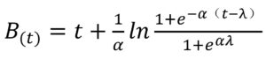
Where μmax is the maximum growth rate at each temperature T, μopt is the optimal growth rate at the optimal temperature (Topt), and Tmin and Tmax are the minimum growth temperature and the maximum growth temperature. The Cardinal model is only suitable for the full temperature range. In Eq. (5), λmax is the maximum lag time at each temperature T, and the others are the same as in Eq. (4).
Full temperature range Ratkowsky square-root model.28
![]() …(7)
…(7)
In this equation, µ s the growth rate (time-1); a and b are coefficients; T is temperature; Tmin is the nominal/notational minimum temperature; Tmax is the estimated maximum growth temperature.
Bf, Af, RMSE values of the models
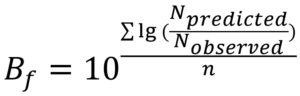
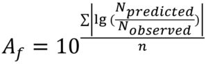
The bias factor (Bf) can be used to determine the structural deviation of a model; that is, the degree of underestimation or overestimation of the estimation. The accuracy factor (Af) can be used to determine the accuracy of the model, and the root mean square error (RMSE) is used to evaluate the change in the data error29.
The expressions are as follows:
 …(8)
…(8)
 …(9)
…(9)
 …(10)
…(10)
Where Npredicted and Nobserved are the predicted and observed growth diameters (mm) of A. flavus, respectively, and n is the number of samples.
Modeling primary growth of A. flavus
Fitting of the first growth model of A. flavus
The experimental data of A. flavus growth on jujube medium showed typical mould growth curves, with clear adaptation time and linear growth phases, absence of stationary growth phase (during the evaluated time), and temperature-dependent growth rates. For the higher temperatures (25,28, 30, 35 and 37 °C), the maximum diameter of the plate was reached by the A. flavus, whereas at 20 °C, no stationary phase was observed over the experimental time.
All the evaluated primary models were able to describe very well the growth of A. flavus on jujube medium. The growth rate (μmax) and lag time (λ) of A. flavus were determined from the primary growth models, as shown in Table 1. The Bf and Af values for the Huang and Baranyi models fitted to the experimental data at 20, 25, 28, 30, 35 and 37 °C are shown in Table 2. The typical mold growth characteristics have important reference significance. The two models all had the highest growth rate at 35°C. And A. flavus grew more vigorously and the lag period shortened as the temperature was increased.
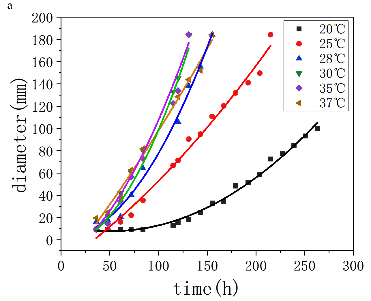

Fig. 1. Growth curve of A. flavus on jujube medium by fitting the model a, Huang model; b, Baranyi model
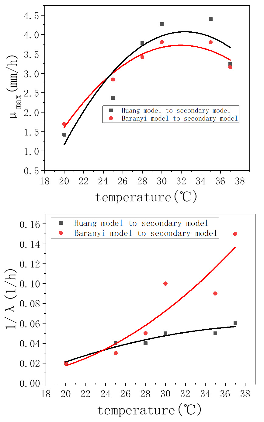
Fig. 2. Effect of temperature (T) of A. flavus on jujube medium on the growth rate (μmax) and the reciprocal lag time (1/λ) by fitting the secondary model fitted by the primary model.
Note: secondary: Cardinal model / Ratkowsky model
According to the analysis in Table 1, the primary Huang model for A. flavus on jujube medium had the best estimation effect, the Bf values of the Huang model were 0.941–0.993, and the Af value were 1-1.124. The fitting effect of the Baranyi model was not as good as that of the Huang model.
Table (1):
Values of parameters µmax and λ for the Huang and Baranyi models fitted to the growth data of A. flavus diameter on jujube at 20, 25, 28, 30, 35 and 37 °C.
| Parameter | Model | Temperature | |||||
|---|---|---|---|---|---|---|---|
| 20 | 25 | 28 | 30 | 35 | 37 | ||
| μmax | Huang | 0.6170 | 1.027 | 1.641 | 1.855 | 1.912 | 1.408 |
| Baranyi | 0.735 | 1.233 | 1.487 | 1.650 | 1.649 | 1.374 | |
| λ | Huang | 118.3 | 57.73 | 57.38 | 43.77 | 50.20 | 38.79 |
| Baranyi | 136.054 | 81.103 | 47.381 | 22.957 | 26.187 | 14.887 | |
Table (2):
Bf and Af values of the fitting of the Huang and Baranyi models to the growth data of A. flavus diameter on jujube at 20, 25, 28, 30, 35 and 37 °C.
| Statistical Index |
Model | Temperature | |||||
|---|---|---|---|---|---|---|---|
| 20 | 25 | 28 | 30 | 35 | 37 | ||
| Bf | Huang | 0.989 | 0.992 | 0.941 | 0.98 | 0.98 | 0.993 |
| Baranyi | 1.014 | 0.988 | 0.916 | 1.053 | 0.959 | 1.002 | |
| Af | Huang | 1.078 | 1.047 | 1.087 | 1.061 | 1.124 | 1.086 |
| Baranyi | 1.173 | 1.204 | 1.222 | 1.12 | 1.346 | 1.107 | |
Secondary modeling of A. flavus
Fitting of the secondary model of A. flavus
Fig. 2 showed the effect of the temperature on the growth of A. flavus on dried jujube medium after fitting the second-level models. According to Fig. 2, the growth rate of A. flavus was maximum at 30–35 °C, the reciprocal lag time tended to increase with increasing temperature.
Table (3):
Tmin, Tmax, Topt, µopt and RMSE values of Aspergillus flavus on jujube medium by fitting the Cardinal model.
| Model | Parameter | ||||
|---|---|---|---|---|---|
| Tmin (℃) | Tmax (℃) | Topt (℃) | μopt (mm/h) | RMSE | |
| fitting of Huang model to Cardinal model | 10.29 | 38.86 | 33.31 | 4.687 | 0.2860 |
| fitting of Baranyi model to Cardinal model | 6.095 | 40.53 | 32.79 | 3.992 | 0.02700 |
Tables 3 and 4 showed the parameters from fitting of the secondary model. According to Table 3, the two different primary Huang and Baranyi models were fitted to obtain the secondary Cardinal and Ratkowsky model. For the fitting of Huang model to Cardinal model, the optimal growth temperatures was 33.31°C, the best growth rates was 4.687. For the fitting of Baranyi model to Cardinal model, the optimal growth temperatures was 32.790°C , the best growth rates was 3.992. According to Table 5, for the fitting of Huang model to Cardinal model, the optimal growth temperatures was 37.941 °C , the best reciprocal lag time was 0.054. For the fitting of Baranyi model to Cardinal model, the optimal growth temperatures was 39.356°C , the best growth rates was 0.032, as shown in Table 5. The smaller the RMSE, and the better the fit. Therefore, the primary model and the secondary model had better fits.
Table (4):
Tmin, Tmax and RMSE values of A. flavus on jujube medium by fitting the Ratkowsky model.
| Model | Parameter | ||||
|---|---|---|---|---|---|
| Tmin(℃) | Tmax (℃) | a | b | RMSE | |
| fitting of Huang model to Ratkowsky model | 15.79 | 38.83 | 0.3020 | 0.3840 | 0.3370 |
| fitting of Baranyi model to Ratkowsky model | 13.05 | 40.38 | 0.2450 | 0.2290 | 0.03000 |
In this study, A. flavus was isolated on dried jujube, and the effects of A. flavus on stored dried jujube at different temperatures (20, 25, 28, 30, 35, and 37 °C) were studied. The growth of A. flavus on stored dried jujube changed with temperature. A. flavus growed fastest in jujube medium at 35 °C. A. flavus displayed the maximum growth rate at 30–35 °C.
The Huang model and the Baranyi model were used to obtain the first-level growth model of A. flavus on the dried jujube medium at different temperatures. The parameters of the first-level growth model were compared to obtain the maximum specific growth rate μmax (mm/h) and the lag period λ (h), fitting factors Af and Bf based on the Huang and Baranyi model at different temperatures. Both the Huang model and the Baranyi model were suitable for describing the growth of A. flavus on dried jujube, and the Huang model fitted the data better, the fitted model was in good agreement with the actual growth curve. The growth rate μmax (mm/h) and lag time λ (h) of A. flavus at different temperatures was fitted with the first-level model through a second-level model, and the effect of temperature on the growth of A. flavus on dried jujube was established. The secondary Cardinal model indicated the optimal growth temperature Topt (°C) and the optimal growth rate μopt (mm/h). The optimal growth temperatures of the secondary Cardinal model fitted by the two primary models (Huang model and Baranyi model) were 33.31 and 32.79 °C, and the optimal growth rates were 4.687 and 3.992, respectively; The secondary Cardinal model indicated the optimal growth temperature Topt (°C) and the optimal inverse lag time 1/λ (1/h). The optimum growth temperatures as indicated by the secondary Cardinal model fitted by the two different primary models (Huang model and Baranyi) were 37.941 and 39.356°C, and the best inverse lag times were 0.054 and 0.166, respectively. The temperature of the model affected the growth rate of A. flavus. The secondary Cardinal model obtained by the primary Huang model had the best fit for the inverse lag time 1/λ in the temperature models. To sum up, A. flavus in dried jujube had an optimum growth temperature of 30–35°C, as predicted by the primary and secondary models, so dried jujube should be avoid storing in this temperature range.
Table (5):
Tmin, Tmax, Topt, 1/λopt and RMSE values of A. flavus on jujube medium by fitting the secondary model.
| Model | Parameter | ||||
|---|---|---|---|---|---|
| Tmin (℃) | Tmax (℃) | Topt (℃) | 1/λopt (1/h) | RMSE | |
| Fitting of Huang model to Cardinal model | 15.199 | 60.683 | 37.941 | 0.054 | 0.008 |
| Fitting of Baranyi model to Cardinal model | 12.029 | 39.354 | 39.356 | 0.166 | 0.032 |
The assessed model and the jujube medium used in this study were suitable to describe the effect of temperature on growth of mold in jujube. Previous studies have isolated and purified molds, but few studies have researched mold in dried jujube or studied mold dynamics by constructing a models for dried jujube. In this study, a kinetic model was constructed by measuring the growth diameters of the fungus. Studies on mold growth and models are just beginning24. In the present study, A. flavus was modeled on dried jujube and verified separately. Performance of the model was good, indicating its suitability for predicting the effect of temperature on growth of mold in dried jujube.
Aspergillus is one of the main pathogens that causes mold on dried jujube, and A. flavus is a toxin-producing species. The aflatoxin produced by A. flavus is extremely toxic and carcinogenic. The kinetic model established in this study will help to understand the growth characteristics of A. flavus, and lay a foundation for evaluating the quality of stored dried jujube and predictions of shelf life, which are conducive to optimizing storage methods for dried jujube. This study may have theoretical and application value to strengthen the safety of jujube storage in the future.
ACKNOWLEDGMENTS
We would like to express our heartfelt thanks to the financial supports by the projects of Innovation and Development Pillar Program for Key Industries in Southern Xinjiang of Xinjiang Production and Construction Corps( No. 2018DB002), the National Natural Science Fund and Shihezi University Achievement Transformation and Technology Promotion Program(CGZH201904). The authors wish to acknowledge Professor of Hua Ji, University of Shihezi, for her help in interpreting the significance of the results of this study.
CONFLICT OF INTEREST
The authors declare that there is no conflict of interest.
AUTHORS’ CONTRIBUTION
Both the authors listed have made a substantial, direct and intellectual contribution to the work, and approved it for publication.
FUNDING
This work was supported by the Projects of Innovation and Development Pillar Program for Key Industries in Southern Xinjiang of Xinjiang Production and Construction Corps(No. 2018DB002); and the Shihezi University Achievement Transformation and Technology Promotion Program (CGZH201904)
ETHICS STATEMENT
This article does not contain any studies with human participants or animals performed by any of the authors.
AVAILABILITY OF DATA
The datasets generated during and/or analysed during the current study are available from the corresponding author on reasonable request.
- Fu W, He R, Kan Z, et al. Experimental Study on Mechanical Properties of Red Jujube. Journal of Shihezi University (Natural Science). 2013;31:518-522.
- He L, Guo K-F, Ainiguli Y, Zhao S-F. Identification of the pathogen causing jujube fruit shrink disease and jujube black spot in Xinjiang. Journal of Shihezi University (Natural Science). 2017;35:312-318.
- Zhang D-H, Li K-F, Zhao S-F. Study on Law and Its Influence Factors of Three Main Jujube Diseases in South Xinjiang Under Dwarf and Close-Planting Managements. Northern Horticulture. 2015;39:105-108.
- Zhao Y, Guo Q-Y, Wang H-K, et al. Investigation of Chinese jujube diseases and their field occurrence in Xinjiang. Xinjiang Agricultural Sciences. 2015;52(3):511-516.
- Liu Y, Ye J-R. Research Progress on Latent Infection of Plant Disease. Journal of Nanjing Forestry University. 2000;24:69-72.
- Xin Y-C, Wang G-X, Cui W-D, Wang H, Guo S-H, Miu J-P. Study of Occurrence and Ecology Correlation of Ziziphus jujube Mill cv. Zhanhuadongzao Disease. Journal of Laiyang Agricultural College. 2003;20:255-257.
- Chen Y, Lu J-X, Fan H-D, Li C-H. Separation, purification and initiative identification of mildew bacteria from mildew dry jujube. Journal of Northwest University for Nationalities (Natural Science). 2012;6:75-80.
- Sha Y-X. Microbial diversity of the jujube (Zizyphusjujube Mill.) fruits surface during harvesting and storage stages. Acta Ecologica Sinica. 2011;31:483-490.
- Li N, Zhang T-F, Rao B, et al. Isolation and characterization of pathogenic fungi strains on mengzi jujube fruit in yunnan mengzi area. Hubei Agricultural Sciences. 2016;55:70-76.
Crossref - Shanawaer S, Yushanjiang M, GUO Q-Y, Bai J-Y. Identification of the pathogen causing jujube fruit mildew (Part I) -isolation and identification of Aspergillus fungus causing jujube fruit mildew. Xinjiang Agricultural Sciences. 2016;53:502-509. http://www.xjnykx.com/CN/
- Hao L, Wang R-F, Hao L-P. Study on main perishable fungi in low temperature storage of fresh jujube. China Fruits. 2000;2:34-35.
Crossref - Lianping W, Hangrong W, Hua S, et al. Study on the mycroflora and the diseases of mulberry (Morus alba) fruits in the storage. Acta Agriculturae Zhejiangensis. 2004;16:329-331.
- Peromingo B, Rodriguez A, Bernaldez V, Delgado J, Rodriguez M. Effect of temperature and water activity on growth and aflatoxin production by Aspergillus flavus and Aspergillus parasiticus on cured meat model systems. Meat Science. 2016;122:76-83.
Crossref - Somjaipeng S, Ta-Uea P. Evaluation of the effect of water activity and temperature on lag phase and growth rate of aflatoxigenic Aspergillus section Flavi strains isolated from stored rice grain. Agriculture and Agricultural Science Procedia. 2016;11:38-45.
Crossref - S Marín,V Sanchis,R Sáenz,A J Ramos,I Vinas,N Magan. Ecological determinants for germination and growth of some Aspergillus and Penicillium spp. From maize grain. Journal of Applied Microbiology. 1998; 84(1): 25-36.
Crossref - Sautour M, Dantigny P, Divies C, Bensoussan M. A temperature-type model for describing the relationship between fungal growth and water activity. Int J Food Microbiol. 2001;67(1-2):63-69.
Crossref - Pitt RE. A descriptive model of mold growth and aflatoxin formation as affected by environmental conditions. J Food Protect. 1993;56(2):139-146.
Crossref - Yue X-Y, Li Z-G, Hao X-Z, Xu J, Liu X-D, Niu T-G. Simulation of effect of main ecological factors on radial growth of Aspergillus flavus during storage period of corn. Transactions of the Chinese Society of Agricultural Engineering (Transactions of the CSAE). 2013; 29:269-276.
- Fang Z-D. Research Methods of Phytophthora (Third Edition). Beijing: China Agricultural Press; 1998.
- Shen P, Fan X-R, Li G-W. Laboratory Exercises in Microbiology (Third Edition). Beijing: Higer Education Press; 1996: 119-120.
- Xue J-B, Mao J, Liu S-P. Comparison of Total Microbial DNA Extraction Methods of Wheat Qu. J Food Sci Biotechnol. 2018;37:217-223.
Crossref - Sun L-F, Zhang Y-H, Pei K-Q. A rapid extraction of genomic DNA from fungi. Mycosystema. 2009;28(2):299-302.
- White TJ, Bruns T, Lee S, Taylor J. Amplification and direct sequencing of fungal ribosomal RNA genes for phylogenetics. PCR Protocols, San Diego: Academic Press; 1990;9:315-322.
Crossref - Huchet V, Pavan S, Lochardet A, Divanac’h ML, Postollec F, Thuault D. Development and application of a predictive model of Aspergillus candidus growth as a tool to improve shelf life of bakery products. Food Microbiology. 2013;36(2):254-259.
Crossref - Huang L. Growth kinetics of Listeria monocytogenes in broth and beef frankfurters-determination of lag phase duration and exponential growth rate under isothermal conditions. Journal of Food Science. 2008;73(5):E235-E242.
Crossref - J Baranyi, TA Roberts. Mathematics of predictive food microbiology.International Journal of Food Microbiology.1995;26(2):199-218.
Crossref - Rosso L, Lobry JR, Flandrois JP. An unexpected correlation between cardinal temperatures of microbial growth highlighted by a new model. Journal of Theoretical Biology. 1993;162:447-463.
Crossref - DA Ratkowsky, RK Lowry, TA McMeekin, AN Stokes, RE Chandler. Model for bacterial culture growth rate throughout the entire biokinetic temperature range.Journal of Bacteriology. 1983;154(3):1222-1226.
Crossref - Ross T. Indices for performance evaluation of predictive models in food microbiology. J Appl Bacteriol. 1996;81(5):501-508.
Crossref
© The Author(s) 2021. Open Access. This article is distributed under the terms of the Creative Commons Attribution 4.0 International License which permits unrestricted use, sharing, distribution, and reproduction in any medium, provided you give appropriate credit to the original author(s) and the source, provide a link to the Creative Commons license, and indicate if changes were made.