


ISSN: 0973-7510
E-ISSN: 2581-690X
In this study, isolation and identification of pathogenic bacteria, with special reference to Ornithobacterium rhinotracheale associated with respiratory diseases complex (RDC), were performed from a total of 60 biomaterials collected from healthy and diseased broilers of commercially reared farms in and around Palanpur, Banaskantha, Gujarat. Prevalence of RDC was 6.67% and 50.00% in apparently healthy and diseased broilers respectively. The incidence of E.coli, Staphylococcus spp., Ornithobacterium rhinotracheale, Pasteurella spp. and Klebsiella spp. were 8 (47.06%), 4 (23.53%), 3 (17.65%), 1 (5.88%) and 1 (5.88%) in broilers respectively. Highest bacteria were isolated from lung (58.33) followed by trachea (41.66). Ornithobacterium rhinotracheale, one of the causative agents of the emerging respiratory diseases complex of broiler could be isolated either singly or concurrently with other bacteria such as Escherichia coli, Klebsiella spp. and Pasteurella spp. indicating its possible etiological role in respiratory disease.
Respiratory diseases complex, isolation, Ornithobacterium rhinotracheale, broilers.
Respiratory diseases complex is the most serious disease affecting poultry and causes heavy economic losses in the poultry industry worldwide. In avian host, several bacteria, Pasteurella (P. multocida, P. gallinarum, P. haemolytica and P. anatipestifer), Klebsiella, Staphylococcus, Bordetella (B. avium) and Haemophilus (H. gallinarum) are involved in respiratory disease complex (Hafez, 2002; Hossain et al., 2013). Escherichia coli are also associated with respiratory infection in chickens (El-Sukhon et al., 2002).
Ornithobacterium rhinotracheale (ORT) has recently been identified as causative agent for respiratory tract infections in poultry and other birds (Saif et al., 2003). Concurrent infection of young poultry with Klebsiella pneumoniae increased the severity of respiratory disease (Saif, 2003). ORT can be a primary or secondary etiological agent, depending on the strain, virulence, environmental factors, immune status of the host and the presence of other infectious agents (Van Empel and Hafez, 1999). ORT causes respiratory infections, such as weakness, pump-handled respiration, gasping, dyspnoea, mucous discharge and mortality, swelling of sinuses, facial oedema, tracheitis, pneumonia, pleuritis, air sacculitis, pericarditis, sinusitis, drop in egg production and poor egg quality, in birds all over the world (Canal et al., 2005; Pan et al., 2012).
Ornithobacterium rhinotracheale is a difficult bacterium to culture. It grows slowly and needs special growth conditions and so attempts at isolation are often negative and plates are overgrown by other bacteria (Hassanzadeh et al., 2010). In the USA, France, Belgium, Spain, Germany, Hungary, Israel, Korea, Japan and Taiwan, the outbreak of respiratory disease associated with ORT has been reported (Van Empel and Hafez, 1999). The aim of the present study was to isolate bacteria with special reference to ORT in broiler flocks associated with RDC in Gujarat and characterize the organisms using cultural characters, morphology and biochemical analyses.
Isolation of Organisms
A total of 60 samples comprising of trachea, larynx, lungs, exudates of infra orbital sinus and air sacs from broiler flocks were separately streaked on Brain-heart infusion medium (BHI) and MacConky Agar (MCA) plates for isolation of organisms. Plates were incubated aerobically at 37°C overnight. The plates were observed after 24 hours for the growth. The isolates were further inoculated on Blood Agar (BA) and Eosin Methylene Blue (EMB) Agar plates for their cultural characteristics.
Morphological and staining characters of isolates
The isolates were subjected to Gram staining for confirming the purity of cultures and morphological characters.
Biochemical identification
Biochemical characterization was performed with Indol, Methyl- Red (MR), Voges-Proskauer (VP), Citrate, Urease, Oxidase, Catalase, Haemolysis on blood agar, Growth on MCA, Growth on BHI, Growth on EMB and Growth on BA for confirmation of isolates. (Barrow and Feltham, 1993; Van Empel et al., 1997).
A total of 60 samples from trachea, larynx, lungs, exudates of infra orbital sinus and air sacs in broiler flocks were cultured on BHI, MCA, EMB and BA plates for isolation of bacteria. Result show that fourteen samples produced growth on BHI agar. Among them, four isolates were found to be morphologically similar to Staphylococcus spp. confirmed by Grams staining revealed typical gram positive cocci in bunch (Fig. 1).
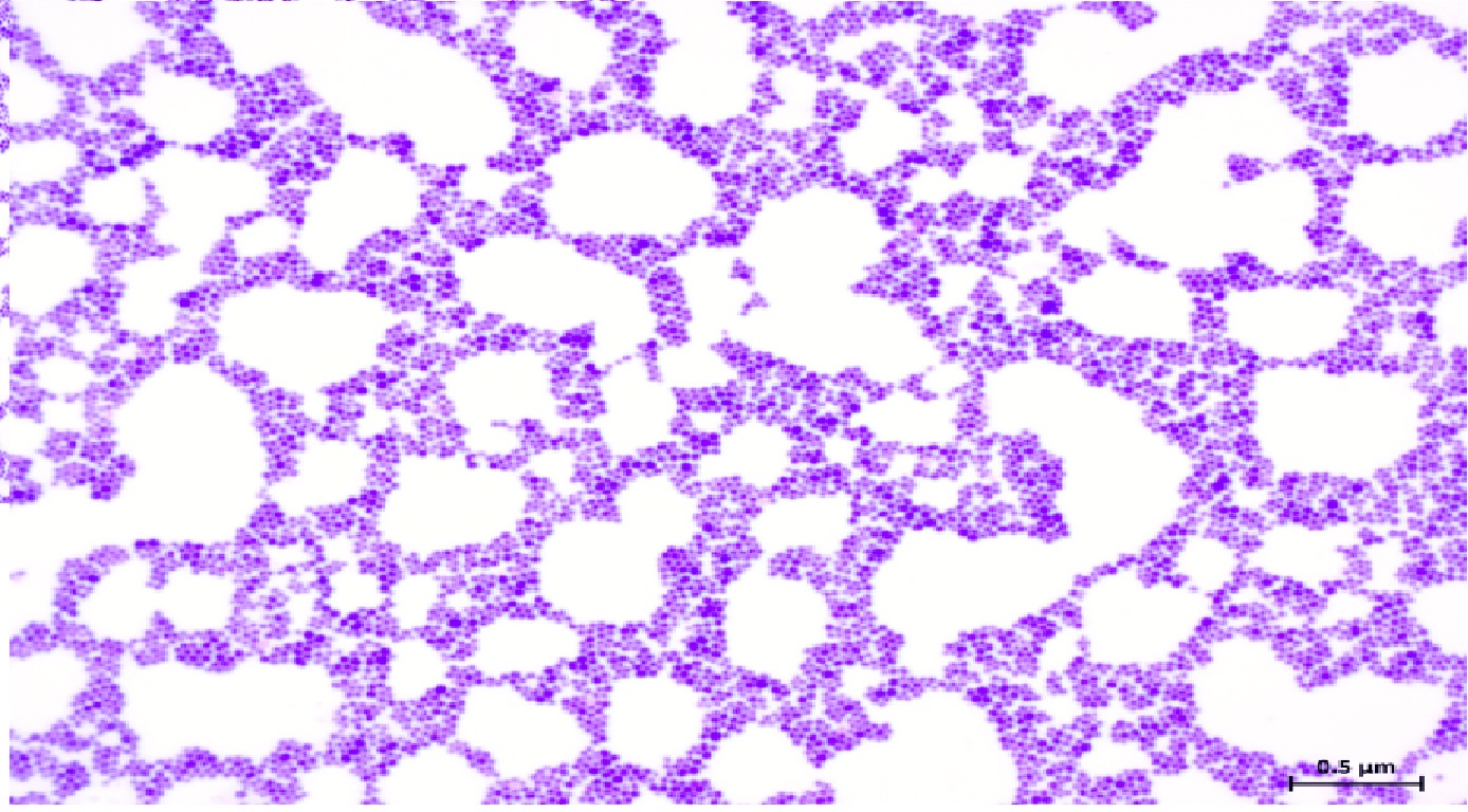
Fig. 1: Grams staining of Staphylococcus isolate showing Gram positive cocci in bunch
Further a total of 9 isolates showing lactose fermenting pink colonies on MacConkey agar (Fig. 2). However, non of the isolate with non lactose fermenting nature was obtained on MacConkey agar. Eight isolates were confirmed as E.coli, which showed typical greenish metallic sheen on EMB agar as well as gram negative rods morphologically similar to E. coli when stained with Gram’s Method of staining (Fig. 3). Similarly, colonies with large, mucoid, pink to purple colonies with no metallic green sheen on EMB agar medium and Gram negative coccobacilli in Gram’s stained smear confirmed as Klebsiella spp.
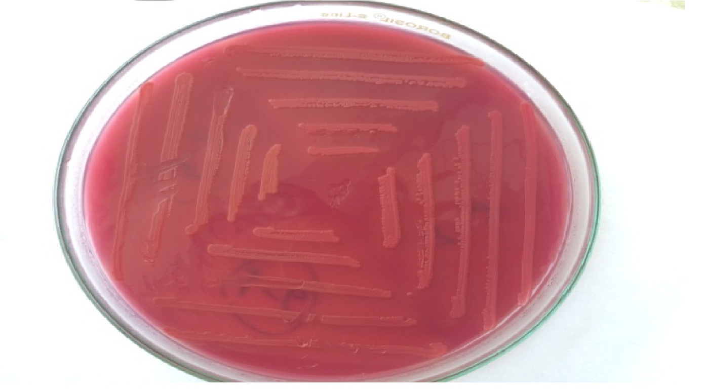
Fig. 2: Lactose fermenting pink coloured colonies on MacConkey agar medium
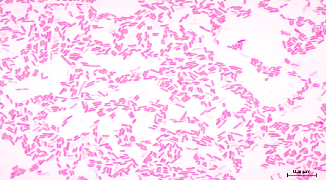 Fig. 3: Grams staining of E. coli isolates showing typical Gram negative rods.
The isolated bacterial colonies on blood agar plates were small, glistening, mucoid, haemolytic and dew drop like, and appeared as gram negative coccobacilli when stained with Gram’s stain stain were identified as Pasteurella spp. Non haemolytic tiny colonies on blood agar and staining by Gram method revealed gram negative, highly pleomorphic, rod shaped bacteria identified as Ornithobacterium rhinotracheale (Fig. 4).
 Fig. 4: Grams staining of O. rhinotracheale isolates showing Gram negative, highly pleomorphic rod shaped bacteria.
All the 17 isolates were characterized by biochemical tests viz. oxidase, catalase, urea and IMViC pattern (indole production, Methyl Red (MR) test, Voges-Proskauer (V.P.) test, citrate utilization on Simmon’s citrate medium). The results have been summarized and presented in Table 1 and Fig. 5-7. ORT could be differentiated from other pathogenic bacteria by biochemical reactions. The present findings were coincided with the findings of Van Empel et al. (1997), Masdooq et al. (2008), Siddique et al. (2008), Hassanzadeh et al. 2010, Itoo et al. (2013) and Ashraf et al. (2015). Yilmaz et al. (2011) during their study of lung lesions of slaughtered broilers in slaughter house reported that E. coli ranked the first among the other bacteria isolated. Gowthman et al. (2012) found E. coli as major secondary invading pathogen in respiratory disease complex.
Table (1):
Various biochemical characters of bacterial isolates.
Organisms/ Biochemical characters |
E. coli (8) |
Staphylococcus spp. (4) |
O. rhinotracheale (3) |
Pasteurella spp. (1) |
Klebsiella spp. (1) |
|---|---|---|---|---|---|
Indol |
+ |
NA |
_ |
+ |
_ |
MR |
+ |
NA |
_ |
_ |
_ |
VP |
_ |
NA |
+ |
_ |
+ |
Citrate |
_ |
NA |
_ |
_ |
+ |
Urease |
_ |
+ |
+ |
_ |
+ |
Oxidase |
_ |
_ |
+ |
+ |
_ |
Catalase |
+ |
+ |
_ |
+ |
+ |
Haemolysis on blood agar |
V |
+ |
_ |
_ |
+ |
Growth on MCA |
LF |
_ |
_ |
_ |
LF |
Growth on BHI |
+ |
+ |
_ |
+ |
+ |
Growth on EMB |
+ |
_ |
_ |
_ |
+ |
Growth on BA |
+ |
+ |
+ |
+ |
+ |
+: Positive, – : Negative, NA: Not applicable, V: Variable and LF- Lactose fermenting colony
The incidence of E. coli, Staphylococcus spp. O. rhinotracheale, Pasteurella spp. and Klebsiella spp. were 8 (47.06%), 4 (23.53%), 3 (17.65%), 1 (5.88%) and 1 (5.88%) in broiler flocks (Table 2). These results were in accordance to that reported by Mustafa and Ali, (2005); Lateef et al., (2006); Murthy et al., (2008); Popy et al., (2011); Bhimani et al., (2014). Concomitant infection with E. coli, Pasteurella spp. and Klebsiella spp. increasing the severity of infections associated with O. rhinotracheale as reported by De Rosa et al. (1996).
Table (2):
Bacterial isolates from commercial broiler showing respiratory disease complex.
Organisms |
No. of isolates |
Percentage (%) |
Healthy broilers |
Diseased broilers |
|---|---|---|---|---|
E. coli |
8 |
47.06 |
2 |
6 |
Staphylococcus spp. |
4 |
23.53 |
0 |
4 |
O. rhinotracheale |
3 |
17.65 |
0 |
3 |
Pasteurella spp. |
1 |
5.88 |
0 |
1 |
Klebsiella spp. |
1 |
5.88 |
0 |
1 |
Total |
17 |
– |
2 |
15 |
The results showed that the prevalence of RDC was 6.67% and 50.00% in apparently healthy and diseased broilers (Table 3). Similar findings have also been documented by Hassan et al., (2014) and Chaudhari, (2017). More number of bacteria were isolated from lung (58.33) followed by trachea (41.66), larynx (16.66), exudates of infra orbital sinus (16.66) and air sacs (8.33) in broiler birds (Table 3). The organism could also be isolated in pure culture in specimens from the trachea, lung or air sac exudates and these findings were in accordance with Murthy et al., (2008) and Pan et al., (2012). The frequency of isolation of ORT from various organs indicated that the isolates were most commonly recovered from the lungs, trachea and air sacs. In some birds, ORT was isolated from the lung, in association with Pasteurella spp. and E. coli. ORT was most frequently isolated from the respiratory tract of poultry, whereas Pasteurella spp. could be isolated from various organs, notably the liver (Murthy et al., 2008).
Table (3):
Prevalence of RDC in apparently healthy and diseased broilers.
Tissues |
No. of sample processed |
No. of isolates |
Percentage (%) |
Healthy broilers |
Diseased broilers |
|---|---|---|---|---|---|
Treachea |
12 |
5 |
41.66 |
1 |
4 |
Larynx |
12 |
2 |
16.66 |
0 |
2 |
Lung |
12 |
7 |
58.33 |
1 |
6 |
Exudates of infra orbital sinus |
12 |
2 |
16.66 |
0 |
2 |
Air sacs |
12 |
1 |
8.33 |
0 |
1 |
Total |
60 |
17 |
– |
2 |
15 |
Prevalence (%) |
– |
28.33 |
6.67 |
50.00 |
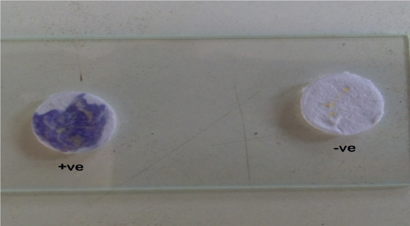
Fig. 5: O. rhinotracheale and Pasteurella isolate showing positive oxidase test.
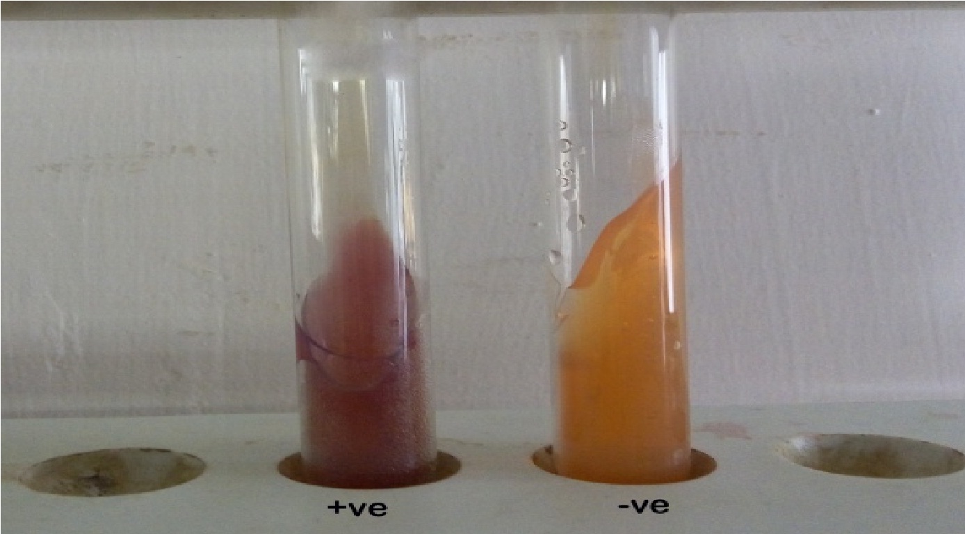
Fig. 6: Figure Staphylococcus, O. rhinotracheale and Klebsiella isolates showing positive urease test.
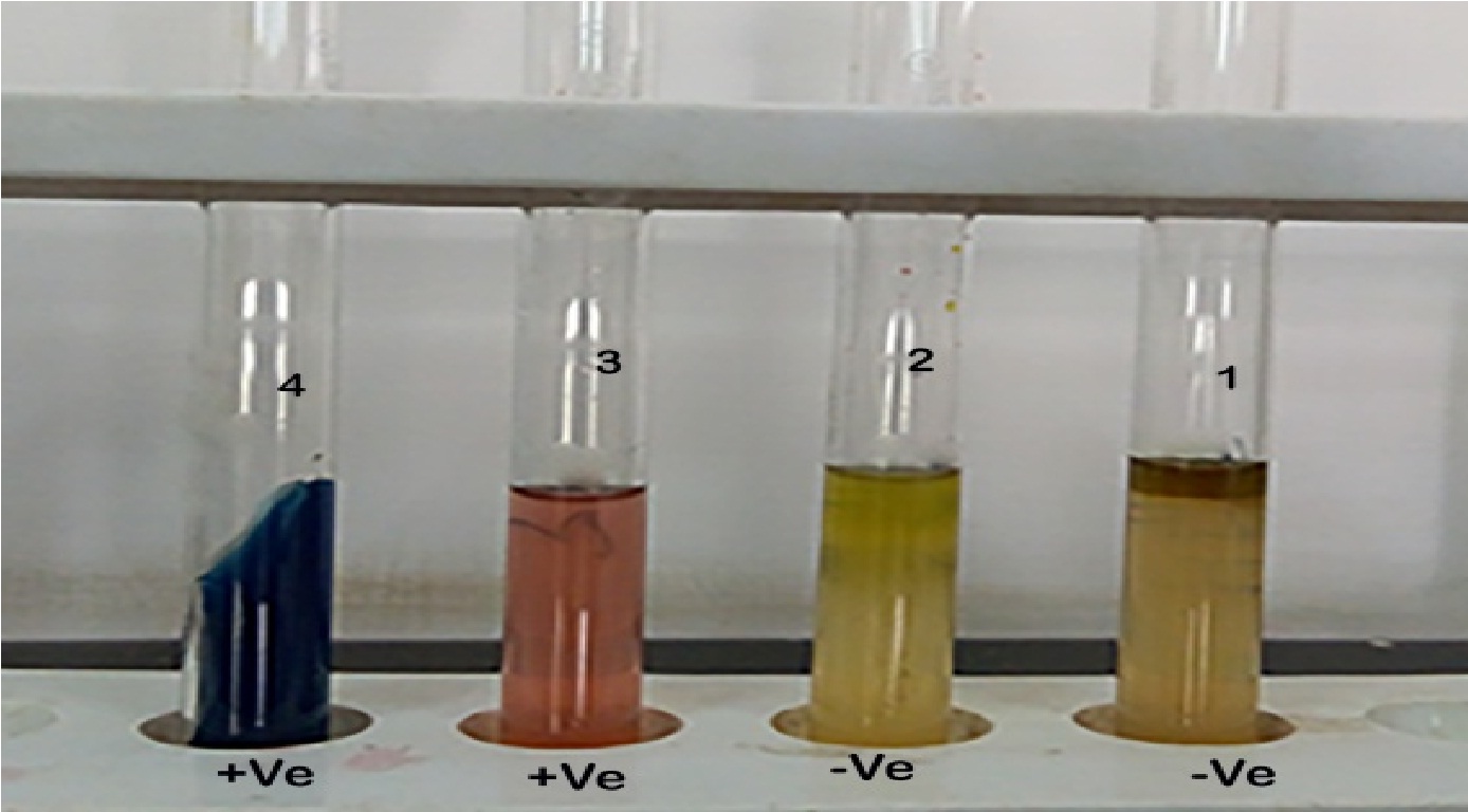 Fig. 7: 1.Indole, 2. Methyl- Red (MR), 3. Voges Proskauer (VP) and 4. Citrate test for Klebsiella spp.
In conclusion, the emerging respiratory diseases complex in broiler caused by O. rhinotracheale, one of the emerged pathogen could be isolated either singly or concurrently with other bacteria, such as E.coli, Pasteurella spp. and Klebsiella spp., indicating its possible etiological role in the respiratory disease complex of broiler. Mixed or concomitant bacterial infections are significant and very severe but the synergistic role between O. rhinotracheale and other bacterial pathogens is yet to be ascertained.
ACKNOWLEDGMENTS
The authors are thankful to Director of Research, Sardar krushi nagar Dantiwada Agricultural University, Sardar krushi nagar for providing funds and facilities to conduct this work.
- Ashraf, A.; Samir, A., Ebtisam, M., Doaa, A. Prevalence of E. coli in broiler chickens in winter and summer seasons by application of PCR with its antibiogram pattern. Benha veterinary medical journal 2015; 29: 119–128.
- Barrow, G. I., Feltham, R. K. A. Cowan and Steels’s Manual for the Identification of Medical Bacteria, 3rd edn., Cambridge University Press, Cambridge, Great Britain, 1993.
- Bhimani, M.P., Roy, A., Bhanderi, B.B., Mathakiya, R. A. Isolation, identification and molecular characterization of Pasteurella multocida isolates obtained from emu (Dromaius novaehollandiae) in Gujarat state, India. Veterinarski Archives 2014; 84: 411–419.
- Canal, C.W., Leao, J.A., Rocha, S.L.S., Macagnan, M., Lima-Rosa, C.A.V., Oliveira, S.D., Back, A. Isolation and characterization of Ornithobacterium rhinotracheale from chickens in Brazil. Research in Veterinary Science 2005; 78: 225–230.
- Chaudhari, S. V. Pathological and molecular studies on upper respiratory tract infections in broilers with special reference to Low Pathogenic Avian Influenza (H9N2), Infectious Bronchitis virus, Escherichia coli and Avian Mycoplasma. In: MVSC, dissertation. Anand Agricultural University, 2017.
- Saif YM, Barnes HJ, Glisson JR, Fadly A M, McDougald LR, Swayne DE (eds.): Disease of Poultry, 11th edn. Iowa State University Press, Ames, Iowa, 2003; pp. 683-690.
- De Rosa, M., Droual, R., Chin, R. P., Shivaprasad, H. L., Walker, R. L. Ornithobacterium rhinotracheale infection in turkey breeders. Avian diseases 1996; 40: 865–874.
- El-Sukhon, S. N., Musa, A., Al-Attar, M. Studies on the Bacterial etiology of airsacculitis of broilers in Northern and Middle Jordan with special reference to Escherichia coli, Ornithobacterium rhinotracheale, and Bordetella avium. Avian Diseases 2002; 46: 605–612.
- Gowthaman, V., Singh, S. D., Dhama, K., Barathidasan, R., Bhatt, P. Infectious bursal disease (IBD) playing a triggering role in E. coli and Mycoplasma induced respiratory disease complex in broilers. Veterinary Practitioner 2012; 13: 223–225.
- Hafez, H. M. Diagnosis of Ornithobacterium rhinotracheale. International Journal of Poultry Science 2002; 1: 114–118.
- Hassan, S., Mukhtar, N., Rahman, S. U., Mian, A. A. Molecular epidemiology of Mycoplasma gallisepticum in different types of chickens. Int J Agric Biol 2014; 16: 165–170.
- Hassanzadeh, M., Karrimi, V., Fallah, N., Ashrafi, I. Molecular characterization of Ornithobacterium rhinotracheale isolated from broiler chicken flocks in Iran. Turkish Journal of Veterinary and Animal Sciences 2010; 34: 373–378.
- Hossain, M. S., Akter, S., Ali, M., Das, P. M., Hossain, M. M. Bacteriological and pathological investigation of nasal passage infections of chickens (Gallus gallus). The Agriculturists 2013; 11: 47–55.
- Itoo, F., Kamil, S., Mir, M., Baba, O., Dar, T., Darzi, M. Occurrence and pathology of diseases with associated respiratory tract affections in commercial broiler chickens reared in Kashmir. SKUAST J Res 2013; 15: 23–34.
- Lateef, M., Rauf, U., Sajid, M. A. Outbreak of respiratory syndrome in Chukar partridge (Alectoris chukar). J Anim Pl Sci 2006; 16: 219–223.
- Masdooq, A. A., Salihu, A. E., Muazu, A., Habu, A. K., Ngbede, J., Haruna, G., Sugun, M. Y. Pathogenic bacteria associated with respiratory disease in poultry with reference to Pasteurella multocida. International Journal of Poultry Science 2008; 7: 674–675.
- Murthy, T. R. G. K., Dorairajan, N., Balasubramaniam, G. A., Dinakaran, A. M., Saravanabava, K. Pathogenic bacteria related to respiratory diseases in poultry with reference to Ornithobacterium rhinotracheale isolated in India. Vet arhiv 2008; 78: 131–140.
- Mustafa, M. Y., Ali, S. S. Prevalence of infectious diseases in local and Fayoumi breeds of rural poultry (Gallus domesticus). Punjab Univ J Zool 2005; 20: 177 – 180.
- Pan, Q., Liu, A., Zhang, F., Ling, Y., Ou, C., Hou, N., Cheng, H. Co-infection of broilers with Ornithobacterium rhinotracheale and H9N2 avian influenza virus. BMC veterinary research 2012; 8: 104.
- Popy, N., Asaduzzaman, M., Miah, M. S., Siddika, A., Sufian, M. A. Pathological study on the upper respiratory tract infection of chickens and isolation, identification of causal bacteria. The Bangladesh Veterinarian 2011; 28: 60–69.
- Saif, Y. M., Barnes, H. J., Glisson, R., Fadly, A. M., Mcdougald, A.R. Diseases of Poultry. Iowa State University Press, Ames, Iowa, USA, 2003.
- Siddique, M. 1, Zia, T., Rehman, S. U. Outbreak of Ornithobacterium rhinotracheale (ORT) infection in chickens in Pakistan. Pakistan Vet J 2008; 31: 117–119.
- Van Empel, P., Van Den Bosch, H., Loeffen, P., Storm, P. Identification and serotyping of Ornithobacterium rhinotracheale. Journal of Clinical Microbiology 1997; 35: 418–421.
- Van Empel, P.C.M. and Hafez, H.M. (1999). Ornithobacterium rhinotracheale/ : A review. Avian Pathology 1999; 28: 217–227.
- Yilmaz, F., Timurkaan, N., Kilic, A., Kalender, H., Kilinc, U. Detection of Mycoplasma synoviae and Mycoplasma gallisepticum in chickens by immunohistochemical , PCR and culture. Revue Med Vet 2011; 162: 79–86.
© The Author(s) 2017. Open Access. This article is distributed under the terms of the Creative Commons Attribution 4.0 International License which permits unrestricted use, sharing, distribution, and reproduction in any medium, provided you give appropriate credit to the original author(s) and the source, provide a link to the Creative Commons license, and indicate if changes were made.