


ISSN: 0973-7510
E-ISSN: 2581-690X
 , Nassima Didouh2,3, Rachid Achek1,4, Ibrahim Nabi5 and Ricardo Araujo6
Staphylococcus aureus colonizes the skin and nasal orifices of both humans and animals. Biofilm-related infections and acquired antimicrobial resistance render this bacterium a major health concern. This study examined the biofilm-forming capacity of eight S. aureus isolates originating from humans, sheep, and food matrices in northern Algeria. The efficiency of sanitizers (povidone–iodine [PVI], peracetic acid [PAA], quaternary ammonium compounds [QACs] and chlorhexidine [CHX]) in inhibiting and detaching biofilms was also assessed. Biofilm-production ability was assessed using a tissue culture plate. The minimal inhibitory concentration (MIC) and minimal bactericidal concentration were measured for each sanitizer, and antibiotic susceptibility profiles were determined by performing the Kirby-Bauer disk diffusion assay. All S. aureus isolates demonstrated the ability to produce biofilms, and among these, food and sheep mastitis isolates exhibited a strong capacity for biofilm formation. CHX exhibited the highest efficacy in bacterial biofilm inhibition and was considered more potent than the other sanitizers (P < 0.0001). Further, it exhibited lower MIC interval values (0.98-1.9 µg/g). However, MIC interval values for PVI, PAA, and QACs were 3125-25000, 39.06-156.25, and 0.98-3.9 mg/L, respectively. PVI did not affect biofilm detachment at different concentrations. All isolates were sensitive to methicillin (with mecA/mecC genes not detected), gentamicin, sulfamethoxazole–trimethoprim, vancomycin, and amikacin; however, two isolates were resistant to erythromycin and clindamycin. These findings demonstrated the biofilm-forming potential of S. aureus isolates and confirmed the effectiveness of sanitizers against S. aureus isolated from different sources.
Staphylococcus aureus, Biofilm, Sanitizers, Antimicrobial Resistance, Chlorhexidine, Peracetic Acid, Quaternary Ammonium
Staphylococcus aureus is a Gram-positive opportunistic pathogen, frequently found as a commensal colonizer of the skin and nares in both humans and animal species.1 It causes different diseases, including minor suppurative skin infections, food poisoning outbreaks, and toxic shock syndrome. Further, it produces several virulence factors, including pathogenic antigens, enzymes, and toxins. Staphylococcal enterotoxins play a crucial role as primary toxins associated with food poisoning.2
Staphylococcus aureus can colonize different surfaces and produce robust biofilms.3 Biofilms consist of microorganisms embedded in an extracellular polymer matrix, and this facilitates strong attachment to surfaces.4 These biofilms, which are composed of polysaccharides, proteins, and organic materials, can adhere to medical devices, industrial equipment, and food processing components, leading to significant issues.5 Biofilms that form on implants within human tissues are associated with a wide range of chronic infections. In the food industry, microbial contamination and biofilm formation are critical threats to food safety and human health.6
The firm adherence ability of S. aureus to living and inert surfaces is due to the production of a polysaccharide extracellular matrix and the presence of proteins that promote adherence to tissues and abiotic surfaces. Consequently, these biofilms exhibit significant resilience to both physical disruption and chemical agents.7 A notable feature of S. aureus is the expression of various adhesins, specifically microbial surface components recognizing adhesive matrix molecules (MSCRAMMs), adhesion proteins that bind collagen (cna), fibronectin-binding proteins (fnb), and other analogous adhesion proteins.8 Importantly, although multiple factors influence biofilm development, polysaccharide intercellular adhesins (PIAs), encoded by the ica operon, play a critical role in synthesis of the extracellular matrix, facilitating cell-to-cell adhesion and structural stability within the biofilm.9
Bacterial biofilms exhibit greater tolerance and resistance to antimicrobial agents than free-floating suspended bacteria.10 The biofilm matrix surrounds the bacterial cells, making antibiotic treatments and chemical agents less effective owing to reduced penetration or complete impermeability.11 Furthermore, previous investigations have established that the S. aureus exopolysaccharide matrix provides the physical integrity of the biofilm, acting as a barrier that increases tolerance to diverse antimicrobial agents, such as antibiotics and sanitizers.12,13 To address this issue, effective disinfectants must be applied at the appropriate concentrations. Low concentrations risk the development of resistance, whereas high concentrations increase costs and environmental impacts.14 Additionally, this inefficiency is facilitated by a highly plastic genome, which allows for the continuous acquisition of resistance determinants through genetic exchange and evasion of the innate host immune system. Three primary approaches are used to eradicate biofilms: (i) altering the surface characteristics to prevent biofilm formation, (ii) modulating cell-to-cell signaling to inhibit biofilm assembly, and (iii) using physical interventions to disrupt established biofilms.15
The use of sanitizers to inhibit bacterial growth and biofilm formation is a well-established method of microbial control. These agents are widely used in healthcare, the food industry, and households to minimize microbial contamination. The choice of sanitizer depends on several factors, including its spectrum of activity, efficacy, cost, surface compatibility, residue formation, and rinsing requirements.16 Selecting an appropriate sanitizer is crucial for ensuring both safety and effectiveness in controlling the presence of bacteria. The most commonly used sanitizers are alcohols, quaternary ammonium compounds, chlorine-based agents, and hydrogen peroxide, each with varying levels of efficacy and applications.17
Povidone–iodine (PVI) is effective against various microorganisms, including antibiotic-resistant bacteria. It targets Gram-negative pathogens such as Klebsiella pneumoniae and Gram-positive strains such as methicillin-resistant S. aureus (MRSA) and Escherichia coli.18 Peracetic acid (PAA) is attracting increasing research interest because of its applications in the medical and food industry.19 As membrane-active agents, quaternary ammonium compounds (QACs) represent a primary class of sanitizers characterized by their strong and broad-spectrum antimicrobial activity, and they are known for their adaptability across consumer and industrial applications. Their broad-spectrum efficacy against bacteria, viruses, and fungi makes them critical tools in public health and infection control strategies.20 Chlorhexidine (CHX) is a potent broad-spectrum sanitizer with strong bactericidal action. It targets the microbial cell membrane, inducing irreversible permeability and the subsequent leakage of cytoplasmic components, disrupting essential protein functions and causing the crystallization of macromolecules within the cytoplasm, ultimately leading to cell death. In addition to its bactericidal effects, CHX also exhibits bacteriostatic properties by interfering with ATPase activity, which inhibits prokaryotic cell division.19,21,22
The aim of this study was to characterize the ability of S. aureus to form biofilms on abiotic surfaces and assess the comparative efficacy of various sanitizers, including PVI, PAA, QACs, and CHX, for the prevention and eradication of these microbial biofilms. Importantly, this study compared the efficacy of these sanitizers against S. aureus isolates of diverse origins, providing critical insights into how environmental and clinical sources influence biofilm-formation and resistance mechanisms. Finally, the susceptibility of S. aureus isolates to various antibiotics was examined to provide insights into their resistance profiles and potential correlations with biofilm formation.
Origins of isolates
This study was carried out on eight S. aureus strains from a collection of staphylococci isolates collected from different samples. Animal isolates (S21 and S22) originated from the milk of sheep with mastitis. Food isolates were obtained from raw milk or minced beef ( F13, F15, and F29). Additional isolates were collected from the nasal swabs of individuals with high occupational animal exposure, including farm staff and veterinary professionals (N1, N9, and N18). Microbial sampling was performed in two provinces in Northern Algeria (Medea and Ain Defla). Bacterial species were identified using matrix-assisted laser desorption/ionization time-of-flight mass spectrometry. DNA microarrays were used to characterize clonal complex (CC) lineages and provide relevant information on various virulence factors, specifically genes associated with exotoxin and enterotoxin production, immune-evasion factors, biofilm formation-associated genes, sanitizer-resistance genes, MSCRAMMs, antibiotic-resistance determinants, and other target genes for typing such as species, SCCmec, capsule, and agr group typing markers.23,24
Assessment of biofilm production
Biofilm formation was induced on polystyrene surfaces using a tissue culture plate. The biofilm-producing capacity of the eight isolates was assessed following a previous protocol.25 Overnight bacterial cultures in tryptic soy broth (TSB)-1% glucose broth (Merck, Germany) were incubated at 37 °C. The cultured suspensions were standardized to an optical density (OD) at 625 nm of 0.08-0.1 (cell concentration ~108 cfu/mL). Sterile microtiter plates (Greiner Bio-One, Germany) were prepared by filling the wells with 100 µL of the bacterial suspension; then, 100 µL of fresh TSB 1% glucose was added to each well. For the negative controls, 200 µL of TSB supplemented with 1% glucose was added to the designated wells. To ensure repeatability, all isolates were tested in triplicate. For each assay, two microtiter plates were prepared, one incubated for 24 hrs and the other incubated for 48 hrs at 37 °C. Following incubation, the culture medium was aspirated and the wells were washed three times with sterile phosphate-buffered saline (PBS, pH 7.4) to eliminate non-adherent cells. Microtiter plates were drained in an inverted position before fixing the attached bacteria. The adherent bacteria were exposed to hot air at 60 °C for 60 min for heat fixation. The adherent biofilm that formed in each well of the microtiter plates was stained with 150 µL of 0.1% crystal violet (Sigma-Aldrich, Merck, Darmstadt, Germany) for 15 min. After the staining procedure, the well contents were aspirated and the plates were rinsed three times with sterile distilled water to eliminate residual stain. The microtiter plate wells were dried at room temperature. To solubilize the crystal violet, each well was treated with 150 µL of 96% ethanol and incubated without agitation for 30 min. Finally, absorbance was measured at 625 nm using a microplate reader (800TS Agilent, BioTek, USA).
The cutoff OD value was defined as three standard deviations above the mean OD of the negative control. Based on the OD625 values calculated previously herein, bacterial isolates were classified into the following biofilm-production categories: non-biofilm producer (OD lower than the negative control), weak biofilm producer (WBP: OD higher than the negative control but lower than twice this value), moderate biofilm producer (MBP: OD between two and four times the negative control), or strong biofilm producer (SBP: OD higher than four times the negative control, as recommended by Stepanović et al.).25
Evaluation of microbial sanitizer efficacy against biofilm formation
The effects of the sanitizers PVI, PAA, QACs, and CHX on limiting bacterial growth (via the minimum inhibitory concentration [MIC] and minimum bactericidal concentration [MBC] of each sanitizer) were investigated. The biofilm-interference and removal effects of various sanitizers were also analyzed. PVI (10%) and QACs (50%) were purchased from a local market. The PAA (5%) sanitizer was provided by the dairy industry in the study region, and CHX (1.6%) was provided by the microbiology laboratory at the Research Center of Biotechnology of Constantine in eastern Algeria.
Determination of MICs
The TCP “tissue culture plate” technique, as previously described (section 2.2), was used in this section to study the effect of various sanitizers on inhibiting biofilm formation and to determine their MICs. The MIC was defined as the lowest concentration of an antimicrobial agent that prevents microbial growth after overnight incubation.26 PVI was tested twice at an initial concentration of 10%, PAA was tested at 5% and 1%, and CHX was tested at initial concentrations of 0.05% and 0.025%. For the QACs, five trials were performed using initial concentrations of 50%, 10%, 1%, 0.5%, and 0.25%. To ensure accuracy and reproducibility, all sanitizer assays were performed in duplicate. Serial dilutions were performed according to the Clinical and Laboratory Standards Institute (CLSI) guideline.27
The concentrations of PVI, PAA, and QACs (in mg/L) were evaluated based on liquid formulations, whereas CHX values were expressed as µg/g) to strictly adhere to the manufacturer’s gravimetric certification of the stock CHX gluconate paste/solution. The microtiter plate was prepared by filling the wells with 100 µL of TSB 1% glucose broth. Serial dilutions were performed, starting with the initial concentrations mentioned previously. Then, 100 µL of bacterial inoculum for each isolate (bacterial concentration of 0.5 McFarland) was added to each well (excluding negative control). The microtiter plate was covered and incubated at 37 °C for 24 hrs. The MICs of the sanitizers were evaluated by visually assessing the turbidity of each well in comparison with the negative and positive controls.
Determination of biofilm MBC
The MBC indicates the lowest concentration of an antimicrobial agent at which only 0.01% of the bacteria remain viable.28 To determine the MBC of the tested sanitizers, aliquots from the wells in the MIC assay were subcultured onto solid growth media.29 Following MIC determinations, a 10 µL aliquot of the bacterial suspension was taken from wells showing no visible growth specifically those with concentrations meeting or exceeding the MIC and spotted onto Mueller-Hinton agar plates. The plates were incubated at 37 °C for 24 hrs. The MBC values were determined as the lowest concentration of sanitizer that resulted in no visible colony growth after subculture on agar plates.
Assessment of inhibitory effect on biofilm formation
After determining the MIC of each sanitizer (Section 2.3.1) and after the treatment period, the well contents were aspirated and the plates were rinsed three times with sterile PBS (pH 7.4) to eliminate any remaining non-adherent bacterial cells. The microtiter plates were drained in an inverted position before the attached bacteria were heat-fixed at 60 °C for 60 min. Then, 150 µL of 0.1% crystal violet was added to each well for 15 min to stain adherent cells. The contents of the wells were then gently aspirated and washed three times with sterile distilled water to eliminate excess stain, and the microtiter plates were dried at room temperature.
For solubilization of the crystal violet, 150 µL of 96% ethanol was added to each well without agitation for 30 min. The OD625 nm was measured using a microtiter plate reader (800TS Agilent, BioTek, USA). The OD results of each well, corresponding to successive sanitizer concentrations, were compared with the control OD values (positive and negative controls).
Evaluation of the detachment effect on preformed biofilms
The detachment evaluation procedure followed the protocol described by Kose and Yapar,30 with slight modifications. After biofilm formation on tissue culture plates, 10 serial two-fold (double-fold) dilutions of each sanitizer were prepared in distilled water. Aliquots of 200 µL of diluted sanitizers (PVI, PAA, QACs, and CHX) were added to the corresponding wells of the microtiter plate, except for the controls. Following a 5-30 min incubation at room temperature, the sanitizer solutions were aspirated from the wells. The wells were then washed three times with sterile distilled water and allowed to dry at room temperature. Staining of the remaining (non-detached) biofilm cells and solubilization with crystal violet were performed as described previously (Section 2.3.3).
Antimicrobial susceptibility testing
Antimicrobial susceptibility was determined for 12 different agents using the Kirby-Bauer disk diffusion method on Mueller-Hinton agar following CLSI protocols.27 The antibiotic disks that were used (Oxoid, Basingstoke, UK) included the following: oxacillin (5 µg), cefoxitin (30 µg), doxycycline (30 µg), gentamicin (10 µg), vancomycin (30 µg), erythromycin (15 µg), trimethoprim-sulfamethoxazole (25 µg), ofloxacin (5 µg), rifampicin (30 µg), clindamycin (2 µg), amikacin (30 µg), and fosfomycin (50 µg). Petri dishes were incubated at 37 °C for 18-24 hrs. Inhibition zones were measured, and the isolates were categorized as susceptible (S), intermediate (I), or resistant (R) according to the established CLSI breakpoints.27
Resistance to vancomycin and oxacillin was confirmed by determining MICs using the E-test method. Briefly, a bacterial suspension standardized to 0.5 McFarland was inoculated onto Mueller-Hinton agar (Liofilchem, Italy). An E-test strip (Liofilchem, Italy) was then placed in the center of the plate, followed by incubation at 37 °C for 24 hrs. Following incubation, the MIC was determined by identifying the point at which the elliptical inhibition zone intersected the graduated E-test strip. The resulting values (µg/mL) were interpreted as S, I, or R, according to CLSI breakpoints.27
Statistical analysis
Statistical analysis was performed using StatView® software (SAS Institute Inc., Cary, NC, USA) (version 5.0). Data are expressed as means ± standard deviations. Differences in optical densities among sanitizer treatments were compared using one-way analysis of variance. Pairwise comparisons between groups were subsequently carried out using Fisher’s Protected Least Significant Difference (PLSD) post-hoc tests. Statistical significance was defined at a P-value < 0.05. Detailed comparisons obtained from Fisher’s PLSD post-hoc analyses are included in Supplementary Tables 1 and 2.
Biofilm formation results (TCP assay)
The TCP results of the eight isolates are presented in Table 1; all isolates exhibited higher OD values than those observed in the negative control. (24 hrs: 0.069; 48 hrs: 0.076). The incubation time affected the development of the biofilm matrix, and the potential for biofilm development after 24 hrs was higher than that after 48 hrs of incubation. After 24 hrs of incubation, 6 of 8 isolates (75%) were classified as SBPs, and the remaining (25%) were classified as MBPs. After 48 hrs of incubation, four of eight isolates (50%) were classified as SBPs, whereas the remaining isolates were classified as WBPs or MBPs. Regarding the origin of isolates, S. aureus isolated from sheep mastitis and the food matrix showed a higher capacity for biofilm formation than the nasal swab isolates after 24 hrs or 48 hrs of incubation. According to the microarray data, S. aureus isolates assigned to clonal complexes CC97, CC5, CC8, and CC398 exhibited the highest OD values and were classified as SBPs. Thus, isolates belonging to CC1, CC15, and CC45 had relatively low OD values.
Table 1. Biofilm producing ability of S. aureus isolates
| Isolate ID | Samples origin | Clonal Complex | Biofilm Production (24 hrs) | Biofilm Production (48 hrs) | Biofilm associated genes | Adhesion matrix genes | |||||||
|---|---|---|---|---|---|---|---|---|---|---|---|---|---|
| OD | Type | OD | Type | icaA | icaC | icaD | Bap | fnbA | fnbB | sasG | |||
| N1 | Nasal swab | CC15 | 0.285 | MBP | 0.139 | WBP | ₊ | ₊ | ₊ | ₋ | ₊ | ₊ | ₊ |
| N9 | Nasal swab | CC45 | 0.261 | MBP | 0.151 | WBP | ₊ | ₊ | ₊ | ₋ | ₊ | ₊ | ₋ |
| N18 | Nasal swab | CC398 | 0.451 | SBP | 0.250 | MBP | ₊ | ₊ | ₊ | ₋ | ₊ | ₊ | ₋ |
| F13 | Food matrix | CC1 | 0.302 | SBP | 0.259 | MBP | ₊ | ₊ | ₊ | ₋ | ₊ | ₊ | ₊ |
| F15 | Food matrix | CC97 | 0.618 | SBP | 0.504 | SBP | ₊ | ₊ | ₊ | ₋ | ₊ | ₊ | ₊ |
| F29 | Food matrix | CC5 | 0.561 | SBP | 0.421 | SBP | ₊ | ₊ | ₊ | ₋ | ₊ | ₊ | ₊ |
| S21 | Sheep mastitis | CC8 | 0.465 | SBP | 0.418 | SBP | ₊ | ₊ | ₊ | ₋ | ₊ | ₊ | ₊ |
| S22 | Sheep mastitis | CC8 | 0.365 | SBP | 0.381 | SBP | ₊ | ₊ | ₊ | ₋ | ₊ | ₊ | ₊ |
WBP: Weakly Biofilm Producer, MBP: Moderately Biofilm Producer, SBP: Strongly Biofilm Producer
Table 1 shows the results of biofilm- and adhesion-related gene detection in S. aureus isolates. All isolates harbored the intercellular adhesion operon (icaACD) and genes encoding fibronectin–fibrinogen-binding adhesins, fnbA and fnbB. However, two of the eight isolates (N9 and N18) did not harbor the adhesin gene sasG, and the biofilm-associated protein gene (bap) was not found in any of the tested isolates. Regulatory genes (agr and sarA) were also detected in all the isolates, independent of their capacity to form biofilms.
Efficacy of microbial sanitizers in reducing biofilm formation
Determination of MIC and MBC concentrations
Tables 2 and 3 show the sanitizer efficacy, MIC, and MBC values. MIC results are summarized as ranging from the lowest to the highest concentrations observed across the eight isolates. The results obtained demonstrate significant variability; CHX showed the highest efficacy in terms of sanitizer potency, and it had a lower MIC concentration (0.98-1.90 µg/g) and MIC90 values (1.9 µg/g), indicating its ability to effectively inhibit bacterial growth. However, the MIC90 values of PVI, PPA, and QACs were 25000, 156.25, and 3.9 mg/L, respectively. The results of MBC show a significant difference between sanitizers, CHX demonstrated the highest potency, with lower MBC (0.98-1.90 μg/g) and MBC90 values (1.9 μg/g). PVI, PPA, and QACs exhibited MBC values of 25000 mg/l, 312, 5 mg/l and 62.50 mg/l, respectively.
Table 2. The MIC values of different sanitizers
| Isolates | Sanitizers | |||
|---|---|---|---|---|
| PVI (mg/l) | PPA (mg/l) | QACs (mg/l) | CHX (μg/g) | |
| N1 | 6250 | 78.12 | 0.98 | 0.98 |
| N9 | 6250 | 39.06 | 1.95 | 1.90 |
| N18 | 25000 | 156.25 | 1.95 | 0.98 |
| F13 | 6250 | 156.25 | 0.98 | 1.90 |
| F15 | 6250 | 78.12 | 1.95 | 1.90 |
| F29 | 6250 | 78.12 | 1.95 | 0.98 |
| S21 | 3125 | 78.12 | 3.90 | 1.90 |
| S22 | 3125 | 156.25 | 3.90 | 1.90 |
| MIC | (3125- | (39.06- | (0.98- | (0.98- |
| interval | 25000) | 156.25) | 3.90) | 1.90) |
Table 3. The MBC values of different sanitizers
| Isolates | Sanitizers | |||
|---|---|---|---|---|
| PVI (mg/l) | PPA (mg/l) | QACs (mg/l) | CHX (μg/g) | |
| N1 | 6250 | 78.12 | 3.90 | 0.98 |
| N9 | 6250 | 39.06 | 3.90 | 1.90 |
| N18 | 25000 | 156.25 | 15.62 | 0.98 |
| F13 | 12500 | 156.25 | 15.62 | 1.90 |
| F15 | 6250 | 78.12 | 15.62 | 1.90 |
| F29 | 6250 | 78.12 | 62.50 | 0.98 |
| S21 | 6250 | 78.12 | 3.90 | 1.90 |
| S22 | 6250 | 156.25 | 3.90 | 1.90 |
| MBC interval | 6250-25000 | 39.06- 312.50 | 3.9-62.50 | 0.98-1.90 |
Assessment of inhibitory effect on biofilm formation
The inhibitory effect of sanitizers on biofilm formation was evaluated by comparing the OD measurements obtained after crystal violet staining to a positive control values (isolates that formed biofilms without sanitizers) (Figure 1). Each sanitizer resulted in a reduction in S. aureus bacterial biofilms compared to those with the untreated isolates. Biofilm formation decreased significantly in a concentration-dependent manner when QACs and CHX were applied at all different concentrations (P < 0.0001), whereas PVI and PAA resulted in no significant decrease in biofilm formation when the concentrations were lower than 1.560·1E3 mg/L and 0.080·1E3 mg/L (P > 0.05), respectively. Regarding the origins, isolates derived from sheep with mastitis demonstrated higher sensitivity to sanitizers than isolates obtained from nasal swabs and food matrices. Therefore, SBP isolates exhibit higher sensitivity to sanitizers than MBPs.
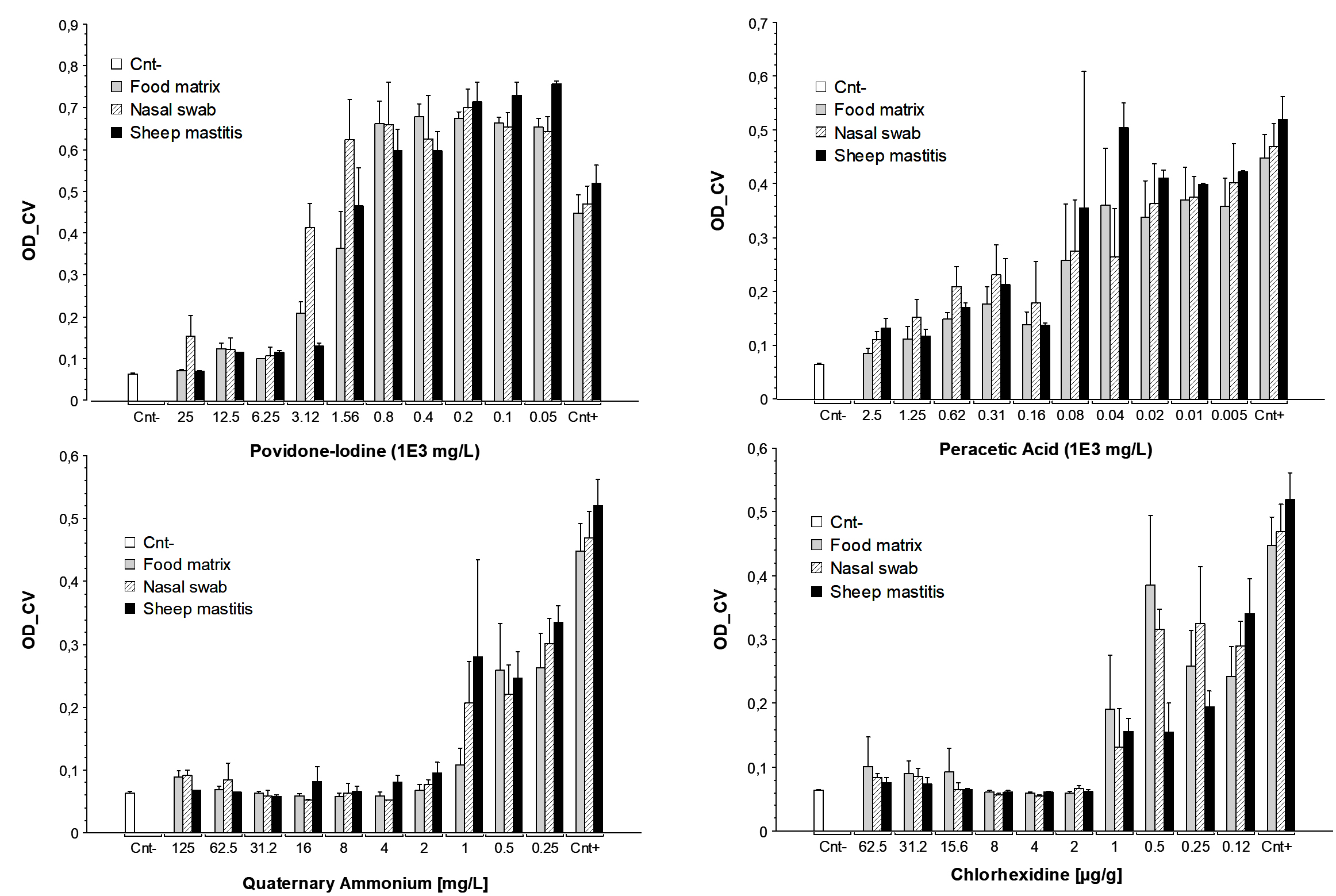
Figure 1. Biofilm inhibition effect of different sanitizers concentrations
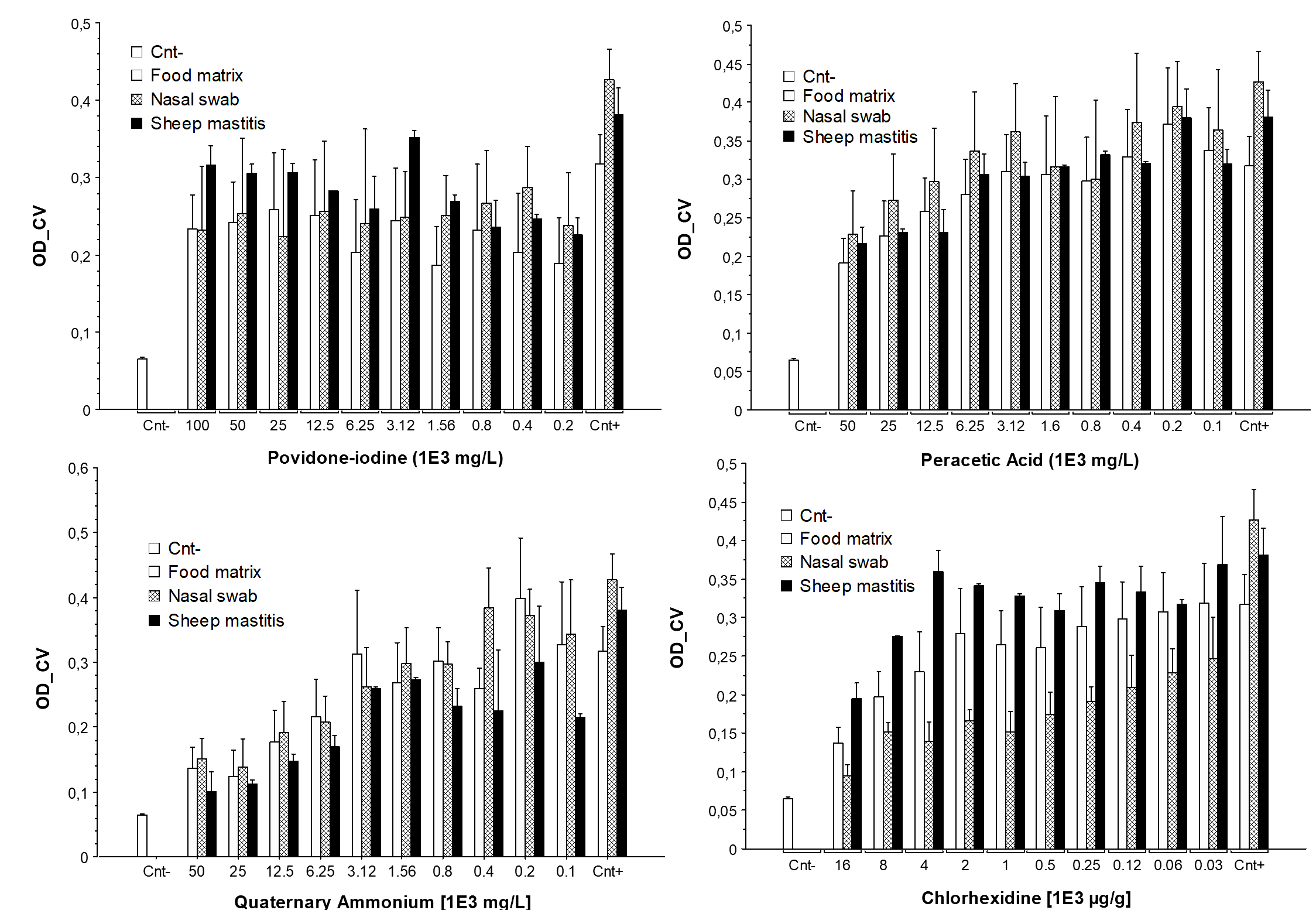
Figure 2. Biofilm detachment effect of different sanitizers’ concentrations
Biofilm detachment effectiveness of sanitizers
Figure 2 illustrates the differences in the efficacy of various sanitizers (PVI, PAA, QACs, and CHX) in detaching preformed S. aureus biofilms, focusing on the influence of the origin of the isolate (sheep, food, or nasal sources). The detachment effects of various sanitizers on the biofilms formed on the microtiter plates were evaluated (Figure 2 and Table 4). QACs demonstrated the greatest biofilm detachment effect, with a reduction in biofilm mass at varying concentrations. Notably, a concentration of 6.25·1E3 mg/L (1/8 of the initial concentration of 50·1E3 mg/L) was able to remove 71% of the preformed biofilm. CHX concentrations between 0.4 mg/g and 1.6 mg/g detached 65%-81% of the bacterial biofilm (P < 0.05); however, lower CHX concentrations resulted in detachment below 50%. In contrast, PAA was less effective with the highest concentration removing only 66% of the biofilm. Lower concentrations of PAA did not significantly affect biofilm detachment
(P > 0.05). PVI showed no significant effect on biofilm detachment across different concentrations, with the undiluted sanitizer (25·1E3 mg/L) achieving only a weak detachment effect (21%, P > 0.8).
Table 4. The OD-CV value ± standard deviation and the percentage on biofilm detachment for all S. aureus isolates
Sanitizer concentrations |
OD-Positive control |
OD-Negative control |
C1 |
C1/2 |
C1/4 |
C1/8 |
C1/16 |
C1/32 |
C1/64 |
C1/128 |
C1/256 |
C1/512 |
|---|---|---|---|---|---|---|---|---|---|---|---|---|
CHX (μg/g) |
0.395 ± 0.096 |
0.060 ± 0.01 |
0.136 ± 0.048 (81%) |
0.200 ± 0.06 (65%) |
0.228 ± 0.107 (58%) |
0.252 ± 0.094 (51%) |
0.238 ± 0.09 (55%) |
0.240 ± 0.081 (54%) |
0.266 ± 0.085 (48%) |
0.274 ± 0.083 (46%) |
0.280 ± 0.07 (44%) |
0.304 ± 0.093 (38%) |
QACs (mg/L) |
0.40 ± 0.135 |
0.084 ± 0.008 |
0.134 ± 0.049 (88%) |
0.127 ± 0.056 (89%) |
0.175 ± 0.066 (77%) |
0.201 ± 0.07 (71%) |
0.280 ± 0.111 (51%) |
0.281 ± 0.079 (51%) |
0.282 ± 0.068 (51%) |
0.298 ± 0.109 (46%) |
0.364 ± 0.114 (30%) |
0.305 ± 0.131 (45%) |
PAA (mg/L) |
0.461 ± 0.118 |
0.054 ± 0.003 |
0.212 ± 0.064 (66%) |
0.245 ± 0.073 (59%) |
0.266 ± 0.083 (54%) |
0.308 ± 0.087 (45%) |
0.328 ± 0.079 (41%) |
0.313 ± 0.11 (44%) |
0.308 ± 0.109 (40%) |
0.344 ± 0.104 (37%) |
0.382 ± 0.09 (30%) |
0.343 ± 0.091 (37%) |
PVI (mg/L) |
0.243 ± 0.082 |
0.061 ± 0.004 |
0.254 ± 0.096 (21%) |
0.262 ± 0.107 (17%) |
0.257 ± 0.13 (19%) |
0.261 ± 0.109 (18%) |
0.231 ± 0.135 (30%) |
0.272 ± 0.098 (13%) |
0.231 ± 0.077 (30%) |
0.246 ± 0.104 (24%) |
0.245 ± 0.095 (24%) |
0.216 ± 0.088 (36%) |
Antimicrobial susceptibly testing
The results of antimicrobial susceptibility testing are presented in Table 5. All isolates showed phenotypic resistance patterns that were different from those of the antimicrobial agents. Eight isolates were sensitive to methicillin (oxacillin was used for MRSA detection), gentamicin, sulfamethoxazole–trimethoprim, vancomycin, or amikacin. The phenotypic susceptibility determined using conventional methods, such as disk diffusion and Escherichia coli testing, was compared with the results of the DNA microarray analysis,24 which provides information about resistance determinants in these S. aureus isolates. No antimicrobial resistance-associated genes, namely mecA/mecC, aacA/aphD, dfrA, vanA/vanB, and aacA, were detected. However, ermB, which confers resistance to erythromycin and clindamycin, was found in one isolate. Phenotypic resistance to doxycycline was found in one isolate without the associated resistance genes tetK/tetM. Interestingly, three isolates that were phenotypically susceptible to fosfomycin harbored fosB-associated genes.
Table 5. Correlation between phenotypic resistance and genes detection in S. aureus isolates
| Antibiotics agents | Phenotypic resistance | Associated resistance genes | Genes carriage | ||
|---|---|---|---|---|---|
| Sensitive | Intermediate | Resistant | |||
| Doxycycline | 12.5% | tetM | 0% | 0% | 0% |
| tetK | 0% | 0% | 0% | ||
| Erythromycin | 25% | msrA | 0% | 0% | 0% |
| ermA | 0% | 0% | 0% | ||
| ermB | 0% | 0% | 12.5% | ||
| ermC | 0% | 0% | 0% | ||
| Gentamicin | 0% | aacA-aphD | 0% | 0% | 0% |
| Oxacillin | 0% | mecA/mecC | 0% | 0% | 0% |
| Trimethoprim- sulfamethoxazole | 0% | dfrA | 0% | 0% | 0% |
| Cefoxitin | 0% | NI | NI | NI | NI |
| Vancomycin | 0% | vanA | 0% | 0% | 0% |
| vanB | 0% | 0% | 0% | ||
| Ofloxacin | 0% | NI | NI | NI | NI |
| Rifampicin | 12.5% | NI | NI | NI | NI |
| Clindamycin | 25% | ermA | 0% | 0% | 0% |
| ermB | 0% | 0% | 12.5% | ||
| ermC | 0% | 0% | 0% | ||
| Amikacin | 0% | aacA | 0% | 0% | 0% |
| Fosfomycin | 12.5% | fosB | 37.5% | 0% | 12.5% |
Staphylococcus aureus is one of the most common pathogens causing foodborne illnesses worldwide.31 It forms biofilms on biotic and abiotic surfaces in natural and clinical environments, making it the leading cause of biofilm-associated infections.32 Biofilm formation protects bacteria against host immune responses, antibiotic treatments, and various external stressors, including resistance to sanitizers.32,33 Notably, the improper use of biocides, such as insufficient exposure times, sublethal concentrations, or inadequately designed equipment, can promote the development and spread of antimicrobial resistance, thereby posing significant risks to public health and food safety.34,35
Given the capacity of S. aureus to persist in various environments and develop resistance, it is essential to regularly assess the efficacy of commercial sanitizers in food processing and healthcare settings.36 Regular testing ensures that the disinfectants are effective against microbial contamination. Furthermore, the continuous surveillance of antimicrobial resistance patterns in Staphylococcus species, against both antibiotics and sanitizers, is crucial for the early detection of resistant strains and for guiding appropriate control measures. This proactive approach helps to prevent the establishment of resistant biofilms, reduces the risk of outbreaks, and protects public health by ensuring the reliability of both chemical and pharmaceutical interventions.37,38
In the present study, the biofilm-forming capacity of eight S. aureus isolates from different sources, namely food matrices, sheep mastitis, and nasal swabs, and the efficacy of four types of sanitizers in inhibiting and detaching biofilms were evaluated using the in vitro TCP method. All tested isolates were capable of forming biofilms on polystyrene surfaces, with the majority (75%) classified as SBPs after 24 hrs of incubation. These results corroborate those of earlier studies showing that S. aureus rapidly colonizes abiotic surfaces, a trait that underpins its persistence in both clinical and industrial environments.39 This short persistence of biofilms reflects their developmental cycle, which involves three sequential stages, attachment, accumulation/maturation, and eventual dispersal, a process that is essential for bacterial survival, dissemination, and adaptation to changing environments.40
The present findings indicate the clear influence of isolate origin on the biofilm-forming capacity. Staphylococcus aureus isolates from sheep mastitis and food matrices consistently demonstrated greater biofilm formation than isolates originating from nasal swabs, regardless of the incubation duration. This is in agreement with the results of Achek et al.,23 indicating that environmental and clinical isolates of S. aureus are exposed to greater selective pressure, driving the evolution of robust adhesion and biofilm-forming phenotypes.41,42 In this study, S. aureus isolates collected from sheep mastitis presented high biofilm-forming capacity, which is in agreement with previous results for other S. aureus isolates from sheep mastitis.43,44
Several studies have highlighted the effect of genetic factors on biofilm production. A key genetic element in this context is the intracellular adhesion (icaA, icaC, and icaD) cluster that encodes proteins essential for polysaccharide intercellular adhesion (PIA) synthesis. PIA plays a critical role in mediating cell–cell adhesion, thereby facilitating S. aureus biofilm formation.45 Some strong biofilm phenotypes are well documented in the literature, including their persistence in livestock environments and their role in potential zoonotic transmission.46,47 The presented findings of the DNA microarray analysis showed that all S. aureus isolates harbored icaACD genes, regardless of their origin. These results are in accordance with several findings from human nasal samples,48 sheep mastitis,49 and isolates from food matrices.42 However, not all icaACD gene-carrying isolates had the capacity to produce biofilms. It has been reported that the correlation between the presence of ica and biofilm-forming ability in S. aureus is unpredictable,50 because the expression of biofilm-dependent genes and adhesion on surfaces comprise a complex process of gene regulation that is dependent on several factors, including nutrients, pH, and surface characteristics.51,52
Regarding adhesion matrix genes, the bap gene was not found in any of the isolates. In general, the bap gene, which promotes inert surface and intracellular adhesion, is rarely detected in S. aureus,53 and its presence has only been reported in a few S. aureus isolates from sheep mastitis.54 FnBPA and FnBPB are involved in biofilm maturation, but not in primary attachment. They are generally involved in ica-encoded PIA.55 This function was confirmed by a report in which a primary attached biofilm was damaged through fnbAB mutation.56 In this study, fnbA and fnbB were detected in all S. aureus isolates, and a previous study reported that fnbB occurred frequently (99.5%) in S. aureus isolated from humans.57 The sasG gene (encoding a surface protein of S. aureus) promotes biofilm formation.46 In the present study, the sasG gene was detected in all strong biofilm-producing S. aureus isolates. Furthermore, sasG promotes strong adhesion affinity, independent of ica-encoded PIA, with its masking properties.13,55
In S. aureus, accessory gene regulator (agr) and staphylococcal accessory regulator (sarA) loci are critical genetic elements that modulate the expression of multiple virulence factors.58 Both sarA and agr serve as key regulatory elements that influence biofilm formation, either by promoting or limiting biofilm development. The sarA gene is strongly associated with polysaccharide poly-N-acetylglucosamine- dependent biofilm formation by S. aureus. In the current study, all isolates carried sarA and agr, independent of their ability to form biofilms. The detection of sarA is likely correlated with the detection of icaA, icaC, and icaD loci, as demonstrated by previous findings.59
In contrast to antibiotics, which are chemotherapeutic drugs typically used internally to control infections and which interact with specific structures or metabolic processes in microbial cells,60 antimicrobial disinfectants are used as primary treatment options against pathogens on surfaces in healthcare settings, thereby helping to prevent healthcare-associated infections.61 They are also widely used on food-contact surfaces62 and play a crucial role in minimizing economic losses across various industries.63 Biocide agents encompass chemical substances used to inhibit growth or kill microorganisms, and they are safe in food manufacturing industries and on surfaces.64 These products may be composed of specific formulations containing one or more active biocidal agents that indiscriminately target bacterial cell structures.62
The enhanced resistance of biofilm-embedded S. aureus cells to sanitizers is a well-established phenomenon that is mainly attributed to the protective barrier formed by the extracellular polymeric matrix, which inhibits the penetration of disinfectants and antibiotics.10,12,13 In our study, CHX and QACs emerged as the most effective sanitizers, exhibiting the lowest MIC (0.98-1.9 µg/g and 0.98-3.9 mg/L, respectively) and MBC (0.98-1.9 µg/g and 3.9-62.5 mg/L, respectively) values against both planktonic and biofilm-associated cells. This aligns with the results of previous studies demonstrating the effectiveness of CHX and QACs against biofilms formed by both MSSA and MRSA isolates.65 Kuznetsova et al. reported the significant performance of CHX and QACs against biofilms, with MICs lower than 1.27 µg/L and 1.5 mg/L, respectively.66 QACs exert their antimicrobial effects by compromising the integrity of bacterial cell membranes, thereby increasing membrane permeability. However, bacterial resistance or tolerance to QACs can arise through several mechanisms, such as alterations in membrane porins, the upregulation of efflux pump systems, enzymatic inactivation, and the acquisition of resistance genes via horizontal gene transfer.67,68 In particular, proton-motive force-driven efflux pumps belonging to the major facilitator superfamily (MFS) can export monocationic QACs from the cytoplasm to the extracellular environment.69 Genes such as qacA and qacB are among the known MFS determinants associated with this resistance.70 In S. aureus, various efflux systems (qacA–G) can actively expel QACs from the membrane, particularly at concentrations below the MIC, thereby diminishing the efficacy of these disinfectants and contributing to reduced biocide susceptibility.71
In this study, DNA microarray analysis showed that genes (qacA and qacC) encoding QAC-resistance proteins A and C were not present in any of the isolates. However, tet efflux genes were identified in all isolates. Although these specific determinants encode tetracycline-specific transport proteins, rather than those involved direct QAC efflux, their presence in mobile genetic elements highlights a significant risk for co-selection. Mobile genetic elements frequently co-harbor both tet alleles and non-specific multidrug or QAC-specific efflux systems (such as qac genes). Consequently, these findings suggest that although tet genes do not directly mediate QAC tolerance, they serve as markers for complex resistance platforms that can facilitate the co-carriage and spread of antiseptic and antibiotic resistance determinants.43,72,73
Similarly, CHX has been widely studied for its effectiveness against biofilms. Wolcott et al. evaluated the activity of CHX gluconate against wound biofilms and reported a reduction in biofilm biomass and bacterial viability.74 CHX also demonstrated excellent activity against S. aureus biofilms with an 84% decrease in biofilm viability.75 The uptake by and interaction of CHX with bacteria were initially investigated by Hugo and Frier (1969), who observed that the absorption of CHX by S. aureus was rapid and dependent on both the concentration and pH level. The antibacterial mechanism of CHX involves crossing the outer membrane, likely through passive diffusion, and subsequently attacking the bacterial cytoplasmic structures.76 This process damages the delicate semipermeable membrane, resulting in the leakage of intracellular constituents.16
Regarding the efficacy of PVI against biofilm formation, our results showed limited anti-biofilm efficacy against S. aureus isolates; its MIC90 and MBC90 values were much higher (25.000 mg/L) than those of other sanitizers, and it did not significantly inhibit biofilm formation at concentrations below 1.560 mg/L or detach established biofilms at any tested concentration. Several studies have investigated the efficacy of PVI against biofilms and found that it may not be effective in combating biofilm formation. Parker et al. showed that PVI did not significantly reduce the biofilm biomass or viability of bacterial cells.77 However, both results are in contrast with those of Oduwole et al., who found that sub-inhibitory concentrations of PVI (0.17%-0.7%) significantly inhibited biofilm formation by S. aureus and S. epidermidis, correlating with the downregulation of icaADBC operon expression and upregulation of the expression of the icaR repressor, suggesting a genetic mechanism for the anti-biofilm effect of PVI at lower doses.78 A study conducted by Hoekstra et al. demonstrated that undiluted and even diluted PVI ointments (down to 10%) could eradicate MRSA biofilms in vitro after 24 hrs, with an efficacy superior to that of several other topical agents, although this effect was less pronounced at lower concentrations and with mixed-species biofilms.79 PVI has been shown to exhibit effective antibiofilm activity in vitro, especially when used at higher concentrations. In contrast, low concentrations did not affect biofilm formation. The variability in study outcomes is likely attributable to differences in the experimental setups, including the microbial species tested, PVI concentration, and exposure time.80
PAA is one of the most widely used sanitizers, owing to its broad-spectrum antimicrobial activity.81,82 PAA has been proven to be highly effective against monospecies and multispecies biofilms and planktonic cells, with MIC and MBC values ranging from 39.06-312.50 mg/L. This finding is supported by other in vitro studies.4,83,84 The antimicrobial activity of PPA can be explained by the non-specific oxidation and disruption of cell wall permeability.85 Chino et al. investigated the activity of PAA against biofilms formed by S. aureus and Pseudomonas aeruginosa and found that it effectively reduced biofilm viability and inhibited biofilm formation.86
The correlation between antibiotic resistance and the extent of biofilm formation in S. aureus has been reported and discussed in several studies.87 The results of this study emphasize that S. aureus isolates derived from food may display a higher rate of biofilm formation and antimicrobial resistance than those from sheep mastitis. This supports previous findings that food-related environments serve as reservoirs for resistant and biofilm-forming S. aureus.88,89 This may be because food-production environments harbor more resistant strains, likely due to repeated exposure to sanitizers and sub-inhibitory antibiotic concentrations, potentially selecting for resistant and biofilm-producing S. aureus. Such environments can facilitate the transmission of resistant strains to humans through the food chain, underscoring their public health significance.39,90
The susceptibility profiles of the isolates in this study are consistent with previously reported findings. Specifically, the observed resistance to erythromycin and clindamycin among the strong biofilm-producing S. aureus isolates is consistent with previous studies that established a correlation between robust biofilm formation and multidrug-resistance.91-93 In the present study, 25% of the isolates exhibited resistance to erythromycin and clindamycin, whereas all isolates were sensitive to methicillin, gentamicin, sulfamethoxazole–trimethoprim, vancomycin, and amikacin. These findings are comparable to those reported by Vitale et al., who observed that human-derived S. aureus isolates with strong biofilm-forming capacities exhibited significant resistance to erythromycin (50%) and clindamycin (42.8%), further supporting the association between biofilm production and resistance to these antibiotics.92 Similarly, Manandhar et al. reported that biofilm-producing S. aureus isolates, particularly methicillin-resistant strains, demonstrated higher resistance to erythromycin (78.9%) and clindamycin (80.7%).91
Biofilms act as physical and biochemical barriers that limit antibiotic penetration and protect embedded bacteria from host immune responses. This phenomenon has been highlighted in multiple studies, including those by Donadu et al. and Peng et al., who emphasized the role of biofilms in enhancing bacterial survival under antimicrobial stress. The observation that isolates classified as SBPs exhibited high resistance to erythromycin concurs with the established relationship between robust biofilm formation and multidrug-resistance, as biofilms act as physical and biochemical barriers limiting antibiotic penetration and protecting embedded bacteria from host immune responses.32,93
Antimicrobial resistance and biofilm formation are escalating and intractable challenges in the health and food safety sectors. This study investigated the ability of S. aureus to form biofilms on polystyrene microplates and assessed the effectiveness of antiseptics and sanitizers (PVI, PPA, QACs, and CHX) against these isolates. Our findings demonstrate that S. aureus strains isolated from various sources, such as food, mastitis, and nasal swabs, exhibit biofilm-forming capabilities, with food-derived isolates being particularly prolific. Susceptibility testing against four sanitizers revealed MIC ranges of 3125-25000 mg/L for PVI, 39.06-156.25 mg/L for PAA, 0.98-3.9 mg/L for QACs, and 0.98-1.9 µg/g for CHX. Significant variability was observed in sanitizer efficacy with regard to biofilm inhibition and detachment, with QACs and CHX markedly reducing biofilm formation and detachment. The results showed significant variability in terms of sanitizer efficacy for biofilm inhibition and detachment, with biofilm formation and detachment being significantly reduced by QACs and CHX. Eight S. aureus isolates were sensitive to methicillin, gentamicin, trimethoprim–sulfamethoxazole, vancomycin, and amikacin; however, some isolates (2/8) were resistant to both erythromycin and clindamycin. Screening for specific sanitizer-resistance genes may help to better understand this complex phenomenon and guide the development of new strategies to control and prevent bacterial resistance to sanitizers.
Additional file: Tables S1 and S2.
ACKNOWLEDGMENTS
None.
CONFLICT OF INTEREST
The authors declare that there is no conflict of interest.
AUTHORS’ CONTRIBUTION
AK and RaA conceptualized and designed the study. ND applied the methodology. RaA performed laboratory analysis. IN performed statistical analysis. RaA performed visualization. RaA and RiA supervised the study. AK wrote the original draft. ND, RaA, IN and RiA wrote, reviewed and edited the manuscript. All authors read and approved the final manuscript for publication.
FUNDING
None.
DATA AVAILABILITY
All datasets generated or analyzed during this study are included in the manuscript and/or in the supplementary files.
ETHICS STATEMENT
Not applicable.
- Tam K, Torres VJ. Staphylococcus aureus secreted toxins and extracellular enzymes. Microbiol Spectr. 2019;7(2):10.1128/microbiolspec. gpp3-0039-2018.
Crossref - Hu D-L, Wang L, Fang R, Okamura M, Ono HK. Staphylococcus aureus Enterotoxins. In: Fetsch A, ed. Staphylococcus aureus. Academic Press. 2018:39-55.
Crossref - Kashi M, Noei M, Chegini Z, Shariati A. Natural compounds in the fight against Staphylococcus aureus biofilms: a review of antibiofilm strategies. Review. Front Pharmacol. 2024;15:1491363.
Crossref - Wang J, Li G, Yin H, An T. Bacterial response mechanism during biofilm growth on different metal material substrates: EPS characteristics, oxidative stress and molecular regulatory network analysis. Environ Res. 2020;185:109451.
Crossref - Muslim SN, Kadmy IMSAL, Ali ANM, et al. Chitosan extracted from Aspergillus flavus shows synergistic effect, eases quorum sensing mediated virulence factors and biofilm against nosocomial pathogen Pseudomonas aeruginosa. Int J Biol Macromol. 2018;107(Pt A):52-58.
Crossref - Abebe GM. The role of bacterial biofilm in antibiotic resistance and food contamination. Int J Microbiol. 2020;2020(1):1705814.
Crossref - Ramasamy M, Lee J-H, Lee J. Direct one-pot synthesis of cinnamaldehyde immobilized on gold nanoparticles and their antibiofilm properties. Colloids Surf B Biointerfaces. 2017;160:639-648.
Crossref - Simpson KH, Bowden MG, Peacock SJ, Arya M, Hook M, Anvari B. Adherence of Staphylococcus aureus fibronectin binding protein A mutants: an investigation using optical tweezers. Biomol Eng. 2004;21(3-5):105-111.
Crossref - Mastoor S, Nazim F, Rizwan-ul-Hasan S, et al. Analysis of the antimicrobial and anti-biofilm activity of natural compounds and their analogues against Staphylococcus aureus isolates. Molecules. 2022;27(20):6874.
Crossref - Felipe V, Breser ML, Bohl LP, et al. Chitosan disrupts biofilm formation and promotes biofilm eradication in Staphylococcus species isolated from bovine mastitis. Int J Biol Macromol. 2019;126:60-67.
Crossref - Idrees M, Sawant S, Karodia N, Rahman A. Staphylococcus aureus biofilm: morphology, genetics, pathogenesis and treatment strategies. Int J Environ Res Public Health. 2021;18(14):7602.
Crossref - Yang L, Liu Y, Wu H, Hoiby N, Molin S, Song Zj. Current understanding of multi species biofilms. Int J Oral Sci. 2011;3(2):74-81.
Crossref - Arciola CR, Campoccia D, Speziale P, Montanaro L, Costerton JW. Biofilm formation in Staphylococcus implant infections. A review of molecular mechanisms and implications for biofilm-resistant materials. Biomaterials. 2012;33(26):5967-5982.
Crossref - Stoodley P, Sauer K, Davies DG, Costerton JW. Biofilms as complex differentiated communities. Ann Rev Microbiol. 2002;56(1):187-209.
Crossref - Yin W, Xu S, Wang Y, et al. Ways to control harmful biofilms: prevention, inhibition, and eradication. Crit Rev Microbiol. 2021;47(1):57-78.
Crossref - McDonnell G, Russell AD. Antiseptics and Disinfectants: Activity, Action, and Resistance. Clin Microbiol Rev. 1999;12(1):147-179.
Crossref - Exner M, Bhattacharya S, Gebel J, et al. Chemical disinfection in healthcare settings: critical aspects for the development of global strategies. GMS Hyg Infect Control. 2020;15:Doc36.
Crossref - Eggers M. Infectious disease management and control with povidone iodine. Infect Dis Ther. 2019;8(4):581-593.
Crossref - Kim M, Weigand MR, Oh S, et al. Widely used benzalkonium chloride disinfectants can promote antibiotic resistance. Appl Environ Microbiol. 2018;84(17):e01201-18.
Crossref - Osimitz TG, Droege W. Quaternary ammonium compounds: perspectives on benefits, hazards, and risk. Toxicology Research and Application. 2021;5:23978473211049085.
Crossref - McDonnell G, Russell AD. Antiseptics and disinfectants: activity, action, and resistance. Clin Microbiol Rev. 2001;14(1):227.
Crossref - Haseeb R, Lau M, Sheah M, et al. Synthesis and characterization of new chlorhexidine-containing nanoparticles for root canal disinfection. Materials. 2016;9(6):452.
Crossref - Achek R, Hotzel H, Nabi I, et al. Phenotypic and molecular detection of biofilm formation in Staphylococcus aureus isolated from different sources in Algeria. Pathogens. 2020;9(2):153.
Crossref - Achek R, El-Adawy H, Hotzel H, et al. Molecular characterization of staphylococcus aureus isolated from human and food samples in northern algeria. Pathogens. 2021;10(10):1276.
Crossref - Stepanović S, Vuković D, Hola V, et al. Quantification of biofilm in microtiter plates: overview of testing conditions and practical recommendations for assessment of biofilm production by staphylococci. Apmis. 2007;115(8):891-899.
Crossref - Andrews JM. Determination of minimum inhibitory concentrations. J Antimicrob Chemother. 2001;48(suppl_1):5-16.
Crossref - CLSI. Clinical and Laboratory Standards Institute. Performance Standards for Antimicrobial Susceptibility Testing. 34th ed. CLSI supplement M100 (ISBN 978-1-68440-220-5 [Print]; ISBN 978-68440-221-2 [Electronic]). Clinical and Laboratory Standards Institute, 950 West Valley Road, Suite 2500, Wayne, Pennsylvania 19087 USA, 2024.; 2024.
- Bury-Mone S. Antibacterial Therapeutic Agents: Antibiotics and Bacteriophages. Reference Module in Biomedical Sciences. Elsevier. 2014.
Crossref - Santos NCdS, Scodro RBdL, Sampiron EG, et al. Minimum bactericidal concentration techniques in Mycobacterium tuberculosis: a systematic review. Microb Drug Resist. 2020;26(7):752-765.
Crossref - Kose H, Yapar N. The comparison of various disinfectants? efficacy on Staphylococcus aureus and Pseudomonas aeruginosa biofilm layers. Turk J Med Sci. 2017;47(4):1287-1294.
Crossref - Narayan KG, Sinha DK, Singh DK. Staphylococcus aureus. In: Veterinary Public Health & Epidemiology. Springer; 2023.
Crossref - Peng Q, Tang X, Dong W, Sun N, Yuan W. A review of biofilm formation of Staphylococcus aureus and its regulation mechanism. Antibiotics. 2022;12(1):12.
Crossref - Bridier A, Briandet R, Thomas V, Dubois-Brissonnet F. Resistance of bacterial biofilms to disinfectants: a review. Biofouling. 2011;27(9):1017-1032.
Crossref - Endale H, Mathewos M, Abdeta D. Potential causes of spread of antimicrobial resistance and preventive measures in one health perspective-a review. Infect Drug Resist. 2023:7515-7545.
Crossref - Li Y, Song Y, Huang Z, et al. Screening of Staphylococcus aureus for disinfection evaluation and transcriptome analysis of high tolerance to chlorine-containing disinfectants. Microorganisms. 2023;11(2):475.
Crossref - Alonso VPP, Furtado MM, Iwase CHT, Brondi-Mendes JZ, Nascimento MdS. Microbial resistance to sanitizers in the food industry. Crit Rev Food Sci Nutr. 2024;64(3):654-669.
Crossref - Timsit JF. Infections liees aux catheters : aspects microbiologiques. Annales Francaises d’Anesthesie et de Reanimation. 2005;24(3):282-284.
Crossref - Kernberger-Fischer IA, Krischek C, Strommenger B, et al. Susceptibility of methicillin-resistant and-susceptible Staphylococcus aureus isolates of various clonal lineages from Germany to eight biocides. Appl Environ Microbiol. 2018;84(13):e00799-18.
Crossref - Liu X, Yao H, Zhao X, Ge C. Biofilm Formation and Control of Foodborne Pathogenic Bacteria. Review. Molecules. 2023;28(6):2432.
Crossref - Periasamy S, Joo H-S, Duong AC, et al. How Staphylococcus aureus biofilms develop their characteristic structure. Proc Natl Acad Sci. 2012;109(4):1281-1286.
Crossref - Cucarella C, Solano C, Valle J, Amorena B, Lasa Ini, Penadeis JR. Bap, a Staphylococcus aureus surface protein involved in biofilm formation. J Bacteriol. 2001;183(9):2888-2896.
Crossref - Vergara A, Normanno G, Di Ciccio P, et al. Biofilm formation and its relationship with the molecular characteristics of food related methicillin resistant Staphylococcus aureus (MRSA). J Food Sci. 2017;82(10):2364-2370.
Crossref - Wales AD, Davies RH. Co-selection of resistance to antibiotics, biocides and heavy metals, and its relevance to foodborne pathogens. Antibiotics. 2015;4(4):567-604.
Crossref - Rodriguez-Lazaro D, Alonso-Calleja C, Oniciuc EA, et al. Characterization of biofilms formed by foodborne methicillin-resistant Staphylococcus aureus. Front Microbiol. 2018;9:3004.
Crossref - Cue D, Lei MG, Lee CY. Genetic regulation of the intercellular adhesion locus in staphylococci. Front Cell Infect Microbiol. 2012;2:38.
Crossref - Kadariya J, Smith TC, Thapaliya D. Staphylococcus aureus and staphylococcal food borne disease: an ongoing challenge in public health. BioMed Res Int. 2014;2014(1):827965.
Crossref - Peton V, Le Loir Y. Staphylococcus aureus in veterinary medicine. Infect Genet Evol. 2014;21:602-615.
Crossref - Piechota M, Kot B, Frankowska-Maciejewska A, Grużewska A, Woźniak-Kosek A. Biofilm Formation by Methicillin-Resistant and Methicillin-Sensitive Staphylococcus aureus Strains from Hospitalized Patients in Poland. BioMed Res Int. 2018;2018:1-7.
Crossref - Tel O, Aslantas O, Keskin O, Yilmaz E, Demir C. Investigation of the antibiotic resistance and biofilm formation of Staphylococcus aureus strains isolated from gangrenous mastitis of ewes. Acta Vet Hung. 2012;60(2):189-197.
Crossref - O’Neill E, Pozzi C, Houston P, et al. Association between methicillin susceptibility and biofilm regulation in Staphylococcus aureus isolates from device-related infections. J Clin Microbiol. 2007;45(5):1379-1388.
Crossref - Abdallah M, Benoliel C, Drider D, Dhulster P, Chihib N-E. Biofilm formation and persistence on abiotic surfaces in the context of food and medical environments. Arch Microbiol. 2014;196(7):453-472.
Crossref - Otto M. Staphylococcal biofilms. Microbiol Spectr. 2018;6(4):10.1128/microbiolspec. gpp3-0023-2018.
Crossref - Azmi K, Qrei W, Abdeen Z. Screening of genes encoding adhesion factors and biofilm production in methicillin resistant strains of Staphylococcus aureus isolated from Palestinian patients. BMC Genomics. 2019;20(1):578.
Crossref - Martins KB, Faccioli-Martins PY, Riboli DFM, et al. Clonal profile, virulence and resistance of Staphylococcus aureus isolated from sheep milk. Braz J Microbiol. 2015;46(2):535-543. doi:
Crossref - O’Neill E, Pozzi C, Houston P, et al. A novel Staphylococcus aureus biofilm phenotype mediated by the fibronectin-binding proteins, FnBPA and FnBPB. J Bacteriol. 2008;190(11):3835-3850.
Crossref - Houston P, Rowe SE, Pozzi C, Waters EM, O’Gara JP. Essential role for the major autolysin in the fibronectin-binding protein-mediated Staphylococcus aureus biofilm phenotype. Infect Immun. 2011;79(3):1153-1165.
Crossref - Arciola CR, Campoccia D, Gamberini S, Baldassarri L, Montanaro L. Prevalence of cna fnbA and fnbB adhesin genes among Staphylococcus aureus isolates from orthopedic infections associated to different types of implant. FEMS Microbiol Lett. 2005;246(1):81-86.
Crossref - Kuai YH, Law JW-F, Ong YS, Letchumanan V, Lee L-H, Tan LT-H. Role of SarA in Staphylococcus aureus: A Virulence Target For Therapeutic Strategies. Progress In Microbes & Molecular Biology. 2024;7(1):444.
Crossref - Beenken Karen E, Blevins Jon S, Smeltzer Mark S. Mutation of sarA in Staphylococcus aureus Limits Biofilm Formation. Infect Immun. 2003;71(7):4206-4211.
Crossref - Meyer B, Cookson B. Does microbial resistance or adaptation to biocides create a hazard in infection prevention and control? J Hosp Infect. 2010;76(3):200-205.
Crossref - Lineback CB, Nkemngong CA, Wu ST, Li X, Teska PJ, Oliver HF. Hydrogen peroxide and sodium hypochlorite disinfectants are more effective against Staphylococcus aureus and Pseudomonas aeruginosa biofilms than quaternary ammonium compounds. Antimicrob Resist InfectControl. 2018;7(1):154.
Crossref - Donaghy JA, Jagadeesan B, Goodburn K, et al. Relationship of sanitizers, disinfectants, and cleaning agents with antimicrobial resistance. J Food Prot. 2019;82(5):889-902.
Crossref - Saverina EA, Frolov NA, Kamanina OA, Arlyapov VA, Vereshchagin AN, Ananikov VP. From antibacterial to antibiofilm targeting: an emerging paradigm shift in the development of quaternary ammonium compounds (QACs). ACS Infect Dis. 2023;9(3):394-422.
Crossref - Verraes C, Van Boxstael S, Van Meervenne E, et al. Antimicrobial resistance in the food chain: a review. Int J Environ Res Public Health. 2013;10(7):2643-2669.
Crossref - Hardy K, Sunnucks K, Gil H, et al. Increased Usage of Antiseptics Is Associated with Reduced Susceptibility in Clinical Isolates of Staphylococcus aureus. mBio. 2018;9(3):10.1128/mbio.00894-18.
Crossref - Kuznetsova MV, Nesterova LY, Mihailovskaya VS, et al. Nosocomial Escherichia coli, Klebsiella pneumoniae, Pseudomonas aeruginosa, and Staphylococcus aureus: Sensitivity to Chlorhexidine-Based Biocides and Prevalence of Efflux Pump Genes. Int J Mol Sci. 2025;26(1):355.
Crossref - Moen B, Rudi K, Bore E, Langsrud S. Subminimal Inhibitory Concentrations of the Disinfectant Benzalkonium Chloride Select for a Tolerant Subpopulation of Escherichia coli with Inheritable Characteristics. Int J Mol Sci. 2012;13(4):4101-4123.
Crossref - Zhang X, Ma J, Chen M, Wu Z, Wang Z. Microbial responses to transient shock loads of quaternary ammonium compounds with different length of alkyl chain in a membrane bioreactor. AMB Expr. 2018;8(1):118.
Crossref - Bjorland J, Steinum T, Sunde M, Waage S, Heir E. Novel plasmid-borne gene qacJ mediates resistance to quaternary ammonium compounds in equine Staphylococcus aureus, Staphylococcus simulans, and Staphylococcus intermedius. Antimicrob Agents Chemother. 2003;47(10):3046-3052.
Crossref - Poole K. Efflux-mediated antimicrobial resistance. J Antimicrob Chemother. 2005;56(1):20-51.
Crossref - Rouch D, Cram D, Di Berardino D, Littlejohn T, Skurray R. Efflux mediated antiseptic resistance gene qacA from Staphylococcus aureus: common ancestry with tetracycline and sugar transport proteins. Mol Microbiol. 1990;4(12):2051-2062.
Crossref - Furi L, Ciusa ML, Knight D, et al. Evaluation of reduced susceptibility to quaternary ammonium compounds and bisbiguanides in clinical isolates and laboratory-generated mutants of Staphylococcus aureus. Antimicrob Agents Chemother. 2013;57(8):3488-3497.
Crossref - Zaki MES, Bastawy S, Montasser K. Molecular study of resistance of Staphylococcus aureus to antiseptic quaternary ammonium compounds. J Glob Antimicrob Resist. 2019;17:94-97.
Crossref - Wolcott RD, Rumbaugh KP, James G, et al. Biofilm maturity studies indicate sharp debridement opens a time-dependent therapeutic window. J Wound Care. 2010;19(8):320-328.
Crossref - Tote K, Horemans T, Berghe DV, Maes L, Cos P. Inhibitory effect of biocides on the viable masses and matrices of Staphylococcus aureus and Pseudomonas aeruginosa biofilms. Appl Environ Microbiol. 2010;76(10):3135-3142.
Crossref - Hiom SJ, Furr JR, Russell AD, Dickinson JR. Effects of chlorhexidine diacetate and cetylpyridinium chloride on whole cells and protoplasts of Saccharomyces cerevisiae. Microbios. 1993;74(299):111-120.
- Parker DM, Koch JA, Gish CG, et al. Hydrogen peroxide, povidone-iodine and chlorhexidine fail to eradicate Staphylococcus aureus biofilm from infected implant materials. Life. 2023;13(6):1230.
Crossref - Oduwole KO, Glynn AA, Molony DC, et al. Anti-biofilm activity of sub-inhibitory povidone-iodine concentrations against Staphylococcus epidermidis and Staphylococcus aureus. J Orthop Res. 2010;28(9):1252-1256.
Crossref - Hoekstra MJ, Westgate SJ, Mueller S. Povidone-iodine ointment demonstrates in vitro efficacy against biofilm formation. Int Wound J. 2017;14(1):172-179.
Crossref - Lux CA, Biswas K, Taylor MW, Douglas RG. The in vitro efficacy of neutral electrolysed water and povidone-iodine against CRS-associated biofilms. Rhinology. 2022;60(1):73-80.
Crossref - Kim J-M, Zhang B-Z, Park J-M. Comparison of sanitization efficacy of sodium hypochlorite and peroxyacetic acid used as disinfectants in poultry food processing plants. Food Control. 2023;152:109865.
Crossref - Lin Y, He Y, Sun Q, et al. Underlying the mechanisms of pathogen inactivation and regrowth in wastewater using peracetic acid-based disinfection processes: A critical review. J Hazard Mater. 2024;463:132868.
Crossref - Fernandes SL, Tartari T, Bronzato JD, et al. Use of peracetic acid as irrigating agent in Endodontics. Dental Press Endod. 2015;5(2):56-60.
Crossref - Lee SHI, Cappato LP, Corassin CH, Cruz AG, Oliveira CAF. Effect of peracetic acid on biofilms formed by Staphylococcus aureus and Listeria monocytogenes isolated from dairy plants. J Dairy Sci. 2016;99(3):2384-2390.
Crossref - Kampf G. Peracetic Acid. In: Kampf G, ed. Antiseptic Stewardship: Biocide Resistance and Clinical Implications. Springer International Publishing. 2024:117-173.
Crossref - Chino T, Nukui Y, Morishita Y, Moriya K. Morphological bactericidal fast-acting effects of peracetic acid, a high-level disinfectant, against Staphylococcus aureus and Pseudomonas aeruginosa biofilms in tubing. Antimicrob Resist Infect Control. 2017;6:122.
Crossref - Senobar Tahaei SA, Stajer A, Barrak I, Ostorhazi E, Szabo D, Gajdacs M. Correlation between biofilm-formation and the antibiotic resistant phenotype in Staphylococcus aureus isolates: a laboratory-based study in Hungary and a review of the literature. Infect Drug Resist. 2021:1155-1168.
Crossref - Rimi SS, Ashraf MN, Sigma SH, et al. Biofilm formation, agr typing and antibiotic resistance pattern in methicillin-resistant Staphylococcus aureus isolated from hospital environments. PLoS ONE. 2024;19(8):e0308282.
Crossref - Jomehzadeh N, Emrani SS. Assessment of biofilm formation, antibiotic resistance patterns, and the prevalence of adhesion-related genes in clinical Staphylococcus aureus isolates. Heliyon. 2025;11(1):e41537.
Crossref - Hamad PA. Phenotypic and molecular detection of biofilm formation in methicillin-resistant Staphylococcus aureus isolated from different clinical sources in Erbil city. Mediterr J Hematol Infect Dis. 2023;15(1):e2023016.
Crossref - Manandhar S, Singh A, Varma A, Pandey S, Shrivastava N. Biofilm Producing Clinical Staphylococcus aureus Isolates Augmented Prevalence of Antibiotic Resistant Cases in Tertiary Care Hospitals of Nepal. Original Research. Front Microbiol. 2018;9:2749.
Crossref - Vitale M, Galluzzo P, Buffa PG, Carlino E, Spezia O, Alduina R. Comparison of antibiotic resistance profile and biofilm production of Staphylococcus aureus isolates derived from human specimens and animal-derived samples. Antibiotics. 2019;8(3):97.
Crossref - Donadu MG, Ferrari M, Mazzarello V, et al. No correlation between biofilm-forming capacity and antibiotic resistance in environmental Staphylococcus spp.: in vitro results. Pathogens. 2022;11(4):471.
Crossref
© The Author(s) 2026. Open Access. This article is distributed under the terms of the Creative Commons Attribution 4.0 International License which permits unrestricted use, sharing, distribution, and reproduction in any medium, provided you give appropriate credit to the original author(s) and the source, provide a link to the Creative Commons license, and indicate if changes were made.