ISSN: 0973-7510
E-ISSN: 2581-690X
To determine the causative pathogens of the patients diagnosed with Ventilator Associated Pneumonia (VAP) and to find out their antibiotic sensitivity by standard Kirby-Bauer disc diffusion method. A prospective cohort study was conducted over a period of one year six months in the ICU of our hospital. All the patients on mechanical ventilation were eligible, among them only clinically suspected VAP cases were enrolled excluding those patients with preexisting pulmonary infection at the time of admission or patients with multiple trauma or evidence of sepsis at the time of admission. Endotracheal aspirate was collected as sample in our study. Quantitative culture threshold of ≥105CFU/ml was considered to diagnose VAP in our study. Out of the total 60 patients enrolled,≥105CFU/ml was obtained from 28 patients who were categorized under VAP group. Growth of <105CFU/ml was obtained from 30 patients and 2 patients showed no growth, both of them being categorized under NO-VAP group. The most common bacteria isolated were Acinetobacter species (12), Klebsiella species (9), Pseudomonas species (5) followed less commonly by Staphylococcus aureus(1), Escherichia coli(1) and Coagulase negative Staphylococcus aureus (1). In our study most of the commonly isolated bacteria were sensitive to imipenem, cefopera zone – sulbactem followed by others.
Ventilator associated pneumonia, quantitative culture.
Pneumonia represents the inflammatory response of the host to the uncontrolled multiplication of microorganisms invading the distal airways.1The magnitude of this response depends on the size and type of inoculum, the virulence of the organism involved and the competence of the host’s immune system.
Ventilator Associated Pneumonia(VAP),an important form of hospital acquired pneumonia specially refers to pneumonia developing in mechanically ventilated patients for more than 48 hours after tracheal intubation or tracheostomy.2Most cases of VAP are caused by aspirated secretions from the oropharynx, although a small number of cases may result from blood stream infection. VAP is the second most common hospital acquired infection among neonatal, pediatric and adult ICU patients on ventilators.3,4
Quantitative cultures obtained by different methods including bronchoalveolar lavage(BAL), protected BAL (pBAL), protected specimen brush(PSB) on tracheobronchial aspirate with diagnostic threshold of 104CFU/ml, 104CFU/ml, 103CFU/ml and 105CFU/ml respectively seem to be rather equivalent in diagnosing VAP.5 Endotracheal aspirate was used as specimen to diagnose VAP in our study.
Most common organisms associated with causing VAP were found to be Gram negative bacilli like Pseudomonas aeruginosa, Klebsiella pneumoniae and other Enterobacter followed by Gram positive cocci like Staphylococcus aureus and Coagulase negative Staphylococcus.6 Most of the organisms isolated showed sensitivity to imipenem, meropenem and cefoperazone-sulbactem in the antibiotic sensitivity pattern done by standard Kirby-Bauer disc diffusion method.7Procalcitonin and soluble triggering receptor expressed on myeloid cells are promising biomarker in diagnosing VAP. An integrated approach should be followed in diagnosing and treating patients with VAP, including early antibiotic therapy and subsequent rectification according to clinical response and bacteriological cultures.5
This study was conducted prospectively to evaluate the bacteriological profile in VAP patients and their antibiotic susceptibility in our hospital ICU. It may be of help in finding out the common bacterias involved and their sensitivity to antibiotics which will rationalise the use of antibiotics and form a local antibiotic policy.
The study was conducted in the Department of Microbiology, J.J.M. Medical College, Davangere, Karnataka. A total number of sixty ventilated cases which fulfilled my study criteria were studied during one year six months period. The study cases were from neonate,pediatric and adult ICU of Chigateri General Hospital and Bapuji Hospital attached to J.J.M. Medical college, Davangere.
Inclusion criteria-All patients on ventilator for more than 48 hours.
Exclusion criteria-Patients with preexisting pulmonary infection at the time of admission/multiple trauma/evidence of sepsis at admission.
Sample collection method-Under strict aseptic precautions endotracheal aspirate (ETA) was collected from all 60 clinically suspected VAP cases by using 5 or 6F infant feeding tubes for neonates, 8F suction catheter for pediatric patients and 12F suction catheter for adults.
Bacteriological examination of the sample was done immediately in the laboratory by Gram staining and quantitative culture by doing standard loop technique.
a) Gram staining-Loopful of ETA was smeared on to a clean, sterile glass slide and subjected to Gram staining. Smear was studied under microscope to look for the presence of pus cells and any bacteria.

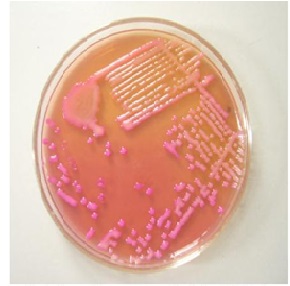
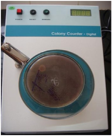
b) Aerobic culture-All samples were plated on Mac Conkey agar and chocolate agar using sterlized standard 4mm bacteriological wire loop, which holds 0.01 ml of ETA. Plates were incubated overnight at 370C and later were checked for growth. Quantitative culture threshold of ≥ 105CFU/ml was considered to diagnose VAP in our study,<105CFU/ml were assumed to be due to colonization or contamination and were categorized under NO-VAP group. Any growth was characterized by colony morphology and Gram stain. Biochemical tests were done to identify the possible causative bacteria.
The antibiotic susceptibility testing of the isolates were done on Muller-Hinton agar using standard Kirby-Bauer disc diffusion method. Bacterial suspension was prepared by inoculating few isolated colonies of similar morphology into 4-5 ml of nutrient broth and was incubated for 2-8 hours. The turbidity of the broth matched with that of 0.5 McFarland turbidity standards.
A total of 60 clinically suspected VAP patients were enrolled for the study who fulfilled our study’s predefined criteria. Among 60 patients,
Table (1):
Spectrum of Cases Studied.
Spectrum of cases |
Number |
Percentage |
|---|---|---|
Poisoning |
21 |
35.0 |
Preterm with low birth weight / hyaline membrane disease |
13 |
21.6 |
Congenital anomaly |
4 |
6.6 |
Post operative cases |
4 |
6.6 |
Meconium aspiration syndrome |
3 |
5.0 |
Dengue haemorrhagic fever |
3 |
5.0 |
Guillain Barre syndrome |
3 |
5.0 |
Hanging |
2 |
3.4 |
Snake bite |
2 |
3.4 |
Others |
5 |
8.4 |
Total |
60 |
100 |
≥105CFU/ml growth indicating pathogenic bacteria causing VAP were seen in only 28 patients (46.6%) and 32 patients (53.4%) with <105CFU/ml classified under NO-VAP group.Our study coincides with the study by Arindam Dey et al8at KMC, Manipal in 2007 and Chiranjay Mukhopadhyay et al9 with an incidence of45.4% and 42% respectively but varies with the study done by T. Rajasekhar et al7 at Nizam Institute, Hyderabad in 2006 and Shalini Tripathi et al10 at CSM Medical University, Lucknow in 2010 with an incidence of 73.3% and 30.6% respectively. The difference could be due to the different methods used for the diagnosis, sample size and the underlying disease state requiring ventilator support.

Out of the 60 clinically suspected VAP cases, 2 showed no growth and the remaining 58 showed a growth of 63 bacterias totally among which 5 multiple growth pattern were seen. In VAP group out of total 29 bacterias isolated, Acinetobacter species-12 isolates(41.4%), Klebsiella species-9 isolates (31.1%), Pseudomonas species-5 isolates(17.3%) were the most common organisms isolated followed by E. coli, methicillin sensitive Staphylococcus aureus and Coagulase negative Staphylococcus aureus accounting for 1 bacteria (3.4%). Thus bacterias considered as high risk pathogens figure prominently in our study. It correlates with various studies done by Rajasekhar et al7, Arinday Dey et al8, K H Raghwendra et al6 and Julio Medine et al11 where similar Gram negative bacterias have been identified as the causative agents of VAP. In the present study multiple isolate growth pattern was appreciated similar to other studies.8,11,12 The isolation of bacteria from clinical specimens may not necessarily mean infection but rather may result from colonization or may be a contaminant to some extent. This is reflected in our study from the bacteria isolated from the ETA of NO-VAP cases.4,7,8,9
Table (2):
Spectrum of Organisms Obtained By Endotracheal Aspiration and Its Association With Vap and No-Vap Cases.
Tracheal Isolate |
VAP (N=29) |
NO-VAP (N=36) |
Total (N=65) |
|---|---|---|---|
Klebsiella Species |
9 (31.1%) |
17 (47.2%) |
26 (40.0%) |
Acinetobacter Species |
12 (41.4%) |
11(30.5%) |
23 (35.8%) |
Pseudomonas Species |
5 (17.3%) |
3 (8.3%) |
8 (12.2%) |
Escherichia Coli |
1 (3.4%) |
2 (5.6%) |
3 (4.5%) |
MSSA |
1 (3.4%) |
1(2.8%) |
2 (3.0%) |
Coagulase Negative Staphylococci |
1 (3.4%) |
0 (0%) |
1 (1.5%) |
No Growth |
0 (0%) |
2(5.6%) |
2 (3.0%) |
N=Total Number Of Cases
Antibiotic susceptibility testing was done by standard Kirby-Bauer disc diffusion method. In our study Acinetobacter species showed sensitivity to imipenem (50%), cephotaxime, cefoperazone-sulbactem, amikacin each accounting for 16.6% and ceftazidime, ciprofloxacin, gentamicin each accounting for 8.3%. Klebsiella species were sensitive to imipenem (77.7%), ceftazidime (66.6%), cefoperazone-sulbactem (33.3%), gentamicin (33.3%), amikacin (22.2%). Pseudomonas species showed sensitivity to imipenem(100%), cefoperazone-sulbactem (80%), ceftazidime (60%). It almost correlates with the studies done by T Rajasekhar et al7,Arindam Dey et al8 and Chiranjay Mukhopadhyay et al.9
Table (3):
Percentage of Antibiotic Sensitivity Pattern of Organisms Isolated from Endotracheal Aspirates of Only Vap Cases.
Organism Drugs |
Acinetobacter Species (N = 12) |
Klebsiella Species (N = 9) |
Pseudomonas Species (N = 5) |
E. coli (N = 1) |
S. aures (N = 1) |
CONS (N = 1) |
|---|---|---|---|---|---|---|
Amikacin |
16.6% |
22.2% |
R |
100% |
– |
– |
Ampicillin |
R |
R |
– |
R |
R |
R |
Cefoperazone-Sulbactam |
16.6% |
33.3% |
80% |
100% |
– |
– |
Ceftazidime |
8.3% |
66.6% |
60% |
100% |
– |
– |
Ceftriaxone |
R |
R |
– |
100% |
– |
– |
Cephotaxime |
16.6% |
R |
– |
100% |
– |
– |
Ciprofloxacin |
8.3% |
R |
R |
R |
R |
R |
Cotrimoxazole |
R |
R |
– |
R |
100% |
100% |
Erythromycin |
– |
– |
– |
– |
100% |
100% |
Gentamycin |
8.3% |
33.3% |
R |
R |
100% |
100% |
Imipenem |
50% |
77.7% |
100% |
100% |
– |
– |
Penicillin G |
– |
– |
– |
– |
R |
R |
Vancomycin |
– |
– |
– |
– |
100% |
100% |
It is very important to have the knowledge of organisms likely to be present and also the local resistance pattern in the respective hospital ICU. Any individual study may not necessarily reflect the same situation in other centres as incriminating organisms vary among hospitals.Injudicious prophylactic use of antibiotics are not recommended in cases of VAP because exposure to antibiotics is a significant risk factor for colonization and infection with nosocomial multidrug resistant pathogens. The rational use of antibiotics may reduce patient colonization and subsequent VAP with multidrug pathogen.As incriminating pathogens vary among hospitals it is very important to know the incidence of VAP and the associated local microbial flora in each setting so as to guide more effective and rational utilization of antimicrobial agents.
- Pingleton SK,Fagon JY,Leeper KV.Patient selection for clinical investigation of ventilator associated pneumonia,criteria for evaluating diagnostic techniques. Chest 1992; 102(5):553S-556S.
- Celis R,Torres A,Gatell JM,Almela M,Rodriguez-Rosin R,Agusti-Vidal A.Nosocomial pneumonia a multivariate analysis of risk and prognosis. Chest 1998; 93(2):318-324.
- Foglia E,Meier MD,Elward A.Ventilator associated pneumonia in neonatal and pediatric intensive care unit patients. Clin Microbiol Rev 2007; 20(3):409-425.
- Koenig SM,Truwit JD.Ventilator associated pneumonia.Diagnosis,treatment and prevention. Clin Microbiol Rev 2006; 19(4):637-657.
- Rea-Neto A,Youssif NCM,Tuche F,Brunkhorst F,Ranieri VM,Reinhart K et al.Diagnosis of ventilator associated pneumonia;a systematic review of the literature. Crit Care 2008; 12(2):R56.
- Raghavendra KH,Xess A,Kumari N,Shashi SK.Microbiological study in patients on mechanical ventilation. Indian J Pathol Microbiol 2002; 45(4):451-454.
- Rajasekhar T,Anuradha K,Suhasini T,Lakshmi V.The role of quantitative cultures of non-bronchoscopic samples in ventilator associated pneumonia. Indian J Med Microbiol 2006; 24(2):107-113.
- Dey A,Bairy I.Incidence of multidrug resistant organisms causing VAP in a tertiary care hospital.A nine months prospective study. Ann Thoracic Med 2007; 2(2):52-57.
- Mukhopadhyay C,Krishna S,Shenoy A,Prakashini K.Clinical,radiological and microbiological corroboration to assess the role of endotracheal aspirate in diagnosing VAP in an ICU of a tertiary care hospital,India. Int J Infect Control 2010; 6(2):1991-1999.
- Tripathi S,Malik GK,Jain A,Kohli N.A study of VAP in neonatal ICU;characteristics,risk factors and outcome. Internet J Med 2010; 5(1):12-19.
- Medina J,Formento C,Pentet J,Curbelo A,Bazet C,Gerez J et al.Prospective study of risk factors for VAP caused by Acinetobacter species. J Crt Care 2007; 22:18-27.
- Rakshit P,Nagar VS,Deshpande AK.Incidence,clinical outcome and risk stratification of VAP-a prospective cohort study. Indian J Crit Care Med 2005; 9(4):211-216.
© The Author(s) 2017. Open Access. This article is distributed under the terms of the Creative Commons Attribution 4.0 International License which permits unrestricted use, sharing, distribution, and reproduction in any medium, provided you give appropriate credit to the original author(s) and the source, provide a link to the Creative Commons license, and indicate if changes were made.


