


ISSN: 0973-7510
E-ISSN: 2581-690X
The emergence of multidrug-resistant bacteria presents a significant global health risk, necessitating the exploration of alternative treatment options. This in vitro study aimed to evaluate the efficacy of Saudi honey Clover and Talh against Gram-negative and positive bacteria, including Escherichia coli, Pseudomonas aeruginosa, Klebsiella pneumoniae, Staphylococcus epidermidis and Methicillin-resistant Staphylococcus aureus. All tested bacteria were collected and two types of Saudi honey were diluted and stored. This is followed by microbiological identification, prolonged exposure to honey (10 times), antibiotic susceptibility testing, and biofilm formation assay. The findings offer valuable insights into potential antimicrobial properties of Saudi honey against bacteria. The results demonstrated that Talh and Clover Honey caused alterations in the antimicrobial sensitivity of the bacteria tested. In Gram-negative bacteria, E. coli treated with honey showed increased sensitivity to imipenem, while P. aeruginosa and K. pneumoniae became increasingly susceptible to cell wall inhibitors. In Gram-positive bacteria, adapted MRSA and S. epidermidis strains exhibited increased sensitivity to ampicillin, amoxicillin/clavulanic acid, and ceftriaxone. The results suggest that honey’s antimicrobial effects vary based on its components and bacterial species. Consequently, further analysis is required to uncover the underlying mechanisms of the observed changes. The complex interactions between honey and pathogens, as shown by varying bacterial responses and limited impact on biofilm formation, highlighting the necessity for alternative therapeutic approaches. This study contributes to the existing evidence on the antimicrobial properties of honey.
MDR, Antimicrobial Activity, Natural Treatment, Honey, Antibiotic Resistance
Multidrug-resistance (MDR) has emerged as a life-threatening health problem that affects millions of patients globally.1 Over the past decades, the number of MDR bacteria has significantly increased, posing a global health challenge.2 In Europe alone, approximately 33,000 individuals die annually due to MDR bacteria.3 A global study has predicted that unless the appropriate prevention protocols are implemented, nearly 10 million lives could be lost by 2050.4
One of the most prevalent MDR bacteria is methicillin-resistant Staphylococcus aureus (MRSA).5 The worldwide prevalence of MRSA infections varies widely, ranging from 13% to 74%. In Europe, the prevalence of MRSA in 2014 ranged from 0.9% to 56%.6 In the United States, around 50% of S. aureus infections acquired in intensive care units were attributed to MRSA.7 Multiple studies conducted in Saudi Arabia have indicated an increasing trend in the prevalence of MRSA over time, emphasizing it as a growing public health concern in the region.8-10
A study conducted in Saudi Arabia at King Fahd Medical City in Riyadh from 2019 to 2020 found that 33.49% of E. coli isolates from urinary tract infections were extended-spectrum β-lactamase-producing strains.10 Further, a previous study was conducted over an eleven-year from January 2011 to December 2021 to inspect the prevalence and antimicrobial profile of Klebsiella pneumoniae (K. pneumoniae).11 A subsequent study conducted at a tertiary hospital in Mecca aimed to evaluate the trends in antibiotic resistance of Klebsiella pneumoniae over this period. During the study, a total of 61,027 bacterial isolates were collected from clinical samples, out of which 14.7% (n = 9014) were identified as K. pneumoniae. The researchers analysed the antibiotic susceptibility pattern of K. pneumoniae isolates to determine their resistance rates. The findings revealed a concerning increase in resistance rates to several commonly used antibiotics over the eleven-year period. Notably, resistance rates for amoxicillin/clavulanate increased from 33.6% in 2011 to 71.4% in 2021, and for piperacillin/tazobactam, the resistance rates rose from 13.6% in 2011 to 84.9% in 2021.11
In Qatar, a study investigated MDR P. aeruginosa isolates and found a prevalence of 5.9% among clinical samples.12 The majority of cases were hospital-acquired, and the isolates displayed high resistance rates to various antibiotics but remained susceptible to colistin.12 The genetic analysis identified different sequence types and highlighted the presence of exotoxin genes, indicating potential virulence.12 Given the challenges posed by MDR bacteria, alternative treatment methods have gained attention. Natural products such as liquorice, chamomile, and honey have shown antimicrobial properties and potential efficacy against MDR bacteria.13-15
The antimicrobial effects of honey result from its high viscosity, acidic nature, osmotic effect, and the presence of hydrogen peroxide, which collectively inhibit bacterial growth and damage bacterial DNA.16 Therefore, in this study, we used two types of Saudi made honey including (Talh and Clover) to investigate their properties against MDR bacteria such as MRSA.
Samples collection
A selection of six bacterial strains including E. coli, P. aeruginosa, extended spectrum beta-lactamase (ESBL) producing K. pneumoniae, S. aureus, MRSA, and S. epidermidis were obtained from King Abdulaziz University Hospital (KAUH), Jeddah, Saudi Arabia. The Clover and Talh honey were ordered from Alabdali Honey Company, Hail, Saudi Arabia. After the collection we diluted the honey by 30 mg/mL with distilled water (dH2O) followed by storage at 10 °C. Antibiotic discs, such as gentamicin (120 µg), ceftazidime (30 µg), ampicillin (10 µg), amoxicillin/clavulanic acid (30 µg), ceftriaxone (30 µg), and imipenem (10 µg), were sourced from Al Wareed Medical Company in Makkah, Saudi Arabia, and stored at 20 °C for further analysis.
Microbiological culture and identification
All bacterial strains were cultured and inoculated on MacConkey agar media for Gram-negative, and blood agar media for Gram-positive followed by aerobic incubation at 37 °C and stored at 4 °C for the next experiment.
Long term exposure to honey (Adaptation process)
Agar-based diffusion was used to repeatedly expose each individual bacterium (ten times) to each tested honey samples (Talh and Colver) to examine the impact of prolonged in vitro exposure of bacterial isolates to different honey samples.17 Three 6-millimeter-diameter wells were created in Mueller-Hinton agar (MHA). Each of these wells was used to culture different bacterial strains by spreading them onto the agar. 200 µL of honey was added and diluted by 30 mg/mL dissolved in sterile (dH2O). In each well, Talh honey and Clover honey were in separate agars. The culture is incubated at 37 °C for 24 hours. The unexposed bacteria P0 were sub-cultured in agar to form a negative control, and to exclude any existing contamination.
Antibiotic Susceptibility Testing (AST)
The first antibiotic sensitivity test was disc diffusion test (DDT) that were used to compare between the (P0) and (P10) Bacteria by applying 6 different antibiotics including gentamicin, ceftazidime, ampicillin, amoxicillin/clavulanic, ceftriaxone, and imipenem.18 DensiChek was used to adjust the bacterial concentration to match the standard turbidity of 0.5 McFarland. Then, using cotton swabs, P0 and P10 bacterial suspensions were applied to the MHA agar’s surface.18 Then added antibiotics disc for the six antibiotics. The MHA agar plates were incubated at 37 °C for 24 hours, and inhibition zones were measured in millimetres (mm) following incubation. To produce more accurate and repeatable results, this testing was done three times for every bacterium. Standard deviations and the mean of the three reads were calculated as well.
Biofilm formation assay
The formation of biofilm formation was evaluated using crystal violet stain by examining bacterial strains that were treated with the honeys (P10) and the untreated strains (P0). The bacterial cultures were diluted (1:100) by using Muller Hinton broth (MHB), followed by transferring to a 96-well microtiter plate and incubated at 37 °C for two days. After incubation the bacterial suspension were discarded, then by using a normal saline it has been washed twice. About 1% of crystal violet satin was added for few minutes. Finally, ethanol was added to each well, then reading the optical density of the staining at wavelength 600 (OD600) by spectrophotometer.16-18
Data analysis
All results were statistically analyzed using GraphPad (Prism 9). The biofilm formation assay was readied to detect any significant changes between the control (P0) (untreated) and (P10) (honey-treated) using both the Student t-test and one-way ANOVA, with p < 0.05 considered statistically significant.
Influence of honey exposure on Gram-negative bacteria to antibiotics
The exposure of E. coli to Talh and Clover honey did not result in any significant changes in antibiotic sensitivity for ceftazidime, ampicillin, amoxicillin/clavulanic acid, or ceftriaxone (Figure 1). However, when gentamicin was combined with Clover honey, there was a slight increase in the zone of inhibition compared to gentamicin alone, although this difference was not statistically significant (p = 0.7016). Alternatively, both Talh and Clover honey had a statistically significant effect on the zone of inhibition for imipenem (p = 0.0457 for Clover and p = 0.0018 for Talh). For P. aeruginosa, the combination of Talh and Clover honey with imipenem and gentamicin significantly enhanced the zone of inhibition across all treated groups. Additionally, a slight increase in the zone of inhibition was observed in the P10 strain treated with Clover honey when combined with ampicillin and amoxicillin/clavulanic acid, and in the P10 strain treated with Talh honey when combined with ceftazidime. However, these increases were not statistically significant. Similarly, for K. pneumoniae, exposure to Talh and Clover honey increased the zone of inhibition for gentamicin and imipenem across all tested groups, but these changes were also not statistically significant. Additionally, a slight increase in the zone of inhibition was observed with Talh honey in P10 strains treated with amoxicillin/clavulanic acid, and with Clover honey in P10 strains treated with ampicillin.
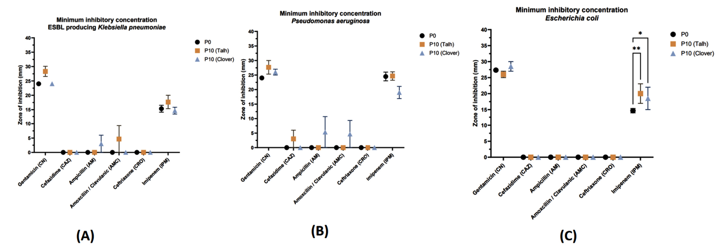
Figure 1. Gram-negative bacterial strains were treated with Clover and Talh honey, and antibiotic sensitivity testing was conducted to determine the minimum inhibitory concentration.
* represent P value ≤ 0.05. ** represent P value ≤ 0.01
Effect of honey exposure on Gram-positive bacteria to antibiotics
Across all Gram-positive strains gentamicin exhibited significant inhibitory effects, with large zones of inhibition observed in the P0 group (Figure 2). However, in both P10 (Talh) and P10 (Clover) groups, a slight reduction in the zone of inhibition was noted, suggesting that honey might reduce the effectiveness of Gentamicin slightly, particularly in MRSA and S. aureus. No significant zones of inhibition were observed across the bacteria for the P0 group, indicating inherent resistance to Cefazidime. Interestingly, a minimal zone of inhibition was detected for S. aureus in the P10 (Clover) group, implying a slight potentiation of Cefazidime effect due to Clover honey. Both P10 (Talh) and P10 (Clover) groups exhibited significant increases in the zones of inhibition across Gram-positve bacteria, compared to the P0 control. This suggests that honey enhances the effectiveness of Ampicillin against Staphylococcus species, including the resistant MRSA. Similar to Ampicillin, the addition of honey (both Talh and Clover) significantly increased the zones of inhibition, indicating that honey enhances the antibacterial activity of Amoxicillin/Clavulanic acid. Ceftriaxone shows that results were consistent with those observed for Ampicillin and Amoxicillin/Clavulanic acid, with significant zones of inhibition in the honey-treated groups compared to the control. This reinforces the idea that honey can potentiate the effectiveness of certain antibiotics.
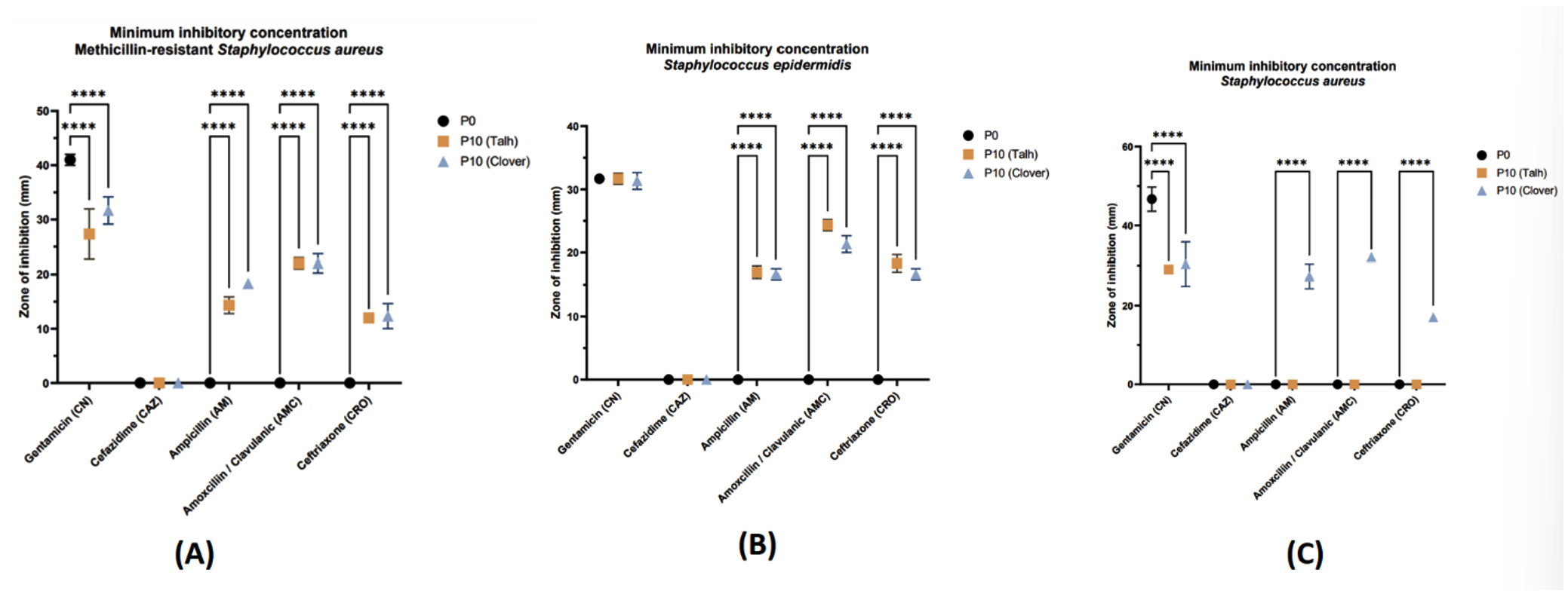
Figure 2. Gram-positive bacterial strains were treated with Clover and Talh honey, and antibiotic sensitivity testing was conducted to determine the minimum inhibitory concentration.
**** represent P value < 0.0001
Biofilm formation of Gram-negative and Gram-positive bacteria
Across both Gram-negative and Gram-positive bacteria, treatment with Talh and Clover honey led to some minor variations in biofilm formation (Figure 3). However, none of these changes were statistically significant, indicating that while honey might have a mild effect on biofilm formation in certain bacterial strains, it does not significantly alter biofilm production under the conditions tested. Further research could explore different concentrations and exposure durations to evaluate whether honey could have a more pronounced effect on biofilm formation of these bacteria.
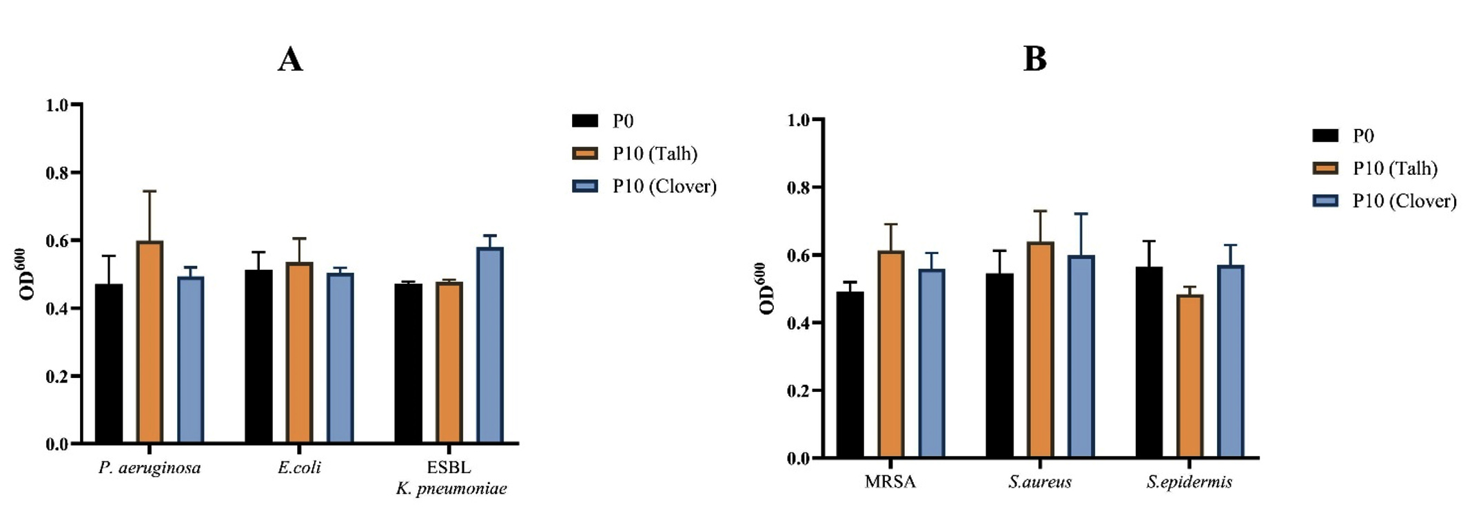
Figure 3. The impact of the adaptation process with Clover and Talh honey on biofilm formation in both Gram-negative and Gram-positive bacteria, including P. aeruginosa, E. coli, ESBL-producing K. pneumoniae, MRSA, S. aureus, and S. epidermidis
Honey is a highly natural substance that has been extensively used across diverse cultures for its medicinal and dietary benefits.17 The remarkable therapeutic potential of honey stems from its distinct properties, including its elevated viscosity, acidic nature, and the presence of hydrogen peroxide.18 These characteristics collectively contribute to its capacity as a potent antimicrobial agent.18,19 The mechanism of antimicrobial activity of honey mainly involved stimulation of bacterial dehydration, inhibition their reproductive capabilities, and facilitate DNA degradation through the generation of radicals and oxygen emphasises its promising role as an alternative treatment option for combating emerged antimicrobial resistance. Although the bacterial adaptation to certain types of honey was previously described,17-20 the adaptation effect of new types of Saudi honey including Talh and Clover has not yet been investigated.
In the current research, a series of clinical Gram-negative bacterial isolates including (E. coli, P. aeruginosa, and K. pneumoniae) and Gram-positive bacterial isolates (S. aureus, MRSA, S. epidermidis) were subjected to a repeated exposure protocol of Talh and Clover honey. The goal was to induce adaptation in the bacterial population, resulting in the emergence of a modified bacteria strain referred to as P10. The antibiotic susceptibility and biofilm formation of the adapted bacterial strains were thoroughly examined. In the case of Gram-negative bacteria, E. coli strain that were treated with Talh and Clover honey exhibited a notable significant increase in the zone of inhibition against imipenem (cell wall inhibitor). This strain showed resistant to ceftazidime, ampicillin, amoxicillin/clavulanic and ceftriaxone. Moreover, P. aeruginosa control strains were resistant to ceftazidime, ampicillin, amoxicillin/clavulanic and ceftriaxone, while the adapted treated strains with Talh and Clover were only sensitive to ceftazidime and ampicillin, amoxicillin/clavulanic (cell wall inhibitors), respectively. Furthermore, ESBL-producing K. pneumoniae strains treated with Talh and Clover were sensitive to gentamicin and imipenem, respectively.
The increase zone of inhibition could be through interfering with bacterial membrane permeability, inhibiting proteins as, Al-Sayaghi explored the antibacterial effects and mechanisms of Yemeni Sidr honey (SH) and Manuka honey (MH) against E. coli. Using the disk diffusion method, both types demonstrated strong inhibitory effects at 700 mg/disk, with similar MIC and MBC values. MH exhibited a stronger bactericidal effect (30%) than SH (50%). SH compromised bacterial membrane integrity, causing potassium and protein leakage, while MH primarily inhibited bacterial protein synthesis.19 Both types of honey also induced bacterial DNA damage.
Regarding Gram-positive bacteria, the adapted S. epidermidis and MRSA strains exposed to Talh and Clover honey showed increased sensitivity to three tested antibiotics (cell wall inhibitors) such as ampicillin, amoxicillin/clavulanic acid, and ceftriaxone. However, only S. aureus adapted with Clover honey exhibited a zone of inhibition to the same antibiotics. This result suggests that the concentration and components of honey, as well as the nature of the bacteria, may have a potential role on its antimicrobial properties and effectiveness. In contrast, previous studies examined the effect of Manuka honey on S. aureus exposed to the same antibiotics, resulted in increased sensitivity.18,20,21
The overall findings of this study are in line with a recent study conducted in Hail, Saudi Arabia where the efficacy of different Saudi honey was analysed.20 The study investigated the effects of Sumra and Sider honey on reference bacterial strains, revealing a more significant impact of in vitro honey exposure on antibiotic sensitivity compared to clinical strains. This suggests that reference strains, typically less exposed to antibiotics, respond differently than clinical strains.20,21
The increased sensitivity of treated bacteria to tested cell wall inhibitors suggests a possible alteration in their cell wall permeabilit22 or efflux system23 after exposure to honey. Furthermore, the repeated in vitro treatment with honey (adaptation process) may affect the bacterial fitness and growth causing an enhanced antimicrobial sensitivity of adapted strains.24,25
Biofilm formation is widely known as one of the essential bacterial virulence factors that can delay wound healing and resist both host immune defence and antimicrobial agents leading to sever infections and treatment complications.26 Previous studies reported the potential antibiofilm activity of various honey types including Manuka, Sumra and Sider that were tested with biofilm former bacteria such as S. aureus and Acinetobacter baumannii.20,27,28 However, there was an observation of an increased tendency of biofilm formation among tested Gram-negative bacteria adapted with Manuka honey.21 Similarly, the current study of Talh and Clover honey treatment showed limited impact on biofilm formation among the tested strains suggesting that honey contains multiple components that may resulted in antimicrobial and antibiofilm activities via various underlying mechanisms, thus limited resistance may be developed as defensive pathway. Therefore, further analysis is required to uncover the underlying mechanisms responsible for the observed changes in antimicrobial sensitivity. The complex interactions between honey and MDR pathogens, as evidenced by varying bacterial responses and limited impact on biofilm formation, highlight the necessity for alternative therapeutic approaches.
Although the current in vitro study exhibits an enhanced antibiotic sensitivity of tested clinical bacteria after exposure to Talh and Clover honey treatment, there were number of limitations faced the study. The virulence of bacteria could be examined using in vivo study to understand the bacterial response and pathogenesis after honey treatment. Furthermore, the effect of honey treatment on bacterial growth and virulence could be examined at the molecular or genomic level to identify any potential variance or mutation. Thus, further investigation is essential to fully comprehend the mechanisms involved and optimize honey-based treatment strategies for addressing antibiotic resistance health issue.
In conclusion, repeated exposure of clinical bacterial pathogens to Saudi Clover and Talh honey led to alterations in their antimicrobial sensitivity profiles and biofilm formation tendencies. While some bacterial strains developed limited resistance to the honey compared to the original strains, the adapted pathogenic bacteria, especially E. coli, generally exhibited increased susceptibility to Saudi Clover honey. Although the precise mechanisms underlying the observed reduction in antibiotic sensitivity remain unclear and require further investigation, this study underscores the potential therapeutic applications of Saudi Honey in treating bacterial infections. These findings highlight the positive impact of Saudi Honey on the sensitivity profiles of various antimicrobial agents against the tested bacteria, offering a promising avenue for future medical research.
ACKNOWLEDGMENTS
The authors would like to acknowledge and appreciate the support from King Abdulaziz University, Saudi Arabia.
CONFLICT OF INTEREST
The authors declare that there is no conflict of interest.
AUTHORS’ CONTRIBUTION
All authors listed have made a substantial, direct and intellectual contribution to the work, and approved it for publication.
FUNDING
None.
DATA AVAILABILITY
All datasets generated or analyzed during this study are included in the manuscript.
ETHICS STATEMENT
Not applicable.
- Catalano A, Iacopetta D, Ceramella J, et al. Multidrug Resistance (MDR): A Widespread Phenomenon in Pharmacological Therapies. Molecules. 2022;27(3):616.
Crossref - Kumar NR, Balraj TA, Kempegowda SN, Prashant A. Multidrug-Resistant Sepsis: A Critical Healthcare Challenge. Antibiotics. 2024;13(1):46.
Crossref - Cassini A, Hogberg LD, Plachouras D, et al. Attributable deaths and disability-adjusted life-years caused by infections with antibiotic-resistant bacteria in the EU and the European Economic Area in 2015: a population-level modelling analysis. Lancet Infect Dis. 2019;19(1):56-66.
Crossref - Garg R, Singh G, Rathore L, Sharma V, Salman M. A review on antimicrobial resistance. Indian J Heal Care Med Pharm Pract. 2024;5(1)50-56.
Crossref - Nandhini P, Kumar P, Mickymaray S, Alothaim AS, Somasundaram J, Rajan M. Recent Developments in Methicillin-Resistant Staphylococcus aureus (MRSA) Treatment: A Review. Antibiotics. 2022;11(5):606.
Crossref - Adam KM, Abomughaid MM. Prevalence of Methicillin-resistant Staphylococcus aureus in Saudi Arabia Revisited: A Meta-analysis. Open Public Health J. 2019;11(1):584-591.
Crossref - Aljeldah MM. Prevalence of Methicillin-Resistant Staphylococcus aureus (MRSA) in Saudi Arabia: A systematic review. J Pure Appl Microbiol. 2020;14(1):37-46.
Crossref - Mahmoud E, Al Mansour S, Bosaeed M, et al. Ceftobiprole for treatment of mrsa blood stream infection: A case series. Infect Drug Resist. 2020;2020(13):2667-2672.
Crossref - Alhunaif SA, Almansour S, Almutairi R, et al. Methicillin-Resistant Staphylococcus aureus Bacteremia: Epidemiology, Clinical Characteristics, Risk Factors, and Outcomes in a Tertiary Care Center in Riyadh, Saudi Arabia. Cureus. 2021;13(5):14934.
Crossref - Abalkhail A, AlYami AS, Alrashedi SF, et al. The Prevalence of Multidrug-Resistant Escherichia coli Producing ESBL among Male and Female Patients with Urinary Tract Infections in Riyadh Region, Saudi Arabia. Healthcare. 2022;10(9):1778.
Crossref - Jalal NA, Al-Ghamdi AM, Momenah AM, et al. Prevalence and Antibiogram Pattern of Klebsiella pneumoniae in a Tertiary Care Hospital in Makkah, Saudi Arabia: An 11-Year Experience. Antibiotics. 2023;12(1):164.
Crossref - Ahmed MAS, Hassan AAI, Jarir SA, et al. Emergence of Multidrug- and Pandrug- Resistant Pseudomonas aeruginosa from Five Hospitals in Qatar. Infect Prev Pract. 2019;1(3-4):100027.
Crossref - El Mihyaoui A, Esteves Da Silva JCG, Charfi S, Castillo MEC, Lamarti A, Arnao MB. Chamomile (Matricaria chamomilla L.): A Review of Ethnomedicinal Use, Phytochemistry and Pharmacological Uses. Life. 2022;12(4):1-41.
Crossref - Combarros-Fuertes P, Fresno JM, Estevinho MM, Sousa-Pimenta M, Tornadijo ME, Estevinho LM. Honey: Another alternative in the fight against antibiotic-resistant bacteria? Antibiotics. 2020;9(11):774.
Crossref - Edo GI, Onoharigho FO, Akpoghelie PO, et al. Natural Honey (Raw Honey): Insights on Quality, Composition, Economic and Health Effects: A Comprehensive Review. Food Sci Eng. 2023;4(2):265-293.
Crossref - Bischofberger AM, Cardozo KRP, Baumgartner M, Hall AR. Evolution of honey resistance in experimental populations of bacteria depends on the type of honey and has no major side effects for antibiotic susceptibility. Evol Appl. 2021;14(5):1314-1327.
Crossref - Samarghandian S, Farkhondeh T, Samini F. Honey and health: A review of recent clinical research. Pharmacognosy Res. 2017;9(2):121-127.
Crossref - Kassem LM, El-Deen AG, Zaki AH. et al. Electrospun manuka honey@PVP nanofibers enclosing chitosan-titanate for highly effective wound healing. Cellulose. 2023; 30:6487-6505.
Crossref - Al-Sayaghi AM, Al-Kabsi AM, Abduh MS, Saghir SAM, Alshawsh MA. Antibacterial Mechanism of Action of Two Types of Honey against Escherichia coli through Interfering with Bacterial Membrane Permeability, Inhibiting Proteins, and Inducing Bacterial DNA Damage. Antibiotics. 2022;11(9):1182.
Crossref - Aldarhami A, Bazaid AS, Qanash H, et al. Effects of Repeated in vitro Exposure to Saudi Honey on Bacterial Resistance to Antibiotics and Biofilm Formation. Infect Drug Resist. 2023;16:4273-4283.
Crossref - Mokhtar JA, McBain AJ, Ledder RG, Binsuwaidan R, Rimmer V, Humphreys GJ. Exposure to a Manuka Honey Wound Gel Is Associated With Changes in Bacterial Virulence and Antimicrobial Susceptibility. Front Microbiol. 2020;11:2036.
Crossref - Brudzynski K, Sjaarda C. Antibacterial compounds of canadian honeys target bacterial cell wall inducing phenotype changes, growth inhibition and cell lysis that resemble action of b-lactam antibiotics. PLoS One. 2014;9(9):106967.
Crossref - Combarros-Fuertes P, Estevinho LM, Teixeira-Santos R, et al. Evaluation of physiological effects induced by manuka honey upon Staphylococcus aureus and Escherichia coli. Microorganisms. 2019;7(8):258.
Crossref - Marro FC, Abad L, Blocker AJ, Laurent F, Josse J, Valour F. In vitro antibiotic activity against intraosteoblastic Staphylococcus aureus: A narrative review of the literature. J Antimicrob Chemother. 2021;76(12):3091-3102.
Crossref - Zur Wiesch PS, Engelstadter J, Bonhoeffer S. Compensation of fitness costs and reversibility of antibiotic resistance mutations. Antimicrob Agents Chemother. 2010;54(5):2085-2095.
Crossref - Gilbert P, Allison DG, McBain AJ. Biofilms in vitro and in vivo: Do singular mechanisms imply cross-resistance? J Appl Microbiol Symp Suppl. 2002;92(1):98-110.
Crossref - Bazaid AS, Aldarhami A, Patel M, et al. The Antimicrobial Effects of Saudi Sumra Honey against Drug Resistant Pathogens: Phytochemical Analysis, Antibiofilm, Anti-Quorum Sensing, and Antioxidant Activities. Pharmaceuticals. 2022;15(10):1212.
Crossref - Kot B, Sytykiewicz H, Sprawka I, Witeska M. Effect of manuka honey on biofilm-associated genes expression during methicillin-resistant Staphylococcus aureus biofilm formation. Sci Rep. 2020;10(1):1-12.
Crossref
© The Author(s) 2025. Open Access. This article is distributed under the terms of the Creative Commons Attribution 4.0 International License which permits unrestricted use, sharing, distribution, and reproduction in any medium, provided you give appropriate credit to the original author(s) and the source, provide a link to the Creative Commons license, and indicate if changes were made.