


ISSN: 0973-7510
E-ISSN: 2581-690X
The enzyme L-asparaginase is commonly used in treatment of acute lymphoblastic leukemia (ALL). Due to its therapeutic importance and growing demands in medical and food industry much of the research have been focused to produce this enzyme with better activity and less disadvantages; for instance microbes have been extensively used to produce L asparaginase at affordable prices, a wide range of bacterial species have been reported that can produce high yields of this enzyme. The present study is an attempt to compare the production of L asparaginases between gram positive and gram negative bacterial species isolated from soil under same fermentation conditions. In this study the productions of L asparaginase from three species of Bacillus namely Bacillus subtilis (LC425423), Bacillus aerophilus (LC425427), Bacillus endophyticus (MG928501) and three strains of Pseudomonas aeruginosa (LC425424, LC425425, LC425426) was compared. The enzyme activity of L asparaginases produced from Bacillus species were noted as 8.5, 14.3, 7.1 (U/ml). Whereas, the enzyme activity of L asparaginase produced from Pseudomonas aeruginosa strains (LC425424, LC425425, LC425426) was noted as, 8.2, 19.4, 19.1(U/ml) respectively. In this study it was found that Pseudomonas aeruginosa strains yields was more in terms of protein concentration and enzyme activity when compared to Bacillus species. In addition this study also reports the use of modified Brain heart infusion media for screening and isolation of L asparaginase producing bacterial species. It was found that the number of colonies producing L asparaginase in modified Brain heart infusion media was comparatively same when compared to modified M9 media which is extensively used in screening of L asparaginase producers. A total of 159 L asparaginase producers were screened by using two fold dilution technique on modified BHI media while modified M9 media screened 163 colonies producing L asparaginase.
Brain heart infusion media (BHI), ALL (acute lymphoblastic leukemia), L asparaginase (ASP).
Acute Lymphoblastic Leukemia is a malignant disease that arises from several cooperative genetic mutations in a single B-or T-lymphoid progenitor, leading to altered blast cell proliferation, survival and maturation and eventually to the lethal accumulation of leukemic cells1. L asparaginase is a wonder drug that is being used extensively in treatment of ALL specially in pediatric cases with a cure rate of 90 %2. A number of microorganisms have been employed to produce L asparaginases on commercial scale, E.coli and Erwinia asparaginases are the most important bacterial asparaginase that are used in ALL treatment3. In recent years search for bacterial asparaginases is more profoundly focused as E.coli asparaginases induce allergic reactions while Erwinia asparaginase is short lived3, 4. L asparaginase during the treatment hydrolyze the freely available aspargine to aspartate and ammonia; depletion of surrounding aspargine results in scarcity of aspargine amino acid, which is an essential amino acid for cancer cells and cannot synthesize it; as they lack enzyme aspargine synthetase which synthesizes aspargine. Therefore leukemic cells cannot proliferate in absence of aspargine; thereby resulting in their death. Besides being used in treatment of ALL the enzyme L asparaginase is also used as acrylamide reducer from fried and baked foods5. Therefore owing to its therapeutic and industrial importance a number of microbial species such as bacteria, fungi, actinomycetes and yeasts have been searched and reported for cost effective production of L asparaginase. Since bacterial species grow rapidly and exhibit easy product recovery; thus have attracted researchers to search and report, novel bacterial species including both gram positive and gram negative that can produce L asparaginases with promising yields and high enzyme activity. The Gram positive bacterial species that were reported to produce L asparaginase are B.cereus, B.subtilis, B.polymyxa, B. licheniformis, B. mesentricus, Coryne bacterium glutamicum etc. Nevertheless gram negative bacterial species such as P aeruginosa, P. stutzeri, E.coli, Erwinia aeroideae, Vibryo cholera etc have been reported to produce L asparaginase enzyme6. However a number of studies have appeared in the past decade pertaining to Pseudomonas aeruginosa and Bacillus species. Hence this study is an attempt to compare the production of L asparaginase from three strains P aeruginosa (LC425424, LC425425,LC425426)and three species of Bacillus namely Bacillus subtilis(LC425423) , Bacillus aerophilus (LC425427) and Bacillus endophyticus (MG928501) under similar fermentation conditions . Besides this the study is also an attempt to compare screening efficiencies of two screening mediums i.e modified M9 media (conventional media) and modified Brain heart infusion media to isolate asparaginase producing bacteria.
Collection of soil samples
In the present study, in an order to isolate promising L-asparaginase producing bacterial species, number of soil samples were collected from places of Hyderabad especially from Gardens from the depth of 10 to 30cm, the samples were labeled. These soil samples were then grinded to fine powder and then sieved to obtain even soil texture powder. All process was carried out in Microbiology research lab in Mumtaz PG College7.
Comparison of modified M9 and BHI media for screening of L asparaginase producing bacterial species
One gram of fine powder of soil was weighed and suspended in 9ml physiological saline kept in a flask. It was then mixed in an orbital shaker set at 100rpm set at about 28°C ± 2°C for one hour. After the given period, flask was allowed to settle the suspended matter. In a next step from the flask series of dilution was made by using physiological saline by taking one ml sample and transferring to 9ml saline to give 10-1 -10-10 dilutions. Further one ml of each dilution was plated on modified brain heart infusion agar (BHA) medium by spread plate technique. Here the 1 liter of BHA medium was previously modified by adding 6gms of KH2PO4, Asparagine -10gms, 4ml of 1M MgSO4, 2ml 0.1MCaCl2, 0.4ml 0.009% phenol red indicator and finally pH was set in as 7 with 1N Hcl. Whereas the 1 liter of modified M9 media was prepared as Na2HPO4.2H20 (0.5g), KH2PO4 (0.75g), NaCl (0.5g); L-asparagine (10gms), 1M MgSO4.7H20- 2 ml; 0.1M CaCl2 2H20- 1 ml; 20% Glucose stock solution- 10 ml, Agar, phenol red indicator (2.5%): 0.04-0.36 ml, pH 7.0. In a control set, plates of BHA and M9 were made without dye and asparagine (Fig. 3). All the inoculated plates were allowed to get incubated at 37°C for 24 hours7-9.
Screening of L-asparaginase producing bacteria
Once the incubation period was over, plates from of BHA and M9 plates with separated colonies were selected. Further only those colonies exhibiting pink zone were considered as L-asparaginase positive colonies (Fig. 1 &2). This pink color zone formed around the colony indicates that the asparagine was hydrolyzed to yield aspartate and ammonia, the release of ammonia is detected by phenol red pH indicator. Phenol red at acidic pH converts to yellow and turns pink when experiences basic pH. Hence in the study isolates showcasing pink zone around isolates were considered as L-asparaginase producers. The isolated colonies with pink zone were sub-cultured followed by Gram staining and processed further to obtain pure culture8, 10.
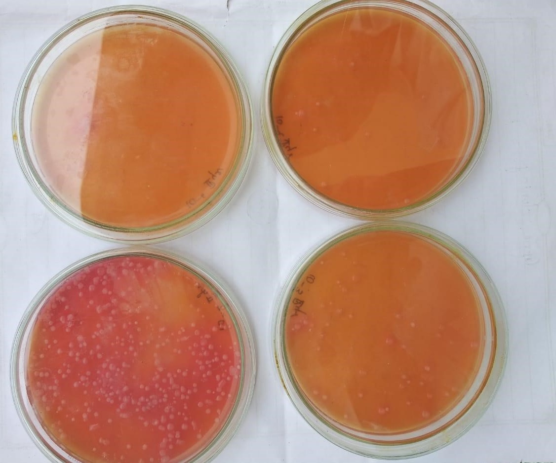 Fig. 1. Colonies producing pink zone (L asparaginase producers) on modified BHI infusion media
 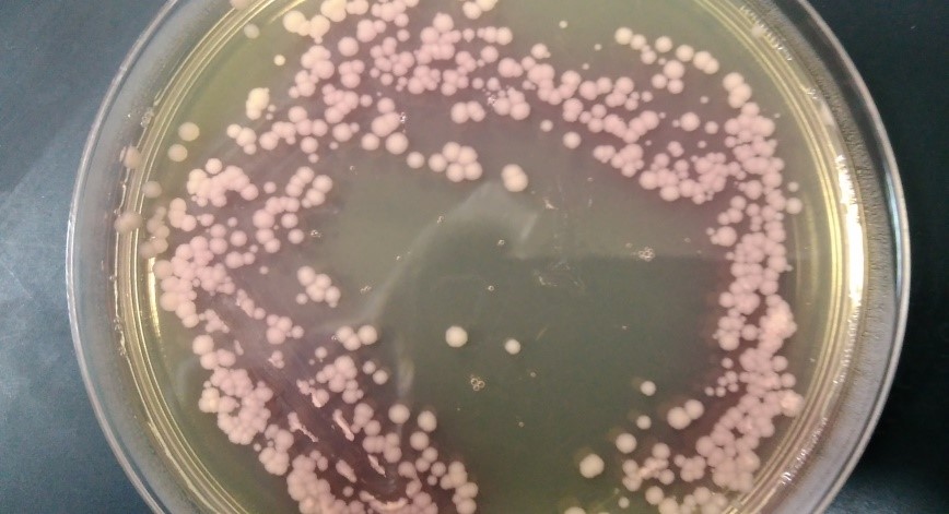 Fig. 2. Colonies producing pink zone (L asparaginase producers) on modified BHI infusion media
 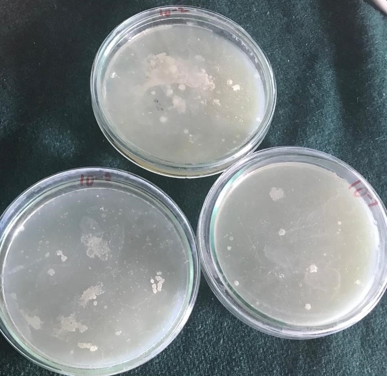 Fig. 3. Control plates of Brain heart infusion media without aspargine and phenol red indicator
 Qualitative screening of l aspaginase producers
Agar well diffusion assay (AWD) was used to select the promising producers of L asparaginase. In this method culture filtrate was screened for the L-asparaginase presence from the selected isolates. In this protocol, 50ml of asparagine tryptone glucose yeast extract broth medium was kept in a 250ml Erlenmeyer conical flask, sterilized, inoculated, and allowed to incubate at 37°C for 48hours in a rotatory incubating shaker at 150rpm. From this medium, 50µl of culture broth was used as an inoculum in a well formed in a modified BHA medium having 1% asparagine and 0.009% phenol red indicator. The well was punctured with 8mm area by using sterile cork borer. In case of control, wells without any culture broth was taken. All these plates were further incubated at 37°C for 24 hours. Upon incubation, appearance of pink zone around the well indicated positive test for L-asparaginase. Zone formed around the well was measured and recorded; the isolates show casing the biggest pink zones where selected for fermentation to produce L asparaginase5, 10, 11.
Enzyme production by submerged fermentation method
Inoculum preparation
In an inoculum built up, loopful of culture of selected isolates from were first allowed to incubate in 50ml of inoculum media having composition Na2HPO4– 0.6gms; KH2PO4-0.3gms; NaCl- 0.5gms; glucose-1gms; 1M MgSO4– 2ml; 0.1M CaCl2– 1ml in 1000ml volume. Here medium pH set at 7.2 and inoculated flasks were incubated at 37°C for 16-18 hours12.
Production of L-Asparaginase
Inoculum of 2 ml volume was inoculated in 250 ml of Flasks containing 50 ml of production media. Here in a production media (50 ml) which is made up of tryptone glucose yeast extract broth along with 1 % asparagine at pH 8.0. These inoculated flasks were kept on shaking orbital incubator at 37°C at 150 rpm for 48 hours incubation along with the control set, where production media was kept un-inoculated11, 13.
L -Asparaginase recovery: After incubation, all flasks were processed by configuring broth at 10,000 rpm for 10 min at 4°C. The supernatant obtained after centrifugation assumes to contain L-asparaginase was used for purification by dialysis method14.
Partial purification of L asparaginase
Activation of nitro cellulose bag
Partial purification of crude enzyme preparation of L-asparaginase was carried out by using dialysis technique; in which 8cm Nitro cellulose membrane was used. The crude supernatant was taken in graduated cylinder and salting out done with 70 % salt, cut-off was carried out for the extract under ice cold conditions with continuous stirring. Finally 10 ml of protein extract was added up with 7g of ammonium sulphate in a pinch to pinch addition and continuous stirring where whole preparation carried out on a cool pack and incubated at 4°C overnight. After 12 hour incubation, precipitated protein was centrifuged at 10,000 rpm for 10 minutes at 4°C. The pellet obtained was dissolved in 10 ml of 10mM tris HCl buffer which was later on subjected to dialysis15.
Dialysis
Dialysis bag of 8cm was activated by boiling in 100ml of distilled water for next 10 minutes so that pores of dialysis membrane gets opened up. It was then boiled in 100ml of 2% sodium carbonate solution for about 10 minutes which ensured removal of glycerol coated in the dialysis membrane. The bag was again kept on boiling in 100ml of distilled water which ensured residual sodium bicarbonate removal. In the next step, the protein dissolved in Tris HCl was centrifuged at 10000 rpm for 10 mins and pellet was collected. Now the mouth of the dialysis bag was gently rubbed to open it. One end of the dialysis membrane was tied with thread and the sample was placed inside the dialysis bag. After addition of sample, the other end of the membrane was tied with thread tightly. The dialysis bag was then suspended in a beaker containing distilled water. This setup was kept in refrigerator overnight. After that the protein present in the bag was dissolved in 10ml Tris HCl. After the overnight the protein present in bag was dissolved in 10ml Tris HCL15, 16.
Quantification of dialyzed protein: by using Lowry’s method amount of protein content present in crude preparation and dialyzed supernatant was determined17.
Enzyme activity
In a first step, 0.5 ml of partially purified enzyme was taken, to which 1 ml of potassium phosphate buffer (0.02 M and pH 8.6) and 0.5 ml of 50mM aspargine was mixed well and incubated at 37°C in a water bath for 15 Minutes. After incubation, 1 ml of 1.5 M trichloroacetic acid was added to the mixture to stop the reaction. Then whole mixture was centrifuged at 12000 rpm for 10 minutes and the supernatant was collected. In a next step, this supernatant was used to determine the ammonia which is liberated due to enzyme action when investigated by Nesslerization8, 18.
Direct Nesslerization: To determine ammonia for each sample, the 0.5 ml of supernatant sample was mixed in 4 ml distilled water and then 0.5ml of Nessler’s reagent Mixture was shaken well and then incubated at 37°C for 15 minutes. The absorbance was measured at wavelength (450 nm). In a blank set, 4.5 ml distilled water was added with 0.5 ml Nessler’s reagent. A stock solution of ammonium sulphate was prepared by adding 2.5 mg of ammonium sulphate in 25 ml distilled water to make a final concentration as 100 µg/ml. Graduated concentrations of ammonium sulphate was prepared by graduating volumes of stock solution to suitable volume of distilled water by which final volume comes to 8 ml. After that, samples were measured for absorbance at 450 nm and relation between concentration and absorbance was plotted8.
Bacterial species Identification
The isolates that produced maximum enzyme activity and protein concentration where identified by using 16S rRNA technique; targeting 1500 bps of 16S rRNA genes located on the bacterial genome. The resultant 16s rRNA gene sequences was searched against nucleotide database by using BLAST program and top five sequence homologs were retrieved from BLAST result to perform multiple sequence alignment via Muscle software. The resulting aligned sequences were cured using program Gblocks 0.9lb. This Gblocks obviates low quality aligned positions and divergent regions. Finally by using phylogenetic programs (PhyML 3.0) a LRT phylogram was created19, 20. Lastly sequences obtained where also registered in DDBJ and NCBI to receive accession numbers for the isolated strains.
On Comparison of modified M9 and BHI media; the number of bacterial colonies producing L asparaginase on modified BHI and M9 mediums for each dilution is presented in table 1. A total of 159 L asparaginase producers were on modified BHI media while modified M9 media screened 163 colonies producing L asparaginase. The enzyme activity and protein concentration of L asparaginase showcased by Bacillus species and Pseudomonas aeruginosa is tabulated in Table 2 &3. The enzyme activity for three strains of Pseudomonas aeruginosa was recorded as 8.2,19.4 and 19.1 (IU/ml) with a protein concentration of 425, 625 and 800 (µg/ml) .On the other hand the three species of Bacillus showed 8.5,14.3,and 7.1IU of enzyme activity; while the protein concentration of the enzyme produced from Bacillus species was estimated to be 550, 525 and 550 (µg/ml). The gram positive cultures expressing promising yields and enzyme activity were identified as Bacillus subtilis , Bacillus aerophilus (LC425427) and B.endophyticus (MG928501). While the three gram negative cultures obtained were identified as Pseudomonas aeruginosa (LC425424, LC425425& LC425426). The phylograms obtained after 16s rRNA typing are presented in Figs. 4-9.
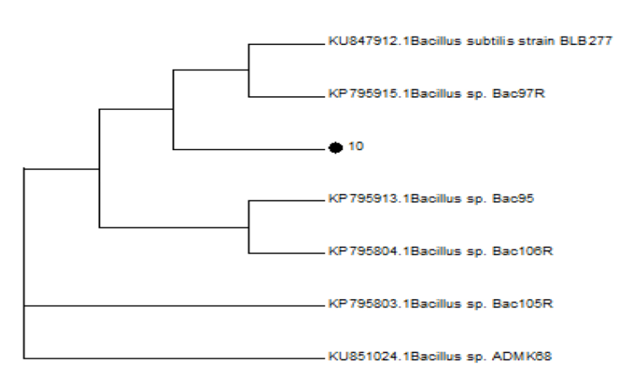 Fig. 4. Phylogram of Pseudomonas aeruginosa LC425424 (31)
 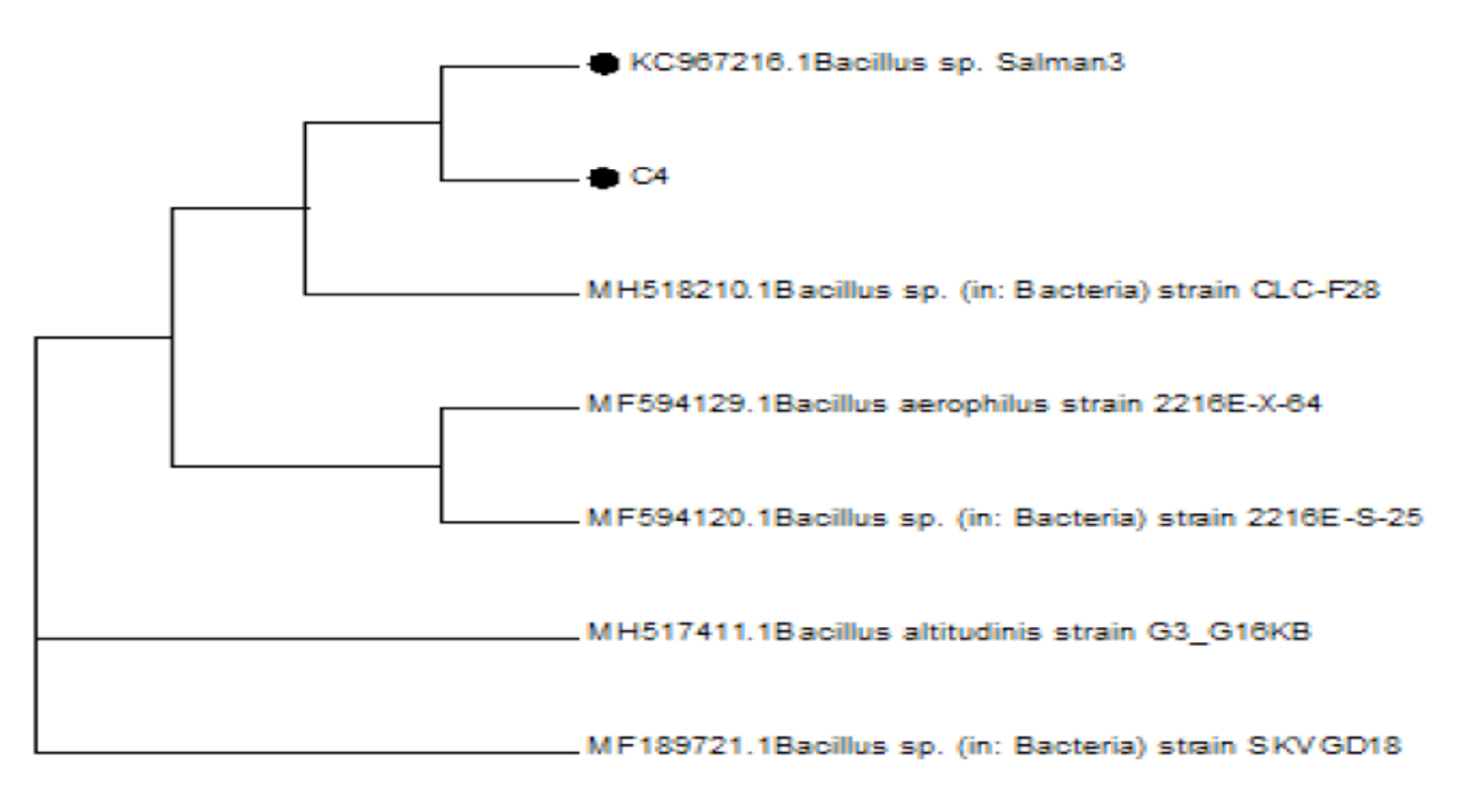 Fig. 5. Phylogram of Pseudomonas aeruginosa LC425425 (32)
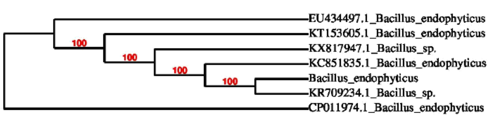 Fig. 6. Phylogram of Pseudomonas aeruginosa LC425424 (26)
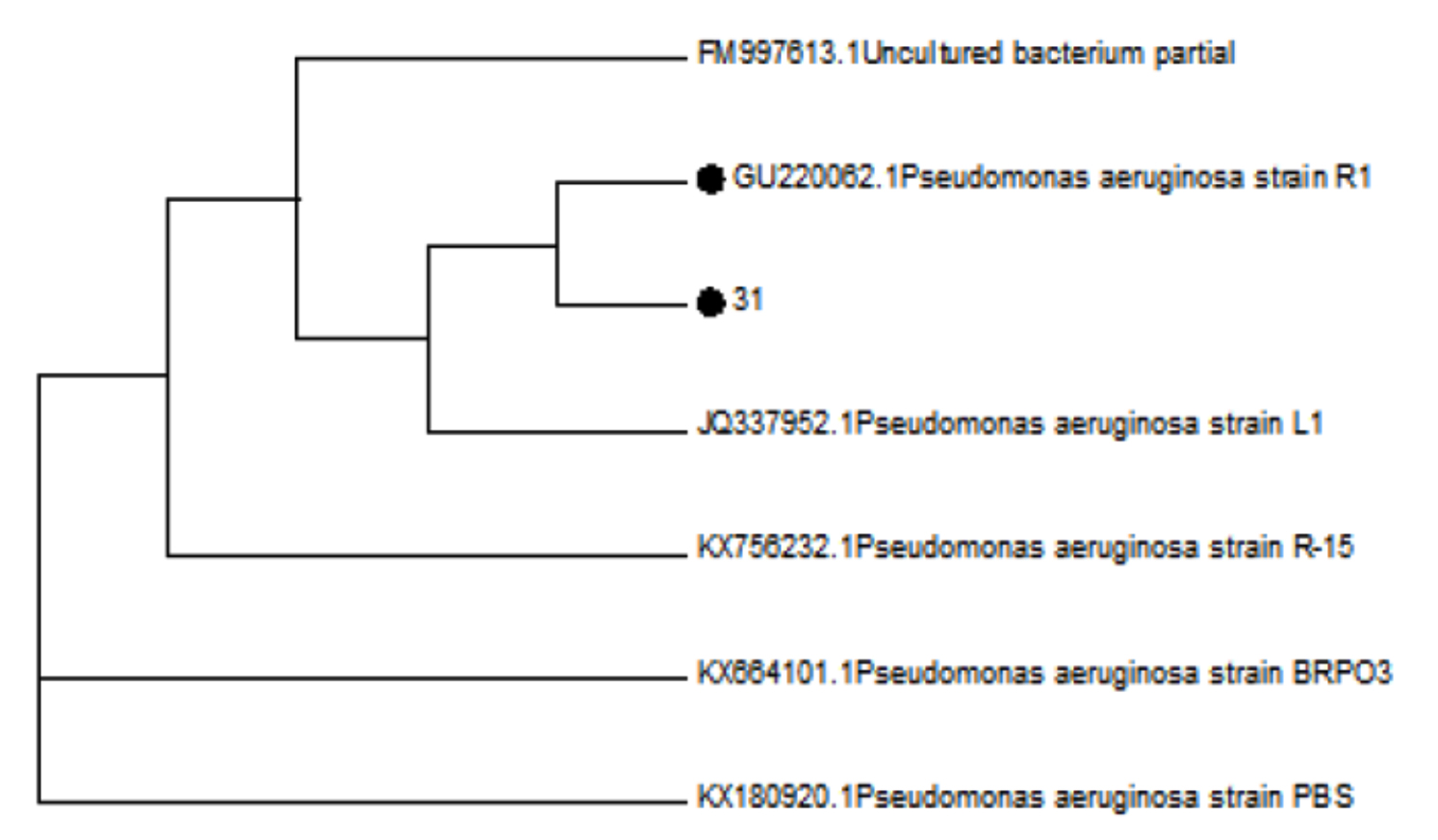 Fig. 7. Phylogram of Pseudomonas aeruginosa LC425424 (31)
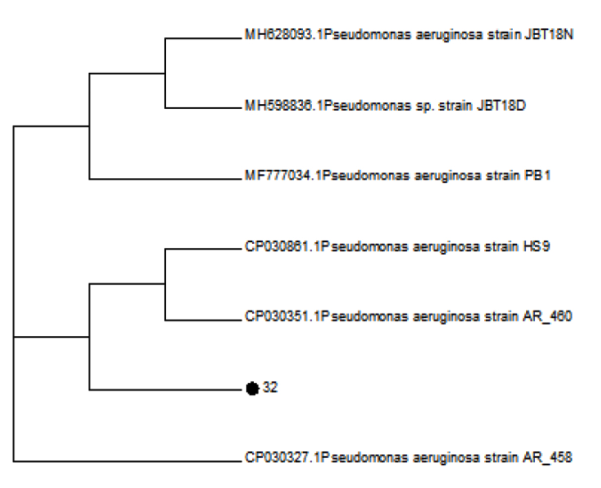 Fig. 8. Phylogram of Pseudomonas aeruginosa LC425425 (32)
 Fig. 9. Phylogram of Pseudomonas aeruginosa LC425424 (34)
Table (1):
Number of colonies producing L asparaginase enzyme in modified BHI and M9 media.
S.No |
Dilution factor |
Number of L asparaginase producing colonies on modified BHI media |
Number of L asparaginase producing colonies on modified M9 media |
|---|---|---|---|
1. |
10-5 |
61 |
75 |
2. |
10-6 |
20 |
35 |
3. |
10-7 |
19 |
26 |
4. |
10-8 |
24 |
12 |
5. |
10-9 |
18 |
08 |
6. |
10-10 |
17 |
07 |
Table (2):
Enzyme activity of the isolates producing L asparaginase (IU/ml).
S.No |
Sample ID |
Name of Bacterial species |
Accession numbers |
Enzyme activity(units/ml) |
|---|---|---|---|---|
1. |
G10 |
B. subtilis |
LC425423 |
8.5 |
2. |
C4 |
B .aerophilus |
LC425427 |
14.3 |
3. |
G1
|
B. endophyticus |
MG928501 |
7.1 |
4. |
31
|
Pseudomonas aeruginosa |
LC425424 |
8.2 |
5. |
32 |
Pseudomonas aeruginosa |
LC425425 |
19.4 |
6. |
34 |
Pseudomonas aeruginosa |
LC425426 |
19.1 |
Table (3):
Protein concentration of L asparaginase produced from Bacillus species and Pseudomonas aeruginosa.
S.No |
Name of Bacterial species |
Accession numbers |
Protein concentration of enzyme (µg/ml) |
|---|---|---|---|
1. |
B. subtilis |
LC425423 |
550 |
2. |
B .aerophilus |
LC425427 |
525 |
3. |
B. endophyticus |
MG928501 |
550 |
4. |
Pseudomonas aeruginosa |
LC425424 |
425 |
5. |
Pseudomonas aeruginosa |
LC425425 |
625 |
6. |
Pseudomonas aeruginosa |
LC425426 |
800 |
L- asparaginase remains pharmaceutically and industrially essential enzyme for cancer treatment and in food industry5. Continuous search of industrially important bacterial species is required to overcome the drawbacks of allergies and expensive production, therefore promising screening media’s are necessary to search for novel microorganisms producing L asparaginase enzyme. Modified M9 media is routinely used in screening and isolation of L asparaginase producers; For instance, 211 bacterial species isolated from aquatic and terrestrial areas was reported to be positive for qualitative assay when studied on modified M9-medium for the isolates from the region of Thane district, Maharashtra, India by Talluri et al. (2013)21. In the current study modified BHI media was used to isolate L-asparaginase producer’s; these isolates successfully yielded aspartate and ammonia from aspargine. The release of ammonia was detected by pH indicator in the BHI media as a pink zone around the colonies. In similar studies, BHI media was also successfully used to isolate Chrseobacterium formosense sp. nov. from the rhizosphere of Lactuca sativa L while Dietzia natronolimnaios sp. nov was also isolated on BHI medium when isolated from East African Soda lake22, 23. All these isolates can produce L- asparaginase successfully. In this study modified BHI media was successful in isolating 159 colonies producing L asparaginase while 163 colonies of L asparaginase producers were screened on modified M9 media.
In the present study the cultures isolated producing L asparaginase enzyme were both gram positive and gram negative organisms. The gram positive cultures producing L asparaginase with good enzyme activity were identified as B. subtilis (LC425423), B. aerophilus (LC425427), B. endophyticus (MG928501) respectively; demonstrating the enzyme activity of 8.5, 14.3 & 7.1 (IU/ml).Whereas the gram negative bacilli were identified as Pseudomonas aeruginosa; the accession numbers allotted for the strains were LC425424, LC425425,LC425426 respectively, the enzyme activity recorded in our study for the three strains of Pseudomonas aeruginosa was 8.2, 19.4 and 19.1(Unit/ml). In the present study it was noted that in comparison with Bacillus species Pseudomonas aeruginosa showed not only high enzyme activity but also high protein concentrations i.e 425, 625, 800 (µg/ml) while the three Bacillus species produced 550,525,550(µg/ml) of protein concentration. Previous studies on Bacillus and Pseudomonas aeruginosa also indicates that the asparaginase produced from Pseudomonas aeruginosa showed high enzyme activity than Bacillus species for example in a study reported by Vidya moorthy et al. (2010), Bacillus species DKMBT10 was reported to produce maximum yields of asparaginase (2.6mg/l) in presence of glucose and maltose. The activity of purified enzyme recorded in this study was 1.12 & 1.05 (units/mg)9. Similarly in a study by Biswa Prakash et al. (2013). demonstrated the production of L asparaginase enzyme (23.8 IU/mL ) from Bacillus subtilis hswx88 isolated from hotsprings24. In another study B. licheniformis (RAM-8) was used to optimize the L asparaginase production in which the maximum production was 32.26 IU/ml25. Similarly a recent study by Pallavi et al. (2015), have tested the effects of parameters like carbon nitrogen, pH and temperature on mangrove isolate of Bacillus subtilis –AVP 18; In this study the maximum yield of the enzyme was noted in presence of dextrose (177.45U/ml), tryptone (84.1 IU/ml), pH 7 & 9 (25.36 IU/ml) and temperature at 37 °C (24.26 IU/ml),This study also showed that after 72 hrs of incubation at 37 °C the enzyme produced was (1.69 IU/ml)13. In our study the isolated Bacillus subtilis (LC425427) produced activity of 8.5 IU/ml, B. aerophilus (LC425427) showed 14.3 (IU/ml), B. endophyticus (MG928501) showed 7.1 IU/ml of enzyme at 37 °C after incubation of 48hrs. Therefore the lower yields of enzyme produced from Bacillus species is in correlation with the enzyme yields reported by other studies on Bacillus species. On the other hand the three strains of Pseudomonas aeruginosa showed an activity of 8.2, 19.4 and 19.1(Units/ml). Likewise a number of studies have reported L asparaginase production from Pseudomonas aeruginosa for example in a study by Ashraf A et al. (2004), reported that Pseudomonas aeruginosa 50071 was used to produce L asparaginase through solid state fermentation; the enzyme produced showed a specific activity of 1900IU/mg16. While a similar study on production optimization in which Pseudomonas aeruginosa 50071 was used for enzyme production in this study the highest enzyme activity of (142.8 IU) was noted when the media was enriched by adding corn steep liquor (3.68%), casein hydrolysate (3.11%) at pH 7.926. The enzyme activity in our study is in similar range to a recent study in which the enzyme activity of asparaginase produced from seven strains of Pseudomonas alcaligenes strains isolated from hot springs is ranging from 20 -240units/ml8. None the less most of the studies have shown optimized production of L asparaginase by using activators, enriched ingredients in media, altering parameters like pH, temperature, fermentation methods etc. Therefore if the isolate is gram positive or gram negative the production can be optimized by altering the production parameters; however type –II asparaginase produced in periplasmic space is of therapeutic importance27. Therefore larger periplasmic space in gram negative bacterial species could be a possible reason for higher yields and activity of L asparaginase enzyme as shown in present and previous studies28. In our study all the three gram positive cultures of B. subtilis (LC425423), B. aerophilus (LC425427), B. endophyticus (MG928501) showed less enzyme activity than three strains of Pseudomonas aeruginosa under same fermentation conditions; here with all the cultures in the study was obtained from soil. Although the enzyme produced in our study showed less activity in comparison with other studies, but the enzyme activity can further be increased by incorporating glucose and metal ions like Cobalt and thiol protecting reagents8, 29-31. In addition the enzyme was produced and its activity was studied at pH 8; which indicates that the produced enzyme can be used anticancerous therapies; as enzyme activity below pH 8 is considered to be therapeutically not effective in tumor patients6. Additionally this study have also reported two new Bacillus species producing L asparaginase i.e B. aerophilus (LC425427), B. endophyticus (MG928501).
This study demonstrates that Gram negative species like Pseudomonas aeruginosa appears to produce good enzyme yield and activity than gram positive Bacillus species under same fermentation conditions .This could be attributed to large periplasmic space in gram negative organisms; however further studies are required inorder to support the finding. Furthermore modified Brain heart infusion media could also provide an alternate screening media besides modified M9 media for isolation of L asparaginase producer.
Acknowledgement
The authors express sincere appreciation to Applied Medical Sciences College, Jazan University, KSA.
Conflicts Of Interest
The authors declares that there is no conflict of interest.
Authors’ Contribution
All authors listed have made a substantial, direct and intellectual contribution to the work, and approved it for publication.
Funding
None.
Data Availability
All datasets generated or analyzed during this study are included in the manuscript.
Ethics Statement
This article does not contain any studies with human participants or animals performed by any of the authors.
- Pui C-H. Acute lymphoblastic leukemia: Springer; 2011.
Crossref - Hunger SP, Mullighan CGJNEJoM. Acute lymphoblastic leukemia in children, 2015; 373(16):1541-52.
Crossref - Egler RA, Ahuja SP, Matloub YJJop, pharmaco-therapeutics. L-asparaginase in the treatment of patients with acute lymphoblastic leukemia. 2016; 7(2):62.
Crossref - Appel IM, Hop WC, Pieters R. Changes in hypercoagulability by asparaginase: a randomized study between two asparaginases. Blood coagulation & fibrinolysis. 2006; 17(2):139-46.
Crossref - Jha SK, Pasrija D, Sinha RK, Singh HR, Nigam VK, Vidyarthi ASJIJoPS, et al. Microbial L-asparaginase: a review on current scenario and future prospects. 2012; 3(9):3076.
- Yadav S, Verma SK, Singh J, Kumar A. Industrial production and clinical application of L-asparaginase: a chemotherapeutic agent. stroke. 2014; 76: 41.
- Kamble K, Bidwe P, Muley V, Kamble L, Bhadange D, Musaddiq MJBd. Characterization of l-asparaginase producing bacteria from water, farm and saline soil. 2012; 3(1):116-9.
Crossref - Badoei-Dalfard A. L-asparaginase production in the pseudomonas pseudoalcaligenes strain JHS-71 isolated from Jooshan Hot-spring. Molecular biology research communications. 2016; 5(1):1.
- Moorthy V, Ramalingam A, Sumantha A, Shankaranaya RT. Production, purification and characterisation of extracellular L-asparaginase from a soil isolate of Bacillus sp. African Journal of Microbiology Research. 2010; 4(18): 1862-7.
- Gulati R, Saxena R, Gupta R. A rapid plate assay for screening l asparaginase producing micro organisms. Letters in applied microbiology, 1997; 24(1):23-6.
Crossref - El-Naggar NE-A, Moawad H, El-Shweihy NM, El-Ewasy SM. Optimization of culture conditions for production of the anti-leukemic glutaminase free L-asparaginase by newly isolated Streptomyces olivaceus NEAE-119 using response surface methodology. BioMed research international. 2015; 2015.
Crossref - Trilokchandran. <media-optimization-and-growth-parameters-for-the-production-of-l asparaginase-enzyme-from-bacterial-isolate.pdf>. Journal of Chemical and Pharmaceutical Research, 2016; 8(8):222-8.
- Rudrapati P, Audipudi AVJIJPSRR. Characterization and bioprocessing of oncolytic enzyme–L-asparaginase isolated from marine Bacillus AVP 14. 2015; 30(2):195-201.
- Scopes RK. Protein purification: principles and practice: Springer Science & Business Media; 2013.
- Amena S, Vishalakshi N, Prabhakar M, Dayanand A, Lingappa K. Production, purification and characterization of L-asparaginase from Streptomyces gulbargensis. Brazilian journal of Microbiology. 2010; 41(1):173-8.
Crossref - El-Bessoumy AA, Sarhan M, Mansour J. Production, isolation, and purification of L-asparaginase from Pseudomonas aeruginosa 50071 using solid-state fermentation. BMB Reports. 2004; 37(4):387-93.
Crossref - Lowry O, Rosebrough N, Farr A, Randall R. Protein estimation by Lowry’s method. J Biol Chem. 1951; 193: 265.
- Wriston JC, Yellin TO. L-asparaginase: a review. Adv Enzymol Relat Areas Mol Biol. 1973; 39:185-248.
Crossref - Talavera G, Castresana J. Improvement of phylogenies after removing divergent and ambiguously aligned blocks from protein sequence alignments. Systematic biology. 2007; 56(4):564-77.
Crossref - Dereeper A, Guignon V, Blanc G, Audic S, Buffet S, Chevenet F, et al. Phylogeny. fr: robust phylogenetic analysis for the non-specialist. Nucleic acids research. 2008; 36(suppl_2):W465-W9.
Crossref - Talluri P, Bhavana M, Rajagopa SJIJoP, Chemical, Sciences B. Isolation And Screening of L-asparaginase Producing Bacteria From Visakhapatnam Soil Samples. 2013; 3(4).
- Young C-C, Kהmpfer P, Shen F-T, Lai W-A, Arun AJIJoS, Microbiology E. Chryseobacterium formosense sp. nov., isolated from the rhizosphere of Lactuca sativa L. (garden lettuce). 2005; 55(1):423-6.
Crossref - Duckworth AW, Grant S, Grant WD, Jones BE, Meijer DJE. Dietzia natronolimnaios sp. nov., a new member of the genus Dietzia isolated from an East African soda lake. 1998; 2(3):359-66.
Crossref - Pradhan B, Dash SK, Sahoo S. Screening and characterization of extracelluar L-asparaginase producing Bacillus subtilis strain hswx88, isolated from Taptapani hotspring of Odisha, India. Asian Pacific journal of tropical biomedicine. 2013; 3(12):936-41.
Crossref - Mahajan RV, Saran S, Kameswaran K, Kumar V, Saxena R. Efficient production of L-asparaginase from Bacillus licheniformis with low-glutaminase activity: optimization, scale up and acrylamide degradation studies. Bioresource technology. 2012; 125:11-6.
Crossref - Abdel-Fattah YR, Olama ZAJPB. L-asparaginase production by Pseudomonas aeruginosa in solid-state culture: evaluation and optimization of culture conditions using factorial designs. 2002; 38(1):115-22.
Crossref - Han S, Jung J, Park W. Biochemical characterization of L-asparaginase in NaCl-tolerant Staphylococcus sp. OJ82 isolated from fermented seafood. J Microbiol Biotechnol. 2014; 24(8):1096-104.
Crossref - Beveridge TJJJob. Structures of gram-negative cell walls and their derived membrane vesicles. 1999; 181(16):4725-33.
- Trilokchandran. media-optimization-and-growth-parameters-for-the-production-of-lasparaginase-enzyme-from-bacterial-isolate.pdf>. Journal of Chemical and Pharmaceutical Research. 2016; 8:223-8.
- Manna S, Sinha A, Sadhukhan R, Chakrabarty S. Purification, characterization and antitumor activity of L-asparaginase isolated from Pseudomonas stutzeri MB-405. Current Microbiology. 1995; 30(5):291-8.
Crossref - Cachumba JJM, Antunes FAF, Peres GFD, Brumano LP, Dos Santos JC, Da Silva SSJbjom. Current applications and different approaches for microbial L-asparaginase production. Braz. J. Microbiol. 2016; 47:77-85.
Crossref
© The Author(s) 2019. Open Access. This article is distributed under the terms of the Creative Commons Attribution 4.0 International License which permits unrestricted use, sharing, distribution, and reproduction in any medium, provided you give appropriate credit to the original author(s) and the source, provide a link to the Creative Commons license, and indicate if changes were made.